-
 Slide 9-1
Feb 25 2019 by Lancaster Course in Ophthalmology
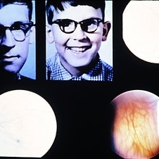
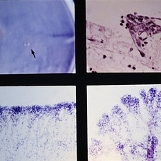
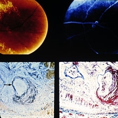
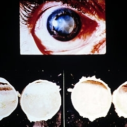
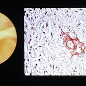
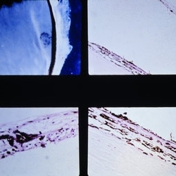
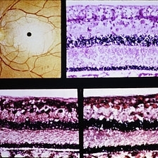
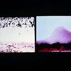
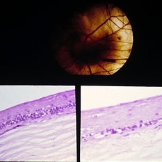
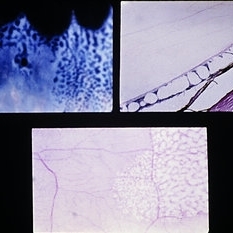
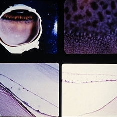
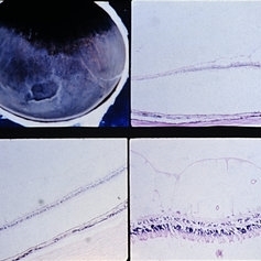
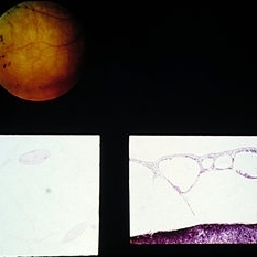
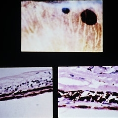
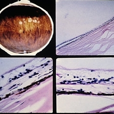
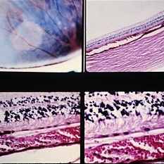
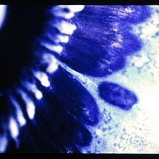
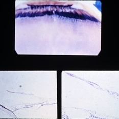
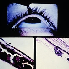
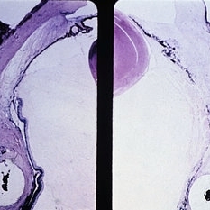
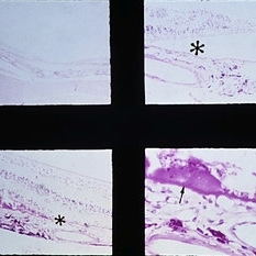
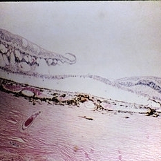
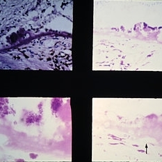
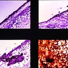
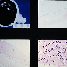
X-linked ocular albinism. The two external views illustrate a normal degree of skin and hair coloring. The upper right view shows a slightly pigmented macular area with no foveal reflex or vascular sparing of the foveola. The lower views illustrate marked absence of pigment, and the choroidal vessels are clearly visible.
Condition/keywords: foveola, large choroidal vessels, ocular albinism, pigment
-
 Slide 9-2
Feb 25 2019 by Lancaster Course in Ophthalmology
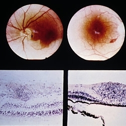
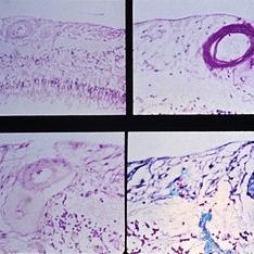
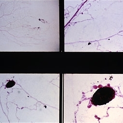
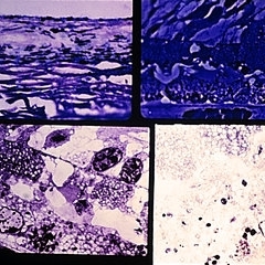
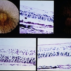
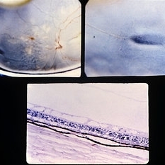
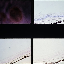
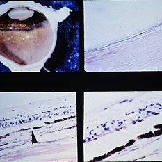
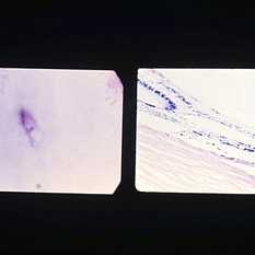
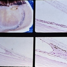
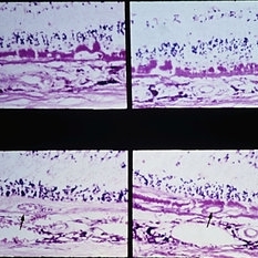
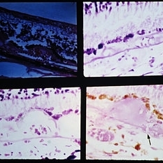
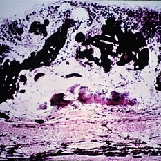
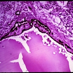
X-linked ocular albinism. Iris pigment epithelium (upper) and retinal pigment epithelium (lower) showing giant pigment granules (arrows) in the eye of an affected male.
Condition/keywords: ocular albinism, pigment epithelium, retinal pigment epithelium
-
 Slide 9-3
Feb 25 2019 by Lancaster Course in Ophthalmology
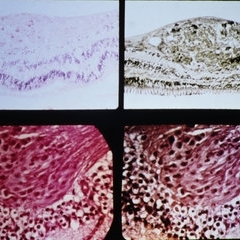
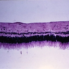
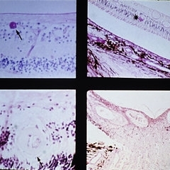
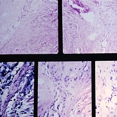
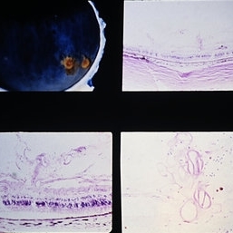
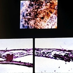
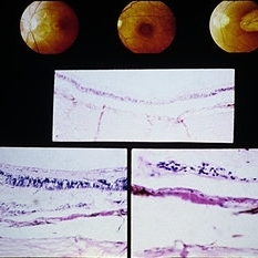
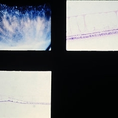
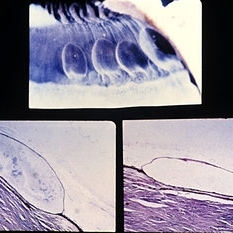
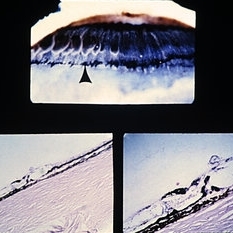
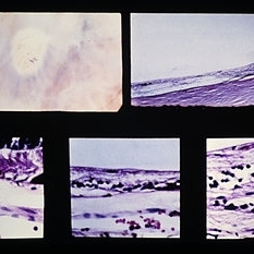
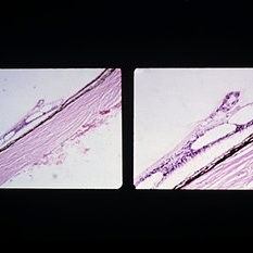
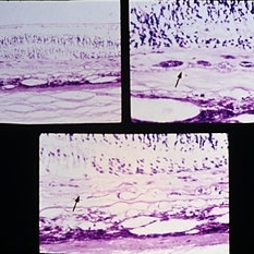
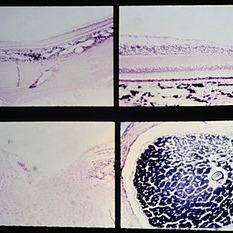
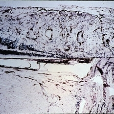
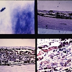
Tyrosinase-positive oculocutaneous albinism. Macular area is shown with normal-appearing retinal pigment epithelium (between asterisks) except for the lack of pigment (upper view). At very high power, fine melanin pigment granules may be seen (arrow, lower view).
Condition/keywords: melanin granules, ocular albinism, retinal pigment epithelium
-
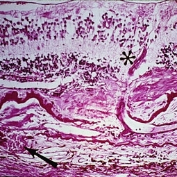
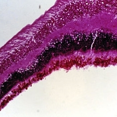
 Slide 9-4
Feb 25 2019 by Lancaster Course in Ophthalmology
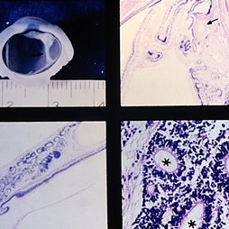
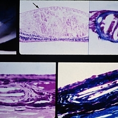
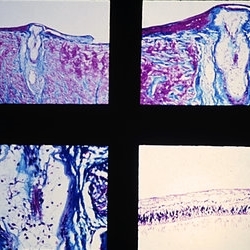
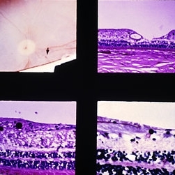
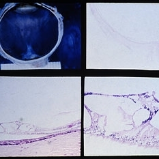
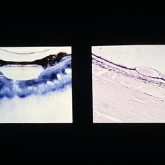
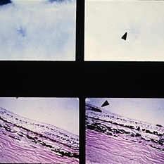
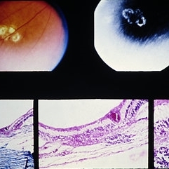
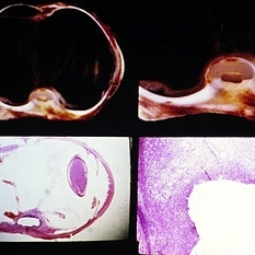
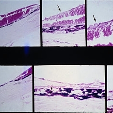
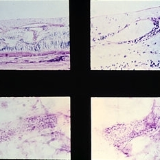
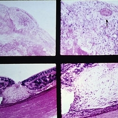
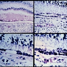
Retinal dysplasia in the Petau syndrome. The microphthalmic eye has a dysplasic retina behind the lens, extending as a strand (arrow) to attach to the retina posteriorly (views on left). The rosettes (asterisks) have a well-developed external limiting membrane (arrow) and photoreceptor outer and inner segments (lower right). There is also a hyperplasic primary vitreous (upper right) with a small nodule of cartilage (arrow).
Condition/keywords: dysplasia, microphthalmos, Patau syndrome, vitreous
-
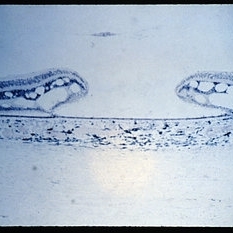
 Slide 9-5
Feb 25 2019 by Lancaster Course in Ophthalmology
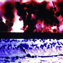
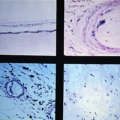
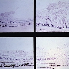
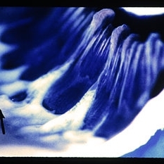
Retrolental fibroplasia. In this acute case, postmortem examination disclosed small areas of retinal neovascularization (arrows) extending into the vitreous (upper left and right). Trypsin digestion of the same case disclosed a line of intense endothelial-cell proliferation at the junction of vascularized and nonvascularized retina (lower left). In some areas these proliferative changes occurred in an intraretinal sea fan-like configuration (lower right).
Condition/keywords: endothelial, retinal neovascularization, retrolental fibroplasia, trypsin digestion
-
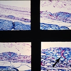
 Slide 9-6
Feb 25 2019 by Lancaster Course in Ophthalmology
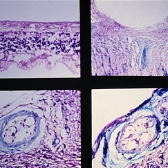
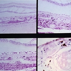
Coats' disease. An exudative detachment of the retina is shown with typical yellowish material and associated vascular abnormalities and some retinal hemorrhage (upper left). Fluorescein angiography shows capillary drop-out of the macroaneurysms and dilated telangiectatic vessels. Venous saccular dilatations also may be present (upper right). With trypsin digestion the background capillaries are acellular, and large, thin-walled telangiectatic vessels (lower left) and microaneurysms are present (lower right).
Condition/keywords: Coats' disease, macroaneurysm, telangiectatic vessels, trypsin digestion
-
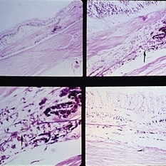
 Slide 9-7
Feb 25 2019 by Lancaster Course in Ophthalmology
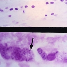
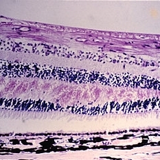
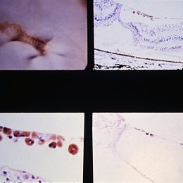
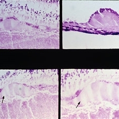
Inner retinal ischemia of central retinal artery occlusion. Early changes with edema and pyknosis of the nuclei of ganglion cells and inner aspect of the inner nuclear layer (left). With time, all of the inner retinal layers supplied by the central retinal artery disappear, including the nerve fiber, the ganglion cell, the inner plexiform layers, and the inner aspect of the inner nuclear layer (right).
Condition/keywords: edema, ganglion cell, pyknosis, retinal ischemia
-
 Slide 9-8
Feb 25 2019 by Lancaster Course in Ophthalmology
Microinfarctions of the nerve fiber layer with cytoid bodies as seen in diabetic retinopathy (above) and hypertension (below). In each there are fusiform areas of swelling of the axons with the accumulation of material forming a pseudonucleus (cytoid body).
Condition/keywords: cystoid, diabetic retinopathy, pseudonucleus
-
 Slide 9-9
Feb 25 2019 by Lancaster Course in Ophthalmology
Localized area of inner ischemic infarction of the nerve fiber layer, presumably the site of a previous cotton-wool exudate. There is loss of the ganglion cell layer and thinning of the inner nuclear layer (between arrows).
Condition/keywords: ganglion cell, inner ischemic infarction
-
 Slide 9-10
Feb 25 2019 by Lancaster Course in Ophthalmology
Deep retinal exudate. There is accumulation of lipid-laden histiocytes in the outer plexiform and inner nuclear layers of the retina in the posterior pole area in diabetic retinopathy.
Condition/keywords: diabetic retinopathy, histiocytes, retinal exudates
-
 Slide 9-12
Feb 25 2019 by Lancaster Course in Ophthalmology
Retinal edema. The retina is thickened by a lightly staining proteinaceous material that accumulates principally in the inner nuclear and outer plexiform layers.
Condition/keywords: retinal edema
-
 Slide 9-13
Feb 26 2019 by Lancaster Course in Ophthalmology
Superficial retinal hemorrhages. These hemorrhages have a flame-shaped appearance and are located just beneath the internal limiting membrane of the retina in a 61-year-old man with severe anemia and thrombocytopenia.
Condition/keywords: retinal hemorrhage, thrombocytopenia
-
 Slide 9-14
Feb 26 2019 by Lancaster Course in Ophthalmology
Deep retinal hemorrhage. A deep retinal hemorrhage is located in the outer plexiform layer.
Condition/keywords: hemorrhage
-
 Slide 9-15
Feb 26 2019 by Lancaster Course in Ophthalmology
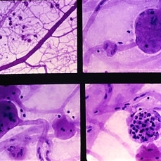
Trypsin digest retinal preparation showing diabetic microaneurysms. Some microaneurysms are relatively acellular (upper right and lower left), and others are filled with neutrophils (lower right).
Condition/keywords: microaneurysms, neutrophils, trypsin digestion
-
 Slide 9-16
Feb 26 2019 by Lancaster Course in Ophthalmology
Retinal neovascularization with extension into the vitreous cavity in diabetic retinopathy. There is direct continuity of a retinal vein with the new vessels in the vitreous cavity (arrow).
Condition/keywords: retinal neovascularization, vitreous cavity
-
 Slide 9-17
Feb 26 2019 by Lancaster Course in Ophthalmology
Retinal and choroidal arteriosclerosis. The arterial walls are slightly thickened and in some areas have a laminated "onionskin" appearance. Specimen is from a 45-year-old urban, but originally rural, black man.
Condition/keywords: choroidal arteriosclerosis, retinal arteriosclerosis
-
 Slide 9-18
Feb 26 2019 by Lancaster Course in Ophthalmology
Malignant hypertension with retinal arterioles that are thickened and have fibrinoid necrosis (arrows). Retinal exudates (asterisk) and papilledema are also present. Papilledema is evidenced by fullness of the optic nerve head and peripapillary crowding of the retina (lower right).
Condition/keywords: fibrinoid, malignant hypertension, papilledema, retinal arteriole, retinal exudates
-
 Slide 9-19
Feb 26 2019 by Lancaster Course in Ophthalmology
Retinal arterial macroaneurysm. A ring of retinal exudate partially surrounds the macroaneurysm (upper left), which is more clearly delineated by fluorescein (upper right). The retinal arteriole is greatly dilated, and the stain for elastic tissue shows a localized area of disruption and loss of the internal elastic membrane (arrow). The surrounding retina is thickened by edema and some hemorrhage. The ectatic area of the vessel wall is greatly thickened by the accumulation of a laminated fibrinous material. (Courtesy of Alan Friedman, M.D.)
Condition/keywords: retinal arterial macroaneurysm, retinal exudates
-
 Slide 9-20
Feb 26 2019 by Lancaster Course in Ophthalmology
Cholesterol emboli to retina and choroid. There is a large microinfarction of the nerve fiber layer (arrows). A cholesterol embolus is lodged in a retinal arteriole (upper right) proximal to the microinfarction. Cholesterol emboli were also found in the choroid, and in one (lower right) erythrocytes (arrow) could be seen in the periphery as they were presumably going around the embolus.
Condition/keywords: choroid, emboli, embolus, erythrocytes
-
 Slide 9-21
Feb 26 2019 by Lancaster Course in Ophthalmology
Septic emboli. The fundus photographs illustrate a man with subacute bacterial endocarditis who presented with branch retinal artery occlusion in the left eye and a single Roth spot in the right eye. The lower views show Roth spots from two different cases. There is an infiltration of neutrophils in the center and extravasated blood in the periphery. (Courtesy of P. Robb McDonald, M.D.)
Condition/keywords: emboli, neutrophils
-
 Slide 9-22
Feb 26 2019 by Lancaster Course in Ophthalmology
Atrial myxoma emboli to eye. The upper left view shows outer ischemic retinal atrophy due to emboli in the choroidal arterioles (upper right and lower left). The atrial myxoma is shown in the lower right views. (Courtesy of T. Smith, M.D.)
Condition/keywords: choroidal arterioles, emboli
-
 Slide 9-23
Feb 26 2019 by Lancaster Course in Ophthalmology
Choroidal infarct (Elschnig spot). There is a localized area of loss of retinal pigment epithelium and outer ischemic retinal atrophy with loss of the photoreceptor cell and outer plexiform layers, and partial loss of the inner nuclear layer without any reparative changes.
Condition/keywords: choroidal infarction, Elschnig's spots
-
 Slide 9-24
Feb 26 2019 by Lancaster Course in Ophthalmology
Atheromatous occlusion of central retinal artery at the level of, and anterior and posterior to, the lamina cribrosa. There is inner ischemic retinal atrophy (lower right) with loss of all layers down to and involving the inner portion of the inner nuclear layer.
Condition/keywords: atherosclerosis, lamina cribrosa
-
 Slide 9-25
Feb 26 2019 by Lancaster Course in Ophthalmology
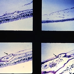
Recanalized thrombus of a branch of the central artery of the retina displaying inner ischemic atrophy (upper left).
Condition/keywords: ischemic atrophy, recanalized thrombus
-
 Slide 9-26
Feb 26 2019 by Lancaster Course in Ophthalmology
Hypotensive retinopathy. This patient presented with a slightly red eye and , aqueous cells, and flare. He developed areas of iris atrophy and cataract. Postmortem examination showed extensive cobblestone (paving-stone) degeneration which extended posteriorly to the equator in the right eye (lower left views). No cobblestone degeneration was present in the right eye (lower right views).
Condition/keywords: cataract, hypertensive retinopathy, iris, keratic precipitates
-
 Slide 9-27
Feb 26 2019 by Lancaster Course in Ophthalmology
Branch retinal vein occlusion. The vein is occluded as it passes under the sclerotic arteriole.
Condition/keywords: branch retinal vein occlusion (BRVO), sclerotic arteriole
-
 Slide 9-28
Feb 26 2019 by Lancaster Course in Ophthalmology
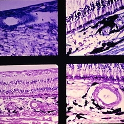
Central retinal vein occlusion. There is a recanalized thrombus in the central retinal vein at the level of the posterior aspect of the lamina cribrosa. Prominent endothelial cell proliferation is present at the proximal area of the thrombus.
Condition/keywords: central retinal vein occlusion (CRVO), endothelial, lamina cribrosa
-
 Slide 9-29
Feb 26 2019 by Lancaster Course in Ophthalmology
Preretinal glial membrane associated with retinal pits (arrow). The glial cells have continuity with the retina through a discontinuity in the internal limiting membrane.
Condition/keywords: epiretinal membrane (ERM), glial cells
-
 Slide 9-30
Feb 26 2019 by Lancaster Course in Ophthalmology
Massive gliosis of the retina (clinicopathologic correlation). Lesion consists of uniform spindle-shaped cells, abundant fibrillar material, and sclerotic blood vessels.
Condition/keywords: clinicopathologic correlation, gliosis
-
 Slide 9-31
Feb 26 2019 by Lancaster Course in Ophthalmology
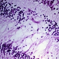
Sarcoidosis with retinal and vitreous involvement. A Daten-Fuchs-like nodule of sarcoid granuloma is present (lower right).
Condition/keywords: Dalen-Fuchs nodules, sarcoid granuloma, sarcoidosis
-
 Slide 9-32
Feb 26 2019 by Lancaster Course in Ophthalmology
Gross and microscopic appearance of leukemic infiltration in the retina in a moundlike configuration. Retinal hemorrhages are present, and leukemic infiltration of the choroid is visible as well.
Condition/keywords: choroid, hemorrhage, leukemic infiltration
-
 Slide 9-33
Feb 26 2019 by Lancaster Course in Ophthalmology
Disseminated intravascular coagulopathy. There is occlusion of the choriocapillaris and some larger choroidal vessels, partial retinal pigment epithelial atrophy, serous retinal detachment, and cystic retinal edema, all in the macular area.
Condition/keywords: choriocapillaris, choroidal vessels, disseminated intravascular coagulopathy, retinal edema, retinal pigment epithelium atrophy, serous retinal detachment
-
 Slide 9-34
Feb 26 2019 by Lancaster Course in Ophthalmology
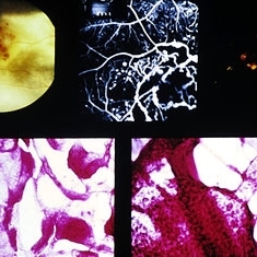
Sickle-cell retinopathy with iridescent spot in the macula. This lesion has an orange-yellow appearance when seen grossly (upper left). Histopathologically, there is an acquired schisis cavity in which hemosiderin-laden macrophages are present. Special staining shows iron deposition in the tissues of the wall of this schisis cavity and in the macrophages within the cavity (lower right).
Condition/keywords: hemosiderin-laden macrophages, retinoschisis, sickle cell
-
 Slide 9-35
Feb 26 2019 by Lancaster Course in Ophthalmology
"Black sunburst" sign of sickle-cell retinopathy. These are localized lesions characterized by hypertrophy, hyperplasia, and migration of the retinal pigment epithelium into the retina in a perivascular location. This latter has given the lesion its spiculate ophthalmoscopic and gross appearance.
Condition/keywords: hyperplasia, hypertrophy, sickle cell, spiculate ophthalmoscopic
-
 Slide 9-36
Feb 26 2019 by Lancaster Course in Ophthalmology
Retinal trypsin digestion preparation of sickle-cell retinopathy. There is abrupt arteriolar obstruction, and at this point arteriolar-venular looping with beading of the vessels occurs.
Condition/keywords: sickle cell, trypsin digestion
-
 Slide 9-37
Feb 26 2019 by Lancaster Course in Ophthalmology
"Sea fans" of sickle-cell retinopathy. At the junction of perfused and nonperfused retina, localized neovascularization erupts into the vitreous cavity, producing the sea-fan lesions.
Condition/keywords: neovascularization (NV), sickle cell
-
 Slide 9-38
Feb 26 2019 by Lancaster Course in Ophthalmology
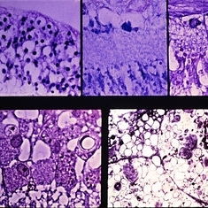
Type III systemic mucopolysaccharidosis (San Filippo syndrome). The retinal ganglion cells are distended (upper left) and contain mucopolysaccharide (middle, top: colloidal iron), lipid (upper right: Sudan-black B). Phase-contrast (lower left) shows numerous vacuoles, and by electron microscopy (lower right) there are numerous inclusions of the fibrillogranular and multimembranous types.
Condition/keywords: mucopolysaccharidoses, Sanfilippo syndrome, Type III systemic mucopolysaccharidosis
-
 Slide 9-39
Feb 26 2019 by Lancaster Course in Ophthalmology
Type Ill systemic mucopolysaccharidosis (San Filippo syndrome). The retinal pigment epithelium contains excessive mucopolysaccharide (upper left: colloidal iron), which corresponds to vacuoles seen by phase-contrast(upper right) and to vacuoles with a fibrillogranular material as seen by electron microscopy (lower views). There is also degeneration of the outer segments of the photoreceptor cells and thinning of the outer nuclear layer of the retina (upper right and lower left}.
Condition/keywords: retinal pigment epithelium, Sanfilippo syndrome, Type Ill systemic mucopolysaccharidosis
-
 Slide 9-40
Feb 26 2019 by Lancaster Course in Ophthalmology
Tay-Sachs disease. Cherry-red spot (upper left). Retinal ganglion cells are distended by material which stains positively with the periodic acid-Schiff reaction (upper right) and with stains for lipid (Nile blue sulfate, lower left) (oil red-O, lower right).
Condition/keywords: ganglion cell, Tay-Sachs
-
 Slide 9-41
Feb 26 2019 by Lancaster Course in Ophthalmology
Traumatic retinopathy with retinal pigment epithelial hypertrophy, hyperplasia, and migration into the retina in a perivascular location, giving a spiculate appearance which is similar to that seen in retinitis pigmentosa. This eye had additional features of trauma.
Condition/keywords: retinal pigment epithelium (RPE) hypertrophy, retinitis pigmentosa, retinopathy
-
 Slide 9-42
Feb 26 2019 by Lancaster Course in Ophthalmology
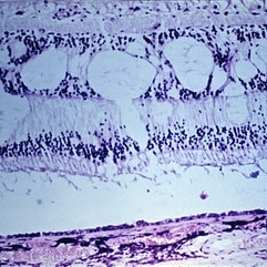
Bassen-Kornzweig syndrome. There is thinning of the outer nuclear layer in the parafoveal area (upper view). In the midperiphery of the retina there is more marked degeneration of the photoreceptor cells, hyperplasia, and migration of the retinal pigment epithelium into the retina in a perivascular location.
Condition/keywords: Bassen-Kornzweig syndrome, hyperplasia, photoreceptor cell, retinal pigment epithelium
-
 Slide 9-43
Feb 26 2019 by Lancaster Course in Ophthalmology
Fundus flavimaculatus. Accumulation of acid mucopolysaccharide along the inner aspect of the retinal pigment epithelium. (Courtesy of B. Klien, M.D.)
Condition/keywords: acid mucopolysaccharide, fundus flavimaculatus, retinal pigment epithelium
-
 Slide 9-44
Feb 26 2019 by Lancaster Course in Ophthalmology
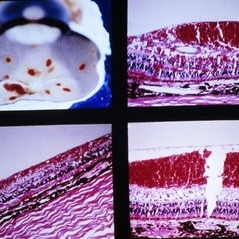
Central areolar choroidal sclerosis. Macular lesions in three generations of the same family. (Upper three views courtesy of R. E. Carr, M.D. ). Histopathologic study shows loss of retinal pigment epithelium and photoreceptor cells but with a preservation of choriocapillaris in some areas. (Middle and lower views courtesy of Andrew Ferry, M.D.)
Condition/keywords: central areolar choroidal dystrophy (CACD), photoreceptor cell, retinal pigment epithelium
-
 Slide 9-45
Feb 26 2019 by Lancaster Course in Ophthalmology
Diffuse choroidal atrophy of right (upper left) and left (upper right) eyes of 54-year-old man. There are rare areas where the and photoreceptor cells and the inner nuclear layer rest against choroid, which is markedly attenuated (lower views).
Condition/keywords: choroidal atrophy, photoreceptor cell, retinal pigment epithelium
-
 Slide 9-46
Feb 26 2019 by Lancaster Course in Ophthalmology
Choroideremia. There is total loss of the choroid and retinal pigment epithelium except for a small area around the optic disc. Sections through the degenerated area show absence of the choroid and retinal pigment epithelium, and the inner nuclear layer of the retina is in juxtaposition to the sclera (lower view). (Courtesy of Clement McCulloch, M.D. )
Condition/keywords: choroideremia, retinal pigment epithelium, sclera
-
 Slide 9-47
Feb 26 2019 by Lancaster Course in Ophthalmology
Typical peripheral cystoid degeneration. A small, round hole within the area of cystoid degeneration is present (upper left). In section the cysts are located mostly in the outer plexiform layer (upper left). A nondigestion flat preparation of the retina shows the cystic spaces, which have coalesced to form meridionally oriented tunnels (lower view).
Condition/keywords: peripheral cystoid degeneration
-
 Slide 9-48
Feb 26 2019 by Lancaster Course in Ophthalmology
Typical peripheral cystoid degeneration (PCD) just posterior to the ora serrata, and reticular peripheral cystoid degeneration located posterior to the typical PCD. Section of reticular peripheral cystoid degeneration showing cystic spaces in the nerve fiber layer (upper right). Lower view shows junction of typical PCD (left} with reticular PCD (right). (Courtesy of Robert Y Foos, M.D. )
Condition/keywords: peripheral cystoid degeneration
-
 Slide 9-49
Feb 26 2019 by Lancaster Course in Ophthalmology
Radial paravascular rarefaction. This eye with moderate background diabetic retinopathy shows an area of retinal thinning along a major retinal vessel in the midperiphery nasally. This oval area has a reticular appearance (upper views). Histologic section through the area shows the cystic spaces within the nerve fiber layer and an intact overlying vitreous (lower view).
Condition/keywords: perivasculitis
-
 Slide 9-50
Feb 26 2019 by Lancaster Course in Ophthalmology
Typical degenerative retinoschisis. The gross appearance of the lesion (upper views) is due primarily to the thicker outer layer which has a beaten-metal appearance. Typical PCD surrounds the area of schisis. Histologic sections (lower views) show splitting in the outer plexiform layer and the irregularity of the thicker outer layer.
Condition/keywords: retinoschisis
-
 Slide 9-51
Feb 26 2019 by Lancaster Course in Ophthalmology
Reticular degenerative retinoschisis. In this case the process extends posteriorly from the equatorial area to the midperiphery. The gross view (upper left) also shows a band of dark pigmentation due to peripheral retinal pigment epithelial (RPE) hypertrophy, a few areas of paving-stone degeneration within the area of RPE hypertrophy, and peripheral retinal thinning with focal areas of RPE hyperplasia and migration into the retina.
Condition/keywords: hyperplasia, hypertrophy, retinal pigment epithelium, retinoschisis
-
 Slide 9-52
Feb 26 2019 by Lancaster Course in Ophthalmology
Cystoid degeneration of the retina in the macular area resulting from localized vitreous traction.
Condition/keywords: cystoid, vitreous
-
 Slide 9-53
Feb 26 2019 by Lancaster Course in Ophthalmology
Macular cystic degeneration and early retinoschsis in eye with chronic iridocyclitis.
Condition/keywords: cystoid macular degeneration, iridocyclitis, retinoschisis
-
 Slide 9-54
Feb 26 2019 by Lancaster Course in Ophthalmology
Coats' disease with retinoschisis. (Clinical view courtesy of Retina Ser vice, Wills Eye Hospital; histopathologic views courtesy of Ophthalmic Pathology Registry, Armed Forces Institute of Pathology, Nos. 82050 [left] and 64551 [right].)
Condition/keywords: Coats' disease, retinoschisis
-
 Slide 9-55
Feb 26 2019 by Lancaster Course in Ophthalmology
Cystic degeneration of retina overlying a small choroidal malignant melanoma. Fluorescein staining of the cystic spaces is present late in transit.
Condition/keywords: degeneration, melanoma
-
 Slide 9-56
Feb 26 2019 by Lancaster Course in Ophthalmology
Acquired retinoschisis (iridescent spot) following subinternal-limiting membrane hemorrhage in sickle-cell-C hemoglobin retinopathy. The lesion has an orange-yellow color (upper left) due to hemosiderin (iron stain) deposition (lower views).
Condition/keywords: hemorrhage, retinoschisis
-
 Slide 9-57
Feb 26 2019 by Lancaster Course in Ophthalmology
Pars plana cysts. Colloidal iron staining without (lower left) and with (lower right) hyaluronidase demonstrates the presence of hyaluronic acid.
Condition/keywords: hyaluronic acid, pars plana
-
 Slide 9-58
Feb 26 2019 by Lancaster Course in Ophthalmology
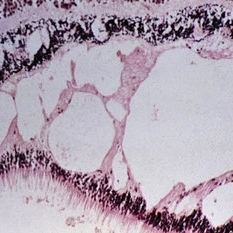
Opaque pars plana cysts and ciliochoroidal effusion in a patient with multiple myeloma.
Condition/keywords: ciliochoroidal effusion, cyst of the pars plana, myeloma
-
 Slide 9-59
Feb 26 2019 by Lancaster Course in Ophthalmology
Two localized areas of RPE hypertrophy. The RPE has melanin granules that are large and spherical (lower views.)
Condition/keywords: melanin granules, retinal pigment epithelium (RPE) hypertrophy
-
 Slide 9-60
Feb 26 2019 by Lancaster Course in Ophthalmology
Diffuse peripheral RPE hypertrophy. There is a band of pigmentation just posterior to the ora serrata (upper left) where the RPE is darker and contains larger, spherical pigment granules(lower right). The junction (arrow) between normal (left) and hypertrophic (right) pigment epithelium is illustrated in the lower left view. A few areas of paving-stone degeneration are present at the equator (upper left).
Condition/keywords: ora serrata, retinal pigment epithelium (RPE) hypertrophy
-
 Slide 9-61
Feb 26 2019 by Lancaster Course in Ophthalmology
Hyperplasia of RPE at ora serrata. Fine-stippled, dark pigmentation at the ora serrata is due to pigment epithelial hyperplasia with migration internally (arrow). Histopathologic sections (lower views) show the strands of hyperplastic epithelium internal to the retina and pars plana.
Condition/keywords: ora serrata, retinal pigment epithelium
-
 Slide 9-62
Feb 26 2019 by Lancaster Course in Ophthalmology
Localized area of RPE hyperplasia with migration into retina around a retinal blood vessel (arrows).
Condition/keywords: retinal pigment epithelium
-
 Slide 9-63
Feb 26 2019 by Lancaster Course in Ophthalmology
Paving-stone degeneration. There is loss of the choriocapillaris, RPE, and outer retinal layers. The thinned inner nuclear layer of the retina rests against Bruch's membrane, and there is no reparative proliferation. Adjacent RPE is hypertrophic.
Condition/keywords: Bruch's membrane, choriocapillaris, retinal pigment epithelium
-
 Slide 9-64
Feb 26 2019 by Lancaster Course in Ophthalmology
Peripheral punched-out lesion from choroidal infarction. The lesion is surrounded by hypertrophic RPE which gives the lesion a dark halo. There is loss of the choriocapillaris, RPE, and outer retinal layers with no reparative proliferation.
Condition/keywords: choroidal infarction, retinal pigment epithelium (RPE) hypertrophy
-
 Slide 9-65
Feb 26 2019 by Lancaster Course in Ophthalmology
Elschnig spot. Localized choroidal infarction with loss of choriocapillaris, RPE, and outer layers of the retina. The thinned inner nuclear layer of the retina rests against Bruch's membrane. At the anterior margin (lower left view) there is an abrupt transition (arrow) between the normal area (left) where the choriocapillaris and RPE are intact and the area of post-ischemic atrophy of the structures (right). A similar but reversed configuration is observed at the posterior margin (lower right view).
Condition/keywords: Bruch's membrane, choroidal infarction, Elschnig's spots
-
 Slide 9-66
Feb 26 2019 by Lancaster Course in Ophthalmology
Midperipheral punched-out lesions in the presumed ocular histoplasmosis syndrome. There is scarring in the choroid and retina, discontinuity in Bruch's membrane, and loss of the RPE. An infiltrate of lymphocytes is present in the subjacent choroid (lower middle and right).
Condition/keywords: Bruch's membrane, ocular histoplasmosis syndrome (OHS), retinal pigment epithelium, scar
-
 Slide 9-67
Feb 26 2019 by Lancaster Course in Ophthalmology
Peripheral albinotic spot. This discrete lesion is due to the reduction or absence of pigment granules in an otherwise normal RPE. The choriocapillaris and photoreceptor cells are normal. The lower left view shows the abrupt transition from pigment-containing RPE to that which is void of melanin pigment. The lower right view is from the center of the lesion.
Condition/keywords: albinism, choriocapillaris, photoreceptor cell, retinal pigment epithelium
-
 Slide 9-68
Feb 26 2019 by Lancaster Course in Ophthalmology
Meridional complex with an enclosed ora bay. A peripheral retinal excavation is present posterior to the enclosed ora bay (arrow).
Condition/keywords: excavation, meridional complex, ora bay
-
 Slide 9-69
Feb 26 2019 by Lancaster Course in Ophthalmology
Noncystic peripheral retinal glial tuft.
Condition/keywords: Morning Glory Syndrome
-
 Slide 9-70
Feb 26 2019 by Lancaster Course in Ophthalmology
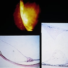
Peripheral retinal zonular traction tuft. A strand of fibroglial tissue extends anteriorly over the pars plana from the peripheral retina. A zonular fiber or condensed vitreous strand (arrow) is attached to the apex of the tuft.
Condition/keywords: fibroglial tissue, pars plana, retinal zonular traction tuft
-
 Slide 9-71
Feb 26 2019 by Lancaster Course in Ophthalmology
Meridional folds of retina, one of which (arrow) is associated with a meridional complex and enclosed ora bay.
Condition/keywords: meridional fold, ora bay
-
 Slide 9-72
Feb 26 2019 by Lancaster Course in Ophthalmology
Ora pearl. In one example (lower left) the lesion is surrounded by hyperplasic pigment epithelium. The other example has a laminated appearance, is calcified, and is not surrounded by pigment epithelium. (Clinical view courtesy of the Retina Service, Wills Eye Hospital.)
Condition/keywords: ora pearl, pigment epithelium
-
 Slide 9-73
Feb 26 2019 by Lancaster Course in Ophthalmology
Inferior retinal dialysis with a localized area of long-standing retinal detachment and demarcation line (upper left). There is total atrophy of the photoreceptor cell layer (upper right). The demarcation line (lower views) is an area of RPE hypertrophy and hyperplasia with nodular basement membrane production and retinal adhesion.
Condition/keywords: photoreceptor cell, retinal dialysis, retinal pigment epithelium (RPE) hypertrophy
-
 Slide 9-74
Feb 26 2019 by Lancaster Course in Ophthalmology
Gross appearance of an eye with a diffuse, flat, choroidal metastatic carcinoma that had been operated on for retinal detachment. Indentation of the sclera from the polyethylene tube is present at the equator.
Condition/keywords: sclera
-
 Slide 9-75
Feb 26 2019 by Lancaster Course in Ophthalmology
Ischemic necrosis of iris and ciliary body (right) in eye following scleral buckling procedure with the use of a polyethylene tube.
Condition/keywords: ciliary, retinal necrosis
-
 Slide 9-76
Feb 26 2019 by Lancaster Course in Ophthalmology
Internal erosion of silicone plate and silastic band. An intense acute and chronic inflammatory reaction surrounds the eroded silastic band (lower right). It was believed that the erosion of the encircling band and plate occurred as a result of its having been tied too tightly.
Condition/keywords: silastic band, silicone plate
-
 Slide 9-77
Feb 26 2019 by Lancaster Course in Ophthalmology
Case of macular pucker following retinal reattachment. There is a thin, hypocellular preretinal membrane that has contracted, producing wrinkling, detachment, and coiling (arrows) of the internal limiting membrane.
Condition/keywords: hypocellular preretinal membrane, macular pucker, retinal pigmentosa
-
 Slide 9-78
Feb 26 2019 by Lancaster Course in Ophthalmology
Senile macular degeneration with hemorrhagic detachment of RPE. Note that there are drusen (upper right) outside the area of detachment, and drusen (arrows) are also detached along with the RPE.
Condition/keywords: hemorrhagic detachment, macular degeneration, retinal pigment epithelium
-
 Slide 9-79
Feb 26 2019 by Lancaster Course in Ophthalmology
Senile macular degeneration. Drusen are shown (arrows), associated with a central area of areolar atrophy (asterisks) in which there is loss of the RPE and photoreceptor cells.
Condition/keywords: central areolar choroidal dystrophy (CACD), drusen, macular degeneration, retinal pigment epithelium
-
 Slide 9-80
Feb 26 2019 by Lancaster Course in Ophthalmology
Senile macular degeneration. Note presence of drusen and diffuse thickening of the inner aspect of Bruch's membrane, areolar RPE and photoreceptor atrophy, and choroidal neovascularization (arrows).
Condition/keywords: Bruch's membrane, choroidal neovascularization (CNV), drusen, macular degeneration, retinal pigment epithelium
-
 Slide 9-82
Feb 26 2019 by Lancaster Course in Ophthalmology
Senile macular degeneration. Hemorrhagic detachment of the RPE is contiguous with sub-RPE neovascularization (arrows, lower middle and lower right). The thickened inner aspect of Bruch's membrane and drusen (upper right, arrows) are detached along with the RPE.
Condition/keywords: Bruch's membrane, hemorrhagic detachment, macular degeneration, retinal pigment epithelium
-
 Slide 9-83
Feb 26 2019 by Lancaster Course in Ophthalmology
Senile macular degeneration. A disciform scar is shown in a typical two component configuration. Arrows indicate the thickened and detached inner aspect of Bruch's membrane. There is one component of the disciform lesion that is located between the retina and the detached inner aspect of Bruch's membrane. RPE hyperplasia is evident in this component. The second component is located between the two layers of Bruch's membrane, and this component has choroidal neovascular tissue in the lower view.
Condition/keywords: Bruch's membrane, choroidal neovascular tissue, disciform scar, macular degeneration, retinal pigment epithelium
-
 Slide 9-84
Feb 26 2019 by Lancaster Course in Ophthalmology
Senile macular degeneration with disciform scar. A retinal arteriole (asterisk) extends into the subretinal component of the scar, through a break in the thickened and detached inner layer of Bruch's membrane, and then into the vascularized intra-Bruch's-membrane component of the scar. Study of serial sections disclosed this retinal vessel to anastomose with the choroidal vessel (arrow) which extends through a branch in Bruch's membrane.
Condition/keywords: Bruch's membrane, disciform scar, macular degeneration, retinal arteriole
-
 Slide 9-85
Feb 26 2019 by Lancaster Course in Ophthalmology
Drusen in the macular area. They are periodic-acid-Schiff-positive (upper and lower right), and stain positive for lipid with the oil red-O technique (lower left). The overlying RPE is effaced, as shown in routine section (upper right) and by flat preparation (lower right).
Condition/keywords: drusen, macular, retinal pigment epithelium
-
 Slide 9-86
Feb 26 2019 by Lancaster Course in Ophthalmology
Calcified drusen.
Condition/keywords: calcified drusen
-
 Slide 9-87
Feb 26 2019 by Lancaster Course in Ophthalmology
Ultrastructural appearance of Bruch's membrane and drusen material in eye with serous detachment of RPE. The RPE basement membrane is intact and normal (arrows). The inner collagenous zone of Bruch's membrane is greatly thickened (between larger arrows) by the accumulation of small vesicles, electron-dense particles, fibrils, and clusters of widely spaced collagen (large circle and inset). The middle-elastic layer of Bruch's membrane (small circle) is essentially normal. The outer collagenous zone (bracket) is mildly thickened with accumulation of material similar to that seen in the inner zone. Splitting of the thickened inner collagenous zone (asterisk) has occurred with the accumulation of a finely granular material, membranous structures, and electron-dense particles (CC =choriocapillaris).
Condition/keywords: Bruch's membrane, drusen, retinal pigment epithelium, serous retinal detachment
-
 Slide 9-88
Feb 26 2019 by Lancaster Course in Ophthalmology
Vascularized drusen in periphery (upper left) and macular area (upper right and lower left). Note the tiny break in Bruch's membrane (arrow) with a choroidal vessel with an erythrocyte extending into a peripapillary druse.
Condition/keywords: Bruch's membrane, drusen, macular
-
 Slide 9-89
Feb 26 2019 by Lancaster Course in Ophthalmology
Nutritional amblyopia. The nerve fiber and ganglion cell layers are absent in the macular area (upper views). The temporal side of the optic nerve head (lower left) is partially atrophy, with marked reduction in the size of the nerve fiber bundles and secondary gliosis.
Condition/keywords: amblyopia, atrophy, gliosis, macular
-
 Slide 9-90
Feb 26 2019 by Lancaster Course in Ophthalmology
lrvine-Gass syndrome. Note presence of cystoid macular edema (upper left), retinal phlebitis in routine section (upper right), and trypsin digestion (lower views).
Condition/keywords: cystoid macular edema (CME), lrvine-Gass syndrome, phlebitis
-
 Slide 9-91
Feb 26 2019 by Lancaster Course in Ophthalmology
Lamellar macular hole in a patient with mild background diabetic retinopathy.
Condition/keywords: lamellar macular hole
-
 Slide 9-92
Feb 26 2019 by Lancaster Course in Ophthalmology
Macular hole following blunt trauma. Note cystic spaces in adjacent retina and rounded margins of the hole.
Condition/keywords: macular hole, trauma
-
 Slide 9-93
Feb 26 2019 by Lancaster Course in Ophthalmology
One side of the ring of chloroquine macular degeneration shows a loss of the photoreceptor cell layer, and pigmented cells cling to and minimally infiltrate the outer aspect of the retina.
Condition/keywords: macular degeneration, photoreceptor cell
-
 Slide 9-94
Feb 26 2019 by Lancaster Course in Ophthalmology
Macular disciform lesion in the ocular histoplasmosis syndrome. Note choroidal scar with vessels (arrow) extending through a break in Bruch's membrane.
Condition/keywords: Bruch's membrane, disciform macular lesion, ocular histoplasmosis syndrome (OHS)
-
 Slide 9-95
Feb 26 2019 by Lancaster Course in Ophthalmology
Macular disciform lesion in a 73-year-old man with angioid streaks and pseudoxanthoma elasticum. The streak is subjacent to the scar and is the point at which choroidal vessels (arrows) extend internally. The same streak closer to the disc is shown in the lower right view.
Condition/keywords: angioid streaks, disciform macular lesion, pseudoxanthoma elasticum (PXE)
-
 Slide 9-96
Feb 26 2019 by Lancaster Course in Ophthalmology
Hypertrophy of peripapillary RPE associated with a vascularized druse with the vessel extending from the choroid through a very small break in Bruch's membrane (arrow).
Condition/keywords: Bruch's membrane, retinal pigment epithelium
-
 Slide 9-97
Feb 26 2019 by Lancaster Course in Ophthalmology
Disciform macular lesion with marked hyperplasia of RPE (A.F.I.P. No. 797119).
Condition/keywords: disciform macular lesion, hyperplasia, retinal pigment epithelium
-
 Slide 9-98
Feb 26 2019 by Lancaster Course in Ophthalmology
Coats' disease with disciform macular lesion. There is accumulation of periodic acid-Schiff-positive staining material within and under the retina peripherally (upper left), in association with telangiectatic retinal vessels (arrow , upper right) . A fibrous nodule is present under the retina in the macular area (lower views). The laminated appearance and partial pigmentation suggest that this fibrous nodule is in part derived from hyperplastic retinal pigment epithelium (lower right).
Condition/keywords: Coats' disease, disciform macular lesion
-
 Slide 9-99
Feb 26 2019 by Lancaster Course in Ophthalmology
Hamartoma of retinal pigment epithelium of optic nerve head (A.F.I.P. No. 334475).
Condition/keywords: hamartoma, retinal pigment epithelium
-
 Slide 9-100
Feb 26 2019 by Lancaster Course in Ophthalmology
Preretinal RPE proliferation. Note the laminated appearance of alternating RPE and basement membrane (upper right) and nodules of basement membrane (lower left). The lower right is a flat preparation of the preretinal membrane.
Condition/keywords: retinal pigment epithelial proliferation
-
 Slide 9-101
Feb 26 2019 by Lancaster Course in Ophthalmology
Retroretinal RPE proliferation forming a membrane.
Condition/keywords: retinal pigment epithelial proliferation
-
 Slide 9-102
Feb 26 2019 by Lancaster Course in Ophthalmology
Pseudoadenomatous hyperplasia of RPE.
Condition/keywords: retinal pigment epithelium
-
 Slide 9-103
Feb 26 2019 by Lancaster Course in Ophthalmology
Acute leukemia with focal areas of RPE defects (lower left), and areas of double-row (upper right) and nodular (lower right) RPE hyperplasia.
Condition/keywords: acute leukemia, retinal pigment epithelium
-
 Slide 9-104
Feb 26 2019 by Lancaster Course in Ophthalmology
Ocular reticulum-cell sarcoma with tumor detachment of the RPE.
Condition/keywords: reticulum cell sarcoma, retinal pigment epithelium, tumor
A project from the American Society of Retina Specialists