 Initializing download.
-
 By Lancaster Course in Ophthalmology
1980 - Uploaded on Feb 26, 2019.
- Last modified by Caroline Bozell on Mar 8, 2019.
- Rating
- Appears in
- Unit 09 Pathology of the Retina
- Condition/keywords
- macular degeneration, disciform scar, Bruch's membrane, retinal pigment epithelium, choroidal neovascular tissue
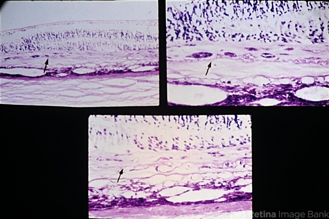
- Description
- Senile macular degeneration. A disciform scar is shown in a typical two component configuration. Arrows indicate the thickened and detached inner aspect of Bruch's membrane. There is one component of the disciform lesion that is located between the retina and the detached inner aspect of Bruch's membrane. RPE hyperplasia is evident in this component. The second component is located between the two layers of Bruch's membrane, and this component has choroidal neovascular tissue in the lower view.


