Search results (48 results)
-
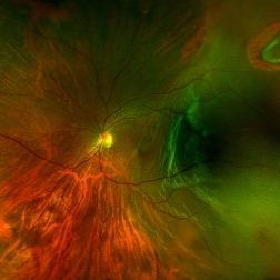
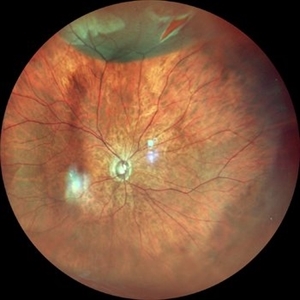
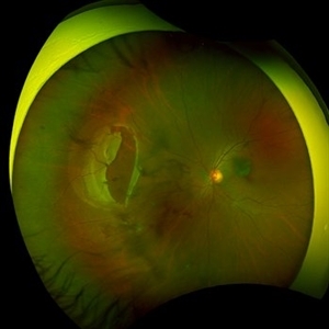 Large Retinal Tear from a Shuttlecock Injury
Oct 11 2024 by Ahmad B. Tarabishy, MD
27 year old woman presenting with floaters and a shadow in her temporal visual field OS. Approximately one week earlier, she was injured in her left eye by a shuttlecock while playing badminton. Fundus exam reveals mild vitreous hemorrhage and a large retinal tear with a small cuff of surrounding SRF.
Photographer: Angela Rico, M.D.
Imaging device: Optos
Condition/keywords: blunt trauma, ocular trauma, retinal tear
-
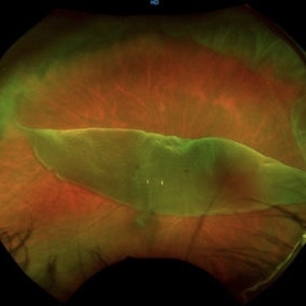
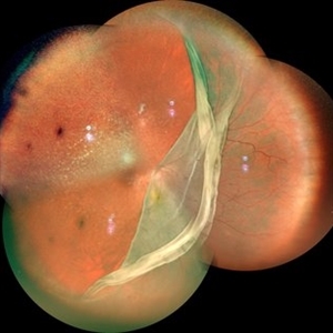
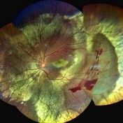
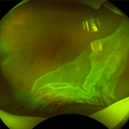
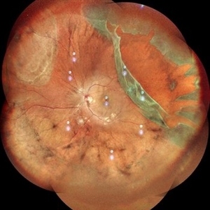 Giant Retinal Tear
Oct 11 2024 by Anjana Mirajkar, MS Ophthalmology
Fundus photograph montage of LE showing a giant retinal extending from 12 to 4 o clock.
Photographer: Dr. Anjana Mirajkar -Retina Foundation, Ahmedabad
Imaging device: Mirante-Nidek
Condition/keywords: GIANT RETINAL TEAR
-
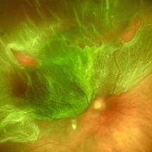
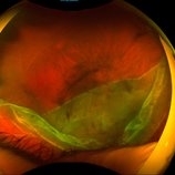
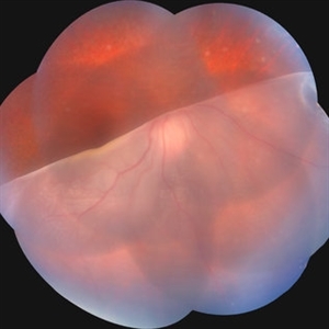 Giant Retinal Tear
Jul 15 2024 by Arthi Mohankumar , MS,MRCS ED, FICO,FAICO
Fundus montage of a 15 year old boy with Marfans syndrome who presented with defective vision in the right eye.
Photographer: Arthi Mohankumar
Condition/keywords: giant retinal tear, Retinal detachment
-
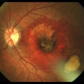
 Failure of Macular Hole Surgery
Jul 2 2024 by Abel Ramírez-Estudillo, MD
Fundus photograph of a 67-year-old woman with failed macular hole surgery, now referred to our clinic with 8 holes.
Photographer: Berenice Palafox, Centro Oftalmológico Mira, Mexico City
Imaging device: Zeiss
Condition/keywords: iatrogenic retinal tear, internal limiting membrane (ILM) peeling, macular hole, vitrectomy
-
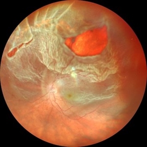
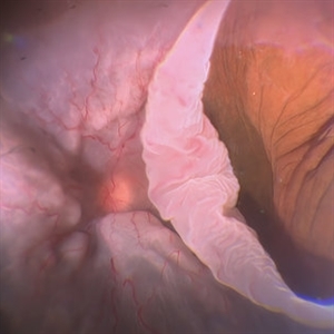 Giant Retinal Tear with Choroidal Detachment
Jun 12 2024 by Anand Temkar
Intra operative still of a 34 year old male showing Giant Retinal Tear with Choroidal Detachment.
Photographer: Dr.Anand Temkar- Retina Foundation, Ahmedabad
Condition/keywords: choroidal detachment, giant retinal tear
-
 Giant Retinal Tear
Oct 24 2023 by Ivan J. Suner, MD, MBA
Fundus photograph of 49-year-old man with a giant retinal tear in the right eye.
Photographer: Norelys Alexander Jimenez, Retina Associates of Florida, Tampa, FL
Imaging device: Optos California
Condition/keywords: GIANT RETINAL TEAR
-
 Retinal Fold
Sep 26 2023 by Mauricio Bayram-Suverza, MD
A 38-year-old man underwent vitrectomy in the left eye due to a giant tear in the upper retina. SF6 gas was used as endotamponade. During the post-surgical check-up, it was identified that the patient developed a full-thickness retinal fold due to retinal slippage during fluid-air exchange. As the fold was away from the macular area, it was decided to observe the patient. Three weeks after the surgery, his best-corrected visual acuity was 20/30.
Photographer: Mauricio Bayram-Suverza, Fundación Hospital Nuestra Señora de la Luz
Imaging device: TRC-50DX
Condition/keywords: giant retinal tear, retina surgery complications, Retinal slippage, vitreoretinal surgery
-
 Rhegmatogenous Retinal Detachment
Sep 4 2023 by Kayne Michael McCarthy, MD, MPH
Fundus photograph of a 59-year-old man with a rhegmatogenous retinal detachment and multiple retinal tears.
Photographer: Gaurav Shah MD, West Coast Retina, San Francisco
Imaging device: Optos p200dtx
Condition/keywords: Retinal Detachment, rhegmatogenous retinal detachment, tears
-
 Retina Detachment
May 1 2023 by RAKESH SHAH, MS DNB FACS FRF FICO MBA
39 year-old female came with sudden loss of vision, on examination rhegmatogenous retinal detachment with large horse shoe tear and linear tear noted
Photographer: Dr.Rakesh shah
Imaging device: Nidek Mirante machine
Condition/keywords: rhegmatogenous retinal detachment
-
 Macula on Retinal Detachment with large Horseshoe Tear
Apr 26 2023 by Kelli Nyenhuis
Optos photograph of a 61-year-old male with a macula on retinal detachment and large horseshoe tear. Patient had no visual changes.
Photographer: Kelli Nyenhuis, COA
Imaging device: Optos California
-
 Total retinal Detachment multiple holes
Sep 26 2022 by Denica Rodriguez
60 year old Male presented with two week old Macula off Retinal detachment with multiple tears.
Photographer: Denica Rodriguez
Imaging device: Optos California
Condition/keywords: color fundus photograph, color photo, macula-off, optos, pseudocolor, Retinal detachment, retinal holes, retinal tear, Retinal tear with detachment, superior arcade, superior field, superior retina, total retinal detachment
-
 Giant Retinal Tear
Apr 21 2022 by Vaidehi Sathaye
Fundus photograph of a 16 year old male with a Rhegmatogenous Retinal Detachment secondary to a Giant Retinal Tear in the right eye.
Photographer: Dr. Vaidehi Sathaye, Retina Foundation
Condition/keywords: giant retinal tear
-
 Giant retinal Tear
Apr 26 2022 by Jeffrey Barker
Giant retinal Tear
Photographer: Jeffrey P. Barker B.S.
Condition/keywords: retinal tear
-
 Traumatic Retinal Tear
Dec 5 2021 by Aditya S Kelkar, MS, FRCS, FASRS,FRCOphth
Color fundus photograph of a 34-year old man's left eye, 2 hours after a tennis ball injury, showing commotio retinae with Berlin's edema and cherry red spot in the fovea along with linear retinal tears in the temporal equatorial zone.
Photographer: Dr Sukanya Mondal. National Institute of Ophthalmology, Pune. India.
Imaging device: Zeiss Clarus 500
Condition/keywords: Berlin's edema, cherry red spot, commotio retinae, retinal tear
-
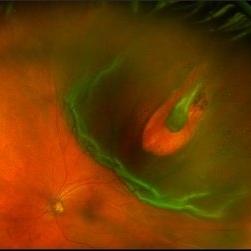
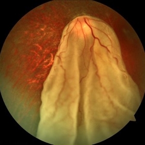 When the Curtain Falls
Jun 12 2021 by Shyamal K Dwivedi, MD
52-year-old male presented with sudden painless vision drop. Giant retinal tear discovered which was about 270 degrees anchored at the disc. Title courtesy: Dr.Aditya Sudhalkar
Photographer: Dr.Aditya Sudhalkar
Imaging device: Zeiss
Condition/keywords: giant retinal tear
-
 Retinal Detachment with Giant Tear
Jul 6 2021 by Lucas Zago Ribeiro, MD
Fundus composite of an 50-year-old man with macula-off retinal detachment due to temporal giant retinal tear.
Photographer: Lucas Zago Ribeiro, Federal University of São Paulo (UNIFESP)
Imaging device: Zeiss Visucam 524
Condition/keywords: giant retinal tear, retinal detachment with retinal defect
-
 Rhegmatogenous Retinal Detachment
Jul 2 2021 by RUSHIK PATEL
Fundus photograph of an 50-year-old female with rhegmatogenous retinal detachment, horseshoe tear.
Photographer: Rushik Patel, Netralaya Super Speciality Eye Hospital, Ahmedabad, Gujarat
-
 RD Montage
Jul 3 2021 by Somnath Chakraborty, MD
Fundus photo montage of the left eye of a 56-year-old male showing subtotal retinal detachment with macular involvement and a large circumlinear tear extending from 1 o' clock to 3 o' clock hours.
Photographer: Pulak Roy
Condition/keywords: acute retinal detachment, retinal detachment of the macula, retinal tear, retinal tear with detachment
-
 Total RRD with GRT with Choroidal Detachment
Apr 11 2021 by Dinesh Rungta, MBBS, DNB
Optos image of a 55-year-old male with history of sudden decrease of vision in right eye associated with flashes showing total RRD with GRT and CD.
Photographer: Dr Dinesh Rungta, Giridhar Eye Institute, Kerala, India.
Imaging device: Optos UWF Daytona Plus
Condition/keywords: detachment, giant retinal tear, re-attached retinal detachment (RRD)
-
 RPE Tear After Anti-VEGF Injection
Mar 17 2021 by RAFAEL REIS PEREIRA, MD
Retinal pigment epithelium (RPE) tear is a rare devastating complication of age-related macular degeneration (AMD). The believed mechanism lies in an adherence of the neovascularization to the undersurface of the RPE creating a contractile force that increases after VEGF therapy and causes the tear.
Photographer: Rafael Reis, Retina Clinic, São Paulo
Condition/keywords: retinal pigment epithelium (RPE) contracture
-
 Giant Tear and Vitreous Abnormalities in Stickler Syndrome
Feb 12 2021 by Anfisa Ayalon, MD
Fundus photograph of a 16-year-old male with Stickler Syndrome and giant tear rhegmatogenous retinal detachment. Note multiple vitreous veils and bands.
Photographer: Anfisa Ayalon, MD., Meir Medical Center, Kfar Saba, Israel.
Imaging device: California, Optos
Condition/keywords: empty vitreous, giant retinal tear, Stickler Syndrome, vitreous veils
-
 Rhegmatogenous Retinal Detachment
Mar 3 2021 by Patrik Rajs
A 51-year-old female patient presented with inferior nasal scotoma and 5/10 vision in the right eye due to a retinal detachment with a giant retinal horseshoe tear.
Photographer: Patrik Rajs, EYE CLINIC of Jan Evangelista Purkyne University and Masaryk Hospital, Czech Republic, Ústí nad Labem
Imaging device: Clarus 700
Condition/keywords: giant retinal tear
-
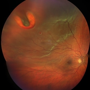
 Retinal Detachment with Horseshoe Tear
Jun 10 2020 by Manish Nagpal, MD, FRCS (UK), FASRS
Localized superior retinal detachment with horseshoe tear and minimal fluid.
Photographer: gayathri mohan
Imaging device: nidek slo mirante
-
 Blunt Ocular Trauma Due to Firework Injury
Jun 9 2020 by Brittany Rota
Ultra- widefield pseudocolor image of an 18-year-old male with blunt ocular trauma in the right eye due to a firework injury. The patient presented with commotio retinae (sclopteria), an acute vitreous hemorrhage, choroidal rupture, and a subretinal hemorrhage. The referring physician performed surgery on the lateral rectus muscle which was macerated but not severed, and several orbital fibrous foreign bodies were removed from the posterior orbit. The globe was intact. There is no evidence of retinal tear in the region of sclopetaria; however, there is complete necrosis of the temporal peripheral choroid and retina. The vitreous hemorrhage was slowly clearing on his exam 6-9-2020. The patient is developing subretinal fibrosis. The physician is concerned about the choroidal rupture that is visible through the submacular hemorrhage. There is one rupture that appears to course directly under the fovea. The physician states that if this is the case, his vision most likely will be 20/200 or worse. His vision was hand motion in all fields except nasally, which he was unable to see hand motion at his visit on 6-9-2020.
Photographer: Brittany Rota
Imaging device: Optos California
Condition/keywords: blunt trauma, choroidal rupture, commotio retinae, fibrosis, firework injury, fundus photograph, hand motion, necrotizing retina, Optos, pseudocolor, subretinal hemorrhage, vitreous hemorrhage
-
 Retinal Tear
Apr 30 2020 by Giselle DeOliveira
Fundus photograph montage of 32-year-old male with retinal tear after repair.
Photographer: Giselle DeOliveira, University of Miami, Bascom Palmer Eye Institute
Imaging device: Topcon
Condition/keywords: retinal tear
 Loading…