Search results (628 results)
-
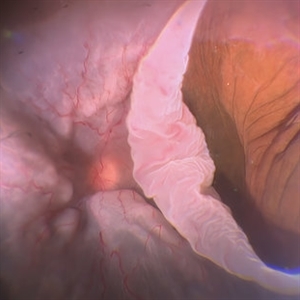
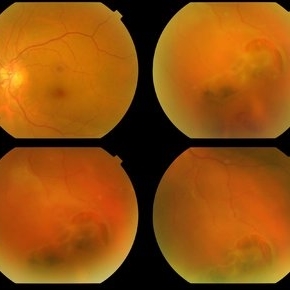
 Total Rhegmatogenous Retinal Detachment With Severe PVR
May 27 2015 by Darin R. Goldman, MD
63-year-old pseudophakic male with hand motion vision in the left eye due to a total retinal detachment with severe proliferative vitreoretinopathy.
Condition/keywords: proliferative vitreoretinopathy (PVR), retinal tear
-
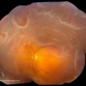
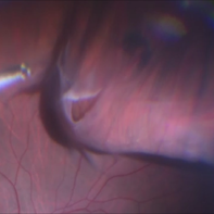
 When the Curtain Falls
Jun 12 2021 by Shyamal K Dwivedi, MD
52-year-old male presented with sudden painless vision drop. Giant retinal tear discovered which was about 270 degrees anchored at the disc. Title courtesy: Dr.Aditya Sudhalkar
Photographer: Dr.Aditya Sudhalkar
Imaging device: Zeiss
Condition/keywords: giant retinal tear
-
 Giant Retinal Tear
Feb 20 2024 by Soobien Lee
Optos color fundus photograph of a 40-year-old caucasian male who is a UFC fighter with a total retinal detachment in his right eye secondary to a giant retinal tear from 10 o'clock to 2 o'clock.
Photographer: Trinity Wolf, Elman Retina Group
Imaging device: Optos Ultra-Widefield Imaging
Condition/keywords: giant retinal tear, optos, Retinal Detachment, Retinal tear with detachment, trauma
-
 Retinal Tear
Sep 16 2021 by Stefanie Palmer
Retinal Tear with a bridge vessel.
Photographer: Stefanie Palmer, CRA
Condition/keywords: detachment, tear
-
 Retinal Detachment With Multiple Retinal Tears
May 18 2017 by Kamal Kishore, MD, MBBS
77-year-old female presented with a report of gradual decreased vision over the span of one week. Vision finger count. Examination showed retinal detachment with multiple retinal tears and vitreous hemorrhage present.
Photographer: Lindsay Shepard, Illinois Retina and Eye Associates, Peru, IL
Imaging device: Topcon TRC- 50 EX
Condition/keywords: retinal tear
-
 Retinal Tear
Apr 30 2020 by Giselle DeOliveira
Fundus photograph montage of 32-year-old male with retinal tear after repair.
Photographer: Giselle DeOliveira, University of Miami, Bascom Palmer Eye Institute
Imaging device: Topcon
Condition/keywords: retinal tear
-
 Giant Retinal Tear
Aug 12 2021 by Stefanie Palmer
Giant Retinal Tear of the Right eye.
Photographer: Stefanie Palmer, CRA
Condition/keywords: giant retinal tear
-
 Giant retinal Tear
Apr 26 2022 by Jeffrey Barker
Giant retinal Tear
Photographer: Jeffrey P. Barker B.S.
Condition/keywords: retinal tear
-
 Giant Retinal Tear
May 15 2014 by Manish Nagpal, MD, FRCS (UK), FASRS
Patient presenting with a acute loss of vision with a giant retinal tear.
Photographer: pooja barot, Optometrist, Retina Foundation, Ahmedabad
Condition/keywords: giant retinal tear
-
 Giant Retinal Tear
May 27 2020 by Jamin S. Brown, MD
Fundus photo montage of 55-year-old male with retinal detachment and giant retinal tear.
Photographer: Stefanie Palmer CRA, Retina-Vitreous Surgeons of CNY
Condition/keywords: giant retinal tear
-
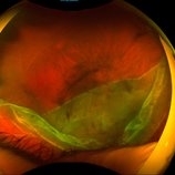
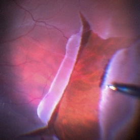
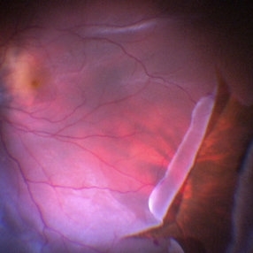
 Giant Retinal Tear with Choroidal Detachment
Jun 12 2024 by Anand Temkar
Intra operative still of a 34 year old male showing Giant Retinal Tear with Choroidal Detachment.
Photographer: Dr.Anand Temkar- Retina Foundation, Ahmedabad
Condition/keywords: choroidal detachment, giant retinal tear
-
 Horseshoe Retinal Break
Apr 3 2018 by Wesam Safwat
Fundus photograph of an 40-year-old woman with lower temporal horseshoe retinal tear associated with lower sub total retinal detachment not involving macula.
Photographer: Wesam Safwat, Elferdaws eye hospital , Zagazig, Egypt.
Imaging device: Topcon
-
 Horseshoe Retinal Tear
Jun 27 2013 by Jason S. Calhoun
Patient came in with retinal detachment. Surgery is scheduled.
Photographer: Jason S. Calhoun, Mayo Clinic Jacksonville, Florida
Imaging device: TOPCON TRC 50-EX
Condition/keywords: retinal tear
-
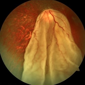
 Intraoperative Photo Taken During Vitrectomy
Jan 26 2017 by Manish Nagpal, MD, FRCS (UK), FASRS
Intraoperative photo while doing vitectomy near a horseshoe tear to clear the adherent vitreous enhanced by peripheral scleral indentation while using chandelier light.
Photographer: Manish Nagpal
Imaging device: Still captured from a 3 chip HD camera on microscope
Condition/keywords: cutter, scleral indentation, vitrectomy, vitreous
-
 Large Retinal Tear
Mar 24 2017 by Manish Nagpal, MD, FRCS (UK), FASRS
Intraoperative photo of a large retinal tear with everted edges.
Photographer: manish nagpal
Imaging device: Still captured from 3 Chip HD camera on microscope
Condition/keywords: retinal tear
-
 Large Retinal Tear
Mar 24 2017 by Manish Nagpal, MD, FRCS (UK), FASRS
Intraoperative photo of a large retinal tear with everted edges.
Photographer: Manish Nagpal
Imaging device: Still captured from a 3 chip HD camera on microscope
Condition/keywords: retinal tear
-
 Macular Tear
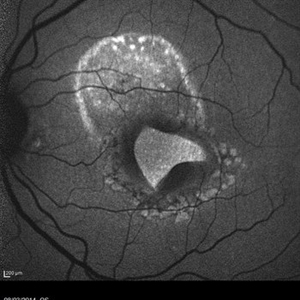
May 14 2014 by Avris Romario Diparaja Siahaan
Blue autofluorescence (BAF) a 40-year-old man with macular tear (had a photocoagulation laser).
Photographer: Avris Romario Diparaja Siahaan
Imaging device: Heidelberg HRA + OCT Spectralis
Condition/keywords: autofluorescence imaging, macular hole
-
 Massive SRH in Patient on Coumadin Being Treated for Exudative AMD
Sep 30 2019 by John S. King, MD
78-year-old white female using 1mg of warfarin for atrial fibrillation, who had a large PED, Type 1 lesion from AMD. Noticed acute darkening of vision one week after anti-VEGF injection. Has very large SRH, subRPE heme, and corrugated retinal appearance post RPE tear. Vision HM (from 20/100). 20/25 in the fellow eye that has dry AMD.
Photographer: Shelly Blair
Imaging device: Optos CA
Condition/keywords: subretinal hemorrhage, wet age-related macular degeneration (wet AMD)
-
 Ozurdex Implant Related Tear
Jan 26 2022 by Tracey Grabowski
Ultra wide-field photograph of a 73-year-old female with an Ozurdex implant causing a retinal tear in the inferior retina. Prompt laser was added to prevent a retinal detachment and patient has been doing well since. Patient had no symptoms following the occurrence.
Photographer: Tracey Grabowski
Imaging device: Optos California
Condition/keywords: fundus photograph, inferior retina, optos, ozurdex, Ozurdex implant, retinal tear, treated retinal tear, ULTRA WIDE FIELD
-
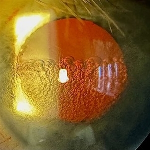 PFO Bubbles
Feb 25 2025 by Parnian Arjmand, MD, MSc, FRCSC, DABO
Post operative day 7 after repair of an RD secondary to a giant retinal tear with temporary PFO tamponade.
Condition/keywords: GRT, PFO
-
 Retinal Detachment with Horseshoe Tear
Jun 10 2020 by Manish Nagpal, MD, FRCS (UK), FASRS
Localized superior retinal detachment with horseshoe tear and minimal fluid.
Photographer: gayathri mohan
Imaging device: nidek slo mirante
-
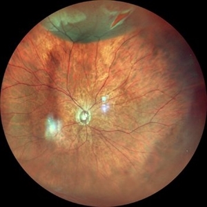
 Retinal Detachment with Giant Retinal Tear
Mar 9 2013 by Young-Gyun Kim, MD
Fundus photograph of a 45-year-old man with retinal detachment and giant retinal tear.
Photographer: Shin Ji-Young, Eulji university, Seoul
Imaging device: Topcon TRC 50 EX
Condition/keywords: retinal tear
-
 Retinal Tear at the Posterior Edge of Lattice Degeneration
Mar 1 2014 by Homayoun Tabandeh, MD, FASRS
Retinal tear at the posterior edge of lattice degeneration.
Condition/keywords: lattice degeneration, retinal tear
-
 Rhegmatogenous Retinal Detachment
Mar 3 2021 by Patrik Rajs
A 51-year-old female patient presented with inferior nasal scotoma and 5/10 vision in the right eye due to a retinal detachment with a giant retinal horseshoe tear.
Photographer: Patrik Rajs, EYE CLINIC of Jan Evangelista Purkyne University and Masaryk Hospital, Czech Republic, Ústí nad Labem
Imaging device: Clarus 700
Condition/keywords: giant retinal tear
-
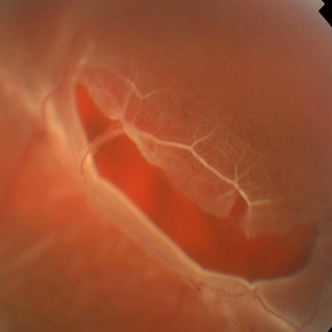
 Sudden Posterior Vitreous Detachment
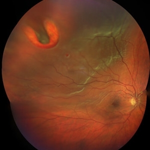
Nov 9 2012 by Norman Byer
This 60-year-old man suffered a sudden posterior vitreous detachment which produced a large tractional retinal tear at 11:30 o’clock in this eye. This white cystic retinal tuft located at 9:30 also suffered minor injury at the same time as revealed in the next slide pair.
Condition/keywords: posterior vitreous detachment, white retinal tuft
 Loading…