Search results (628 results)
-
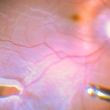
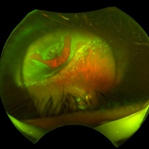
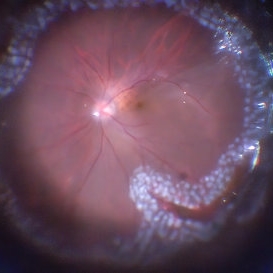 360 Endolaser Barrage
Feb 2 2022 by Manish Nagpal, MD, FRCS (UK), FASRS
Intraoperative photo of a 360 laser barrage done for a case of retinal detachment with a large superior tear.
Photographer: Manish Nagpal, Retina Foundation, Ahmedabad, India
Imaging device: Sony PMW -10 MD surgical camera
Condition/keywords: laser, laser photocoagulation, tear
-
 Choroid Detachment
Jul 7 2021 by Patrik Rajs
This eye was a tough one. The patient underwent PPV twice, the second one with silicone oil (SO) for retinal re-detachment. Due to the development of secondary glaucoma, silicone oil evacuation and lavage of the anterior chamber were performed. Because of the high IOP even after the evacuation, the XEN was implanted. The surgery was followed by choroidal detachment presented in the picture on the left side along with the residual silicone bubble superiorly. The retinal tear is captured inferiorly surrounded by laser spots. The second image (on the right) was taken only 7 days later and it shows that choroidal detachment in the eye resolved completely.
Photographer: Patrik Rajs, EYE CLINIC of Jan Evangelista Purkyne University and Masaryk Hospital, Czech Republic, Ústí nad Labem
Condition/keywords: choroid, detachment, glaucoma, retina, silicone oil, tear
-
Flattening a bullous retinal detachment
Oct 24 2022 by Manish Nagpal, MD, FRCS (UK), FASRS
This surgical clip shows the way a bullous retinal detachment reattaches at the time of endodrainage
Photographer: Manish Nagpal
Condition/keywords: bullous detachment, endodrainage, tear, video, vitrectomy
-
 Horseshoe Tear with Vitreous Hemorrhage
Oct 5 2022 by Vishal Agrawal, MD, FRCS,FACS,FASRS
Fundus picture of a 54 year old female presenting with acute onset of floaters . On examination nasal HST was noted & lasered.
Photographer: Pankaj
Imaging device: CLARUS 700
Condition/keywords: hemorrhage, tear
-
 Retinal Detachment and Retinal Tear Treated with Laser
Feb 2 2022 by Manish Nagpal, MD, FRCS (UK), FASRS
Intraoperative photo of a lasered tear noted in a case of retinal detachment with large superior tear.
Photographer: Manish Nagpal, Retina Foundation, Ahmedabad, India
Imaging device: Sony PMW -10 MD surgical camera
Condition/keywords: laser, tear
-
 Retinal Detachment with a Large Superior Tear
Feb 2 2022 by Manish Nagpal, MD, FRCS (UK), FASRS
Intraoperative photo of a retinal detachment with a large superior retinal tear.
Photographer: Manish Nagpal, Director, Retina Foundation, Ahmedabad
Imaging device: Sony PMW -10 MD surgical camera
Condition/keywords: retina, tear
-
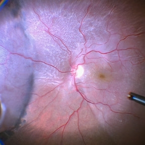
 Retinal Detachment with a Posterior Tear
Jan 11 2022 by Manish Nagpal, MD, FRCS (UK), FASRS
Intraoperative photo of a retinal detachment with a posterior tear with everted edges.
Photographer: Manish Nagpal, Retina Foundation, Ahmedabad, Gujarat
Imaging device: Sony PMW -10 MD surgical camera
Condition/keywords: tear
-
 Retinal Detachment with a Superonasal Tear
Feb 4 2022 by Manish Nagpal, MD, FRCS (UK), FASRS
Intraoperative photo of a retinal detachment with a large supero nasal tear.
Photographer: Manish Nagpal, Director, Retina Foundation, Ahmedabad
Imaging device: Sony PMW -10 MD surgical camera
Condition/keywords: tear
-
 Retinal Tear
Sep 16 2021 by Stefanie Palmer
Retinal Tear with a bridge vessel.
Photographer: Stefanie Palmer, CRA
Condition/keywords: detachment, tear
-
Vitrectomy for bullous retinal detachment with superior tear
Jan 2 2023 by Manish Nagpal, MD, FRCS (UK), FASRS
Vitrectomy for bullous Retinal detachment with superior tear| In this case vitrectomy is being done for a retinal detachment with superior tear. Once the vitreous is removed, air fluid exchange is carried out. Perfluorocarbon heavy liquid is injected to flatten the posterior pole and push the fluid to the periphery for endo drainage. This is followed by endo drainage from the superior break. Once the retina flattened endolaser was carried out.
Condition/keywords: air fluid exchange, bullous retinal detachment, endo drainage, endolaser, holes, Prophylaxis, RD, reattachment of retinal detachment, tear, video, vitrectomy
-
Vitrectomy for Myopic Retinal Detachment with multiple tears
Jan 2 2023 by Manish Nagpal, MD, FRCS (UK), FASRS
Vitrectomy for Myopic Retinal detachment with multiple tears and lattice degenerations | Vitrectomy is carried out and triamcinolone staining is used to stain the hyaloid attachment. The hyaloid attachment is extremely adherent. With high vacuum the cutter engages the stained hyaloid and gradually peels it off the mobile retina. After this Perfluorocarbon heavy liquid is injected to flatten the posterior pole and push the fluid to the periphery till the edge of the tear. This is followed by endo drainage from the tear. Once the retina flattened endolaser was carried out.
Condition/keywords: air fluid exchange, endo drainage, endolaser, holes, lattice degeneration, myopia, myopic retinal retachment, RD, reattachment of retinal detachment, tear, video, vitrectomy
-
Vitrectomy for Myopic Retinal Detachment with multiple tears status post prophylaxis
Jan 3 2023 by Manish Nagpal, MD, FRCS (UK), FASRS
Vitrectomy for Myopic Retinal detachment with multiple tears and lattice degenerations status post prophylaxis laser done | Vitrectomy is carried out. Once the vitreous is removed the retina is freely mobile. After this Perfluorocarbon heavy liquid is injected to flatten the posterior pole and push the fluid to the periphery till the edges of the tear. This is followed by endo drainage from infero nasal tear. Scars of laser marks are seen around it. Once the retina flattened endolaser is carried out.
Condition/keywords: air fluid exchange, endo drainage, endolaser, holes, lattice degeneration, myopia, myopic retinal detachment, prophylaxis, RD, reattachment of retinal detachment, tear, video, vitrectomy
-
Vitrectomy for retinal detachment with multiple large tears
Oct 20 2022 by Manish Nagpal, MD, FRCS (UK), FASRS
Vitrectomy for retinal detachment with multiple large tears
Condition/keywords: tear, video, vitrectomy
-
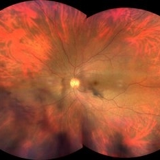
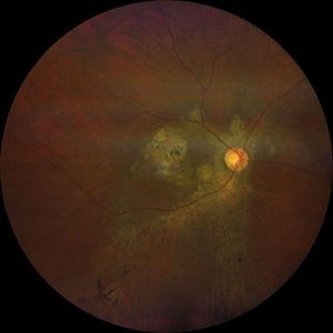
 Tear drop CSCR
Jun 20 2022 by T. P . VIGNESH, MBBS,MS
Ultra wide field fundus photograph of left eye of 47 year old male patient, revealing pigmentary changes and RPE degeneration at the macula as well as inferiorly suggestive of old tear drop CSCR .
Photographer: Shivanath
Imaging device: OPTOS
Condition/keywords: chronic central serous chorioretinopathy (CSCR)
-
 Tear Drop CSCR
Apr 10 2023 by T. P . VIGNESH, MBBS,MS
Fundus photograph of a 45-year-old man with Tear Drop CSCR.
Photographer: Bharathi S
Imaging device: ZEISS CLARUS
Condition/keywords: central serous chorioretinopathy (CSCR)
-
 Tear drop CSCR
Aug 28 2024 by T. P . VIGNESH, MBBS,MS
Fundus photograph of an 68-year-old man with old tear drop CSCR.
Photographer: Bharathi
Imaging device: Zeiss Clarus
Condition/keywords: chronic central serous chorioretinopathy (CSCR)
-
 Tear Drop CSCR
Aug 28 2024 by T. P . VIGNESH, MBBS,MS
Fundus photograph of an 68-year-old man with old tear drop CSCR.
Photographer: Bharathi
Imaging device: Zeiss Clarus
Condition/keywords: chronic central serous chorioretinopathy (CSCR)
-
 Tear Drop-CSCR
May 17 2024 by T. P . VIGNESH, MBBS,MS
Fundus photo of the right eye of a 59 year old man with history of defective vision in the right eye for the past 11 years .
Photographer: Bharathi
Imaging device: Zeiss Clarus
Condition/keywords: central serous chorioretinopathy (CSCR)
-
 Teardop CSCR
May 24 2023 by T. P . VIGNESH, MBBS,MS
Fundus photograph of a 45 year old man revealing chronic CSCR with tear drop sign .
Photographer: Bharathi S
Imaging device: ZEISS CLARUS
Condition/keywords: central serous chorioretinopathy (CSCR)
-
 "Hang in There"
Apr 20 2021 by Tomas Minelli, MD
Fundus wide field photograph of a 50-year-old man with a macular detachment associated with a big temporal superior tear. The laser is firmly holding the progression of the tear in the 14th day post- laser. BCVA 20/20
Photographer: Livia Conci, Universtity of São Paulo
Imaging device: Optos Daytona
Condition/keywords: giant retinal tear
-
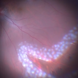
 24 Hours Post Scleral Wound Closure+ Scleral Buckle+25 g Vitrectomy+Silicon Oil
Jan 23 2015 by Carlos Quezada-Ruiz, MD, FASRS
24 hours post op fundus photograph of a 43-year-old man who had perforating injury to the right eye with a small piece of plastic while he was hammering. OD LP, subconjunctival hemorrhage, clear cornea, hyphema, irido and ciclodyalisis as well as a luxated lens with traumatic cataract and a dense vitreous hemorrhage. B-US showed rhegmatogenous retinal detachment with a tear and a big inferior hemorrhagic choroidal detachment. 360 peritomy revealed 2-entry scleral wounds were found in zone II (M V and M VI) and closure was performed. 25 G PPV was performed with the infusion canal placed in the AC through the limbus. Lensectomy and removal of a dense recent vitreous hemorrhage revealed a white detached retina with an exit wound through the temporal inferior segment of the optic nerve with a nasal GRT and sub retinal hemorrhage as well as temporal inferior choroidal, PVD was induced and PFOs helped stabilizing the retina while vitrectomy and sub-retinal hemorrhage was removed through the GRT. Fluid air exchange was made and 360 endolaser over the buckle indentation was done and silicon oil was used as endotamponade. This picture was taken 24 hrs after the surgery.
Photographer: Lilibeth Rodriguez, Instituto de la Visión. Torreon, Mexico.
Condition/keywords: central retinal artery occlusion (CRAO), giant retinal tear, trauma
-
 Acute Posterior Vitreous Detachment
Nov 9 2012 by Norman Byer
This large and complicated retinal tear in a 51-year-old man resulted from an acute posterior vitreous detachment which concentrated its tractional forces around this area of lattice degeneration. Because of the powerful traction, there is an additional central tear splitting the large retinal flap and almost severing one of its arms. The traction was strong enough to completely rupture the blood vessel just to the left of the flap. Marking the ruptured peripheral end of the blood vessel is a yellow depigmented thrombus.
Condition/keywords: acute posterior vitreous detachment, depigmented thrombus, lattice degeneration, retinal tear, tractional retinal detachment
-
 Acute Retinal Detachment
Nov 9 2012 by Norman Byer
This 54-year-old man was referred because of sudden symptoms in his opposite eye in which he had suffered an acute retinal detachment secondary to a horseshoe tear around lattice degeneration. During the examination, the fellow eye shown here was also found to have this large horseshoe tear about 1 o’clock hour (4 disc diameters) in size. A tear occurred around a lattice lesion which is present on the flap but is out of focus. This tear had been asymptomatic even though it was caused by a posterior vitreous detachment and illustrates that even very large tears may produce no symptoms or mild symptoms that are easily overlooked.
Condition/keywords: lattice degeneration, posterior vitreous detachment
-
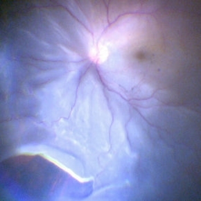
 Acute Retinal Detachment
Oct 12 2012 by Jeffrey G. Gross, MD, FASRS
Acute RD with posterior retinal tear, with hemorrhage.
Condition/keywords: acute retinal detachment, posterior retinal tear
-
 Acute Retinal Detachment with Posterior Tear
Oct 16 2012 by Jeffrey G. Gross, MD, FASRS
Acute RD with posterior tear.
Condition/keywords: acute retinal detachment, posterior tear
 Loading…