Search results (628 results)
-
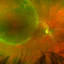
 Cystic Retinal Tuft
Nov 9 2012 by Norman Byer
This is the same lesion as in the previous slide pair but the photograph was taken nine years later when the patient was 58-years-old soon after an acute posterior vitreous detachment. This demonstrates that posterior vitreous detachment can produce large retinal tears at these sites. However, it is important to emphasize that prophylactic treatment of cystic retinal tufts in the absence of a retinal tear would be very ill-advised because several hundred innocence and harmless lesions would have to be treated in order to prevent one tear of the retina.
Condition/keywords: cystic retinal tuft, posterior vitreous detachment, retinal tear
-
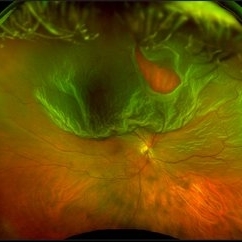
 Total Rhegmatogenous Retinal Detachment With Severe PVR
May 27 2015 by Darin R. Goldman, MD
63-year-old pseudophakic male with hand motion vision in the left eye due to a total retinal detachment with severe proliferative vitreoretinopathy.
Condition/keywords: proliferative vitreoretinopathy (PVR), retinal tear
-
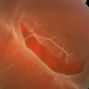
 White Retinal Tuft
Nov 9 2012 by Norman Byer
After six years, the previous lesion looked like this. The former flap has been completely avulsed and is now a free operculum. The white zone around the tear represents the small area of detachment and subretinal fluid. It is still asymptomatic and does not require treatment.
Condition/keywords: does not require treatment, free operculum, operculated retinal hole, subretinal fluid, white retinal tuft
-
 Horseshoe Retinal Tear
Jun 27 2013 by Jason S. Calhoun
Patient came in with retinal detachment. Surgery is scheduled.
Photographer: Jason S. Calhoun, Mayo Clinic Jacksonville, Florida
Imaging device: TOPCON TRC 50-EX
Condition/keywords: retinal tear
-
 Retinal Detachment Right Eye Optomap
Mar 31 2014 by James B. Soque, CRA, OCT-C, COA, FOPS
36-year-old white male presented with non traumatic retinal detachment OD, with six very distinct demarcation lines and isolated tear, and detachment parameters. Patient underwent PPV OD on 12/3/13 with 20% SF6 gas placement and face down in his first 1 month post op period.
Photographer: James Soque, CRA, COA
Imaging device: Optos Daytona
Condition/keywords: Cryopexy, demarcation line, gas pneumatic displacement, Optomap, Optos, pars plana vitrectomy (PPV), retinal tear, scanning laser ophthalmoscope
-
 Peripheral Retinal Lesion
Nov 9 2012 by Norman Byer
This small elevated peripheral retinal lesion in a 48-year-old woman is a cystic retinal tuft. Such tufts are congenital developmental anomalies present from birth and situated behind the vitreous base. They are sites of abnormal vitreoretinal attachment, and can occasionally lead to retinal tears at the time of posterior vitreous detachment. They are present in about 5% of patients.
Condition/keywords: abnormal vitreal retinal attachment, behind the vitreous base, congenital anomaly, cystic retinal tuft, developmental anomaly, peripheral retinal lesion, present from birth
-
 Traumatic Retinal Dialysis-RD
Jan 1 2013 by John T. Thompson, MD
Traumatic retinal dialysis with localized retinal detachment after blunt trauma.
Condition/keywords: acute retinal detachment, retinal dialysis, retinal tear
-
 RD With Posterior Tear
Sep 2 2012 by Jonathan L. Prenner, MD
Relatively Posterior Break in an RD
Photographer: Vivian Chacon, Retina Vitreous Center, UMDNJ
Condition/keywords: retinal detachment with retinal defect
-
 Optos Giant Tear within Retinal Detachment
Apr 30 2019 by Lauren Whaley
Noticed an inferior visual field defect on a patient with history of vitreous hemorrhage. Decided to take an Optos image and this is what we found. Doctor performed pneumatic retinopexy in office and patient recovering well.
Photographer: Lauren R. Whaley
Imaging device: Optos
Condition/keywords: Optos, retinal tear, subretinal fluid
-
 UWF of Retinal Detachment Corrected with Scleral Buckle
Aug 29 2017 by Carolyn Daley
An ultra wide field fundus photograph of a 57-year-old male who has a past history of retinal detachment corrected with scleral buckle and three treated retinal tears.
Photographer: Carolyn Daley
Imaging device: Optos Imaging
Condition/keywords: cryo-retinal tear, cryotherapy, Optos, retinal tear, scleral buckle, ultra-wide field imaging
-
 Retinal Break at Site of Lattice Degeneration with Scleral Indentation
Nov 9 2012 by Norman Byer
This is the same case as the previous photograph. With scleral indentation slightly more posterior, the flap is seen to be associated with a large retinal tear. This is a tractional tear and it is possible that in this case the cryotherapy itself may have increased the vitreoretinal traction at this site and in this way led to this new tear. The age of the tear is unknown because it was asymptomatic, and even though the eye is aphakic the tear has not caused a clinical retinal detachment.
Condition/keywords: retinal flap, scleral indentation, tractional retinal tear, vitreoretinal traction
-
 Retinal Tear at the Posterior Edge of Lattice Degeneration
Mar 1 2014 by Homayoun Tabandeh, MD, FASRS
Retinal tear at the posterior edge of lattice degeneration.
Condition/keywords: lattice degeneration, retinal tear
-
 Horseshoe Tear, In Stereo
Sep 28 2012 by Michael P. Kelly, FOPS
Horse shoe tear, stereo.
Photographer: Michael P. Kelly, FOPS Director, Duke Eye Labs, Duke University Hospital, Duke Eye Center, Durham, NC
Imaging device: Zeiss FF3C
Condition/keywords: stereo pair
-
 Rhegmatous Retinal Detachment
Oct 20 2012 by Hyung-Woo Kwak, MD
Fundus showed a large tear in superior bullous retinal detachment.
Condition/keywords: retinal tear
-
---thumb.JPG/image-square;max$300,300.ImageHandler) Retinal Detachment with Horseshoe Tear
Retinal Detachment with Horseshoe Tear
Jul 11 2013 by Jason S. Calhoun
Fundus photo shows horseshoe tear superior-temporal in the left eye with retinal detachment.
Photographer: Jason S. Calhoun, Department of Ophthalmology, Mayo Clinic Jacksonville, Florida
-
 Sudden Posterior Vitreous Detachment
Nov 9 2012 by Norman Byer
This is the same lesion seen in the previous slide pair. With the scleral indentation performed more posteriorly, a small hemorrhage can be seen on the white tuft. This is proof of the vitreal retinal attachment at this spot. Posterior vitreous detachment can produce a retinal tear at the site of a cystic retinal tuft, but in this case has caused only a small hemorrhage.
Condition/keywords: posterior vitreous detachment, retinal hemorrhage, scleral indentation, vitreoretinal attachment
-
---thumb.JPG/image-square;max$300,300.ImageHandler) Horseshoe Tear With Laser Treatment
Horseshoe Tear With Laser Treatment
Jul 13 2013 by Jason S. Calhoun
Retinal tear which was treated with a laser retinopexy.
Photographer: Jason S. Calhoun, Department of Ophthalmology, Mayo Clinic Jacksonville, Florida
Condition/keywords: laser retinopexy, retinal tear
-
 White Retinal Tuft
Nov 9 2012 by Norman Byer
This white retinal tuft was seen in a 20-year-old man. It is associated with an asymptomatic retinal tear posterior to the tuft and with a tiny adjacent amount of subretinal fluid. It remained just like this for six years and then underwent the change shown in the next slide pair.
Condition/keywords: asymptomatic, subretinal fluid, white retinal tuft
-
 Horseshoe Retinal Tear With Bridging Blood Vessel
Jun 27 2013 by Jason S. Calhoun
Patient came in with curtain appearing in the superior part of vision. Retinal tear superior visible with detachment. Surgery is scheduled.
Photographer: Jason S. Calhoun, Mayo Clinic Jacksonville, Florida
Imaging device: TOPCON TRC 50-EX
Condition/keywords: retinal tear
-
 Giant Retinal Tear
Oct 9 2012 by Audina M. Berrocal, MD FASRS
Teenager with high myopia and a GRT
Photographer: Ditte Hess CRA, BPEI
Imaging device: Fundus Camera
Condition/keywords: high myopia, retinal degeneration, retinal tear
-
 Dialysis of Retina in Upper Nasal Quadrant
Nov 9 2012 by Norman Byer
This 20-year-old wrestler sustained a sharp powerful blow to his right eye from his opponent’s thumb. One hour later he saw hundreds of black specs in his vision and was found to have this dialysis of his retina in the upper nasal quadrant. Note the triangular piece of retina in the center that remained attached to the ora serrata causing the retinal flap to resemble a man’s flared shirt collar.
Condition/keywords: retinal dialysis, retinal tear, upper nasal quadrant
-
 Lattice Degeneration
Nov 9 2012 by Norman Byer
In this 54-year-old woman, lattice degeneration has led to a large horseshoe tractional tear around the posterior side on one end of the lesion resulting in a clinical retinal detachment. Note the very attenuated blood column passing through the white sheath vessel that crosses the tear. This demonstrates that the white blood vessels and a fragment of attached tissue are the only structures which have escaped the tearing effect of the strong vitreoretinal traction which occurred. This usually is true, although, in some cases this bridging vessel may bleed.
Condition/keywords: bridging vessel, lattice degeneration, tractional retinal tear, white sheath vessel
-
---thumb.jpg/image-square;max$300,300.ImageHandler) Cryo-Retinal Tear
Cryo-Retinal Tear
Oct 18 2012 by Larry Halperin, MD
Cryo-retinal tear
Condition/keywords: cryo-retinal tear, retinal tear
-
 Symptomatic Retinal Tear
Nov 9 2012 by Norman Byer
This is another example of a symptomatic retinal tear which occurred at the site of a cystic retinal tuft two days prior to the photograph when an acute posterior vitreous detachment occurred in this 64-year-old woman. Note the horizontal line of vitreous blood along the lower edge of the flap which demarcates the vitreous attachment to the flap.
Condition/keywords: acute posterior vitreous detachment, cystic retinal tuft, retinal flap, retinal tear, vitreous blood
-
 Subclinical Retinal Detachment
Nov 9 2012 by Norman Byer
This 50-year-old man was treated with cryotherapy for two tiny non tractional round holes which had produced a small subclinical retinal detachment at 7:00 o’clock in this eye. Two years later he was seen with this large horseshoe tractional tear just anterior to the treated area and we must assume that it was a complication of that treatment.
Condition/keywords: cryotherapy, non-tractional holes, tractional retinal tear
 Loading…