Search results (628 results)
-
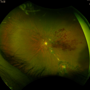
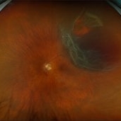
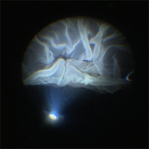
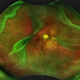 Giant Retinal Tear with Multiple Retinal Breaks
Apr 21 2025 by Hrishikesh Naik, MS
A 28 year old high myope with retinal detachment associated with a supero-temporal giant retinal tear in addition to multiple peripheral horseshoe tears and an additional supero-nasal retinal tear.
Photographer: Hrishikesh Naik
Imaging device: Optos Daytona
Condition/keywords: giant retinal tear, High Myopia, horseshoe tear, retinal break, retinal detachment
-
 Traumatic Hemorrhage
Apr 17 2025 by Virginia Gebhart
60 year old male with vitreous and sub hyaloid hemorrhage from being hit in the eye. No holes, tears, or detachment. Will observe closely, if no improvement will consider surgical repair. Treated melanoma s/p brachytherapy in 2008.
Photographer: Virginia Gebhart, Retina Consultants of Carolina
Imaging device: Optos California
Condition/keywords: blunt trauma, sub hyaloid hemorrhage, vitreous hemorrhage
-
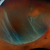
 Blister Retinal Detachment Superotemporal with a Flap Tear
Apr 10 2025 by Daniela Bogenschutz
Autofluorescence of a 70-year-old male with a superotemporal retinal detachment prior to having an OCT with unusual findings. Patient states symptoms were "starburst" in his vision in the location of the retinal detachment with the retinal tear. Surgery was scheduled immediately to avoid further progression.
Photographer: Daniela Bogenschutz, OSC; Retina Consultants of the Carolinas, PA
Condition/keywords: Retinal Detachment, retinal detachment with single break
-
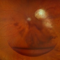
 Superior Rhegmatogenous Retinal Detachment (RRD) in the Right Eye, With a Retinal Tear Located Between the 1 and 2 O'clock Positions
Apr 4 2025 by Cesar Orlando Oviedo Vera
A 45-year-old male patient presented with a sudden onset of decreased visual acuity in the right eye, with a 24-hour progression. Upon examination, Image 1 revealed a superior rhegmatogenous retinal detachment in the right eye, with a retinal tear located between the 1 and 2 o'clock positions. Image 2: Pneumatic retinopexy by intravitreal injection of Sulfur Hexafluoride gas (SF6) at the time of diagnosis with subsequent application of 532 nm laser around the retinal tear.
Photographer: Cesar Orlando Oviedo Vera, Hospital Militar de Especialidades Oftalmológicas
Imaging device: Optos
Condition/keywords: Pneumatic Retinopexy, Retinal tear, Rhegmatogenous retinal detachment, SF6, Superior rhegmatogenous retinal detachment
-
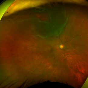
 Pneumatic Retinopexy by Intravitreal Injection of Sulfur Hexafluoride Gas (SF6) at the Time of Diagnosis With Subsequent Application of 532 Nm Laser Around the Retinal Tear
Apr 4 2025 by Cesar Orlando Oviedo Vera
A 45-year-old male patient presented with a sudden onset of decreased visual acuity in the right eye, with a 24-hour progression. Upon examination, Image 1 revealed a superior rhegmatogenous retinal detachment in the right eye, with a retinal tear located between the 1 and 2 o'clock positions. Image 2: Pneumatic retinopexy by intravitreal injection of Sulfur Hexafluoride gas (SF6) at the time of diagnosis with subsequent application of 532 nm laser around the retinal tear.
Photographer: Cesar Orlando Oviedo Vera, Hospital Militar de Especialidades Oftalmológicas
Imaging device: Optos
Condition/keywords: Pneumatic Retinopexy, Retinal tear, Rhegmatogenous retinal detachment, SF6, Superior rhegmatogenous retinal detachment
-
 Large Horseshoe Tear
Apr 4 2025 by Tejaswita Verma
Fundus photo of a 29 year old myopic male with RE 6/12P vision with large ragged tear on the posterior edge of lattice degeneration
Photographer: DR. TEJASWITA VERMA
Imaging device: MIRANTE
Condition/keywords: horseshoe tear
-
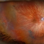
 Retinal Detachment with Retinal Tear
Mar 31 2025 by Kimberly Wakester
Optomap RGB of an 48-year-old woman with a retinal detachment with retinal tear in the left eye. Surgery was recommended. Patient is to continue follow up care post operatively.
Photographer: Kimberly Wakester, COA, OCT-C
Imaging device: Optos California
Condition/keywords: Retinal Detachment, retinal tear
-
 Rhegmatogenous Retinal Detachment with Gd C PVR Changes
Mar 28 2025 by Shrishti mishra
Fundus photograph of a 58 year old male who had undergone a pneumatic retinopexy elsewhere presented to us with a total retinal detachment with a retinal tear in the superotemporal quadrant and grade c pvr changes.
Photographer: Mrs Vinutha
Imaging device: Optos nikon
Condition/keywords: proliferative vitreoretinopathy (PVR), retinal tear with detachment, rhegmatogenous retinal detachment
-
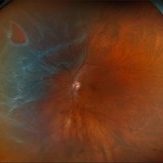
 Rhegmatogenous Retinal Detachment
Mar 24 2025 by DR APOORVA JADHAV, MBBS , DNB
This is a color fundus photograph showing rhegmatogenous retinal detachment with posterior pole retinal tear with macula off.
Condition/keywords: retinal detachment of the macula
-
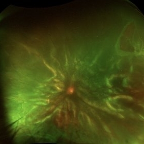
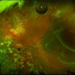
 Hosreshoe Tears on Posterior Pole
Mar 22 2025 by Deepak Bhojwani, MS
A fundus image of an asymptomatic 64 year old male with large horseshoe shaped breaks in inferonasal quadrant on posterior pole, an unusual location for retinal breaks.
Photographer: DR DEEPAK BHOJWANI
Condition/keywords: horseshoe tear, posterior pole break, retinal break
-
 Inadvert Globe Perfuration After Peribulbar Block
Mar 13 2025 by Bruno B Ribeiro
Fundus photograph of a 74-year-old woman who underwent pars plana vitrectomy OS due to rhegmatogenous retinal detachment. A horseshoe retinal tear can be seen at 5h. Intraoperative evaluation revealed a chorioretinal scar with the shape of the needle track at the same location. Despite rare, globe perfuration after peri or retrobulbar block may happen, even by the most experienced anesthesiologist.
Photographer: Bruno Barbosa Ribeiro, Angelina Meireles
Imaging device: Optos California
Condition/keywords: retinal detachment
-
 Inadvert Globe Perfuration After Peribulbar Block
Mar 13 2025 by Bruno B Ribeiro
Fundus photograph of a 74-year-old woman who underwent pars plana vitrectomy OS due to rhegmatogenous retinal detachment. A horseshoe retinal tear can be seen at 5h. Intraoperative evaluation revealed a chorioretinal scar with the shape of the needle track at the same location. Despite rare, globe perfuration after peri or retrobulbar block may happen, even by the most experienced anesthesiologist.
Photographer: Bruno Barbosa Ribeiro, Angelina Meireles
Imaging device: Optos California
Condition/keywords: hemmorhage
-
 Retinal Detachment with Multiple Breaks
Mar 5 2025 by Kimberly Wakester
Optomap RGB image of an 44-year-old man with a retinal detachment with a complex lattice break in the right eye. Surgery was recommended. Patient is to continue follow up care post operatively.
Photographer: Kimberly Wakester, COA
Imaging device: Optos California
Condition/keywords: Retinal Detachment, retinal tear
-
 Retinal Detachment
Mar 5 2025 by Kimberly Wakester
Optomap RGB image of an 9-year-old boy with a retinal detachment with retinal break at 9:00 in the right eye. Surgery was recommended. Patient is to continue follow up care post operatively.
Photographer: Kimberly Wakester, COA
Imaging device: Optos California
Condition/keywords: myopic eye, Retinal Detachment, retinal tear
-
 S/P Vitreo Retinal Surgery
Feb 27 2025 by Angela Rico
Patient referred to our office for Evaluation and Treatment of re- detached retina following previous repair of RD. 10% gas Bubble- Macular detachment- PVR Temporal Star fold super-temporal - Multiple irregular Tears infero nasal
Photographer: Angela Rico M.D.
Imaging device: California Optos
Condition/keywords: PVR, retinal detachment
-
 PFO Bubbles
Feb 25 2025 by Parnian Arjmand, MD, MSc, FRCSC, DABO
Post operative day 7 after repair of an RD secondary to a giant retinal tear with temporary PFO tamponade.
Condition/keywords: GRT, PFO
-
 Retinal Detachment (Mac-Off)
Feb 20 2025 by Virginia Gebhart
63 year old male with a mac-off retinal detachment from 4:00 to 1:30 with a single break at 10:00. Pt schedule for PPV/GFE. Guarded prognosis for visual recovery.
Photographer: Virginia Gebhart, Retina Consultants of Carolina
Imaging device: Optos California
Condition/keywords: horseshoe tear, retinal detachment, retinal detachment of the macula
-
 Retinal Detachment with Horseshoe Retinal Tear
Feb 17 2025 by Kimberly Wakester
Optomap RGB image of a 62-year-old woman with a retinal detachment with a horseshoe retinal tear in the left eye. Patient had emergent surgery same day. She is doing well post operatively. Will continue follow up care as directed.
Photographer: Kimberly Wakester, COA
Imaging device: Optos California
Condition/keywords: horseshoe tear, retinal detachment
-
 Retinal Detachment with Single Break
Feb 5 2025 by Virginia Gebhart
61 year old male with mac-off retinal detachment with single horseshoe tear. Macula has been off for several days and has developed associated cystic edema. Visual prognosis guarded. Pt schedule for PPV/Laser/GFE
Photographer: Virginia Gebhart, Retina Consultants of Carolina
Imaging device: Optos California
Condition/keywords: horseshoe tear, PVD, retinal detachment
-
 Retinal Detachment with Multiple Breaks
Feb 3 2025 by Kimberly Wakester
Fundus photograph of a 67-year-old man with a retinal detachment with multiple breaks in the right eye. Patient is doing well s/p PPV and will continued to be observed during PO period.
Photographer: Kimberly Wakester, COA
Imaging device: Optos California
Condition/keywords: horseshoe tear, multiple retinal tears, retinal detachment
-
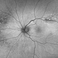
 Central Retinal Vein Occlusion with Macular Edema
Jan 29 2025 by Kimberly Wakester
Fundus photograph of a 62-year-old man with central retinal vein occlusion with macular edema and a new PVD with an operculated retinal tear in the left eye. Laser to retinal tear was completed. Patient will return in 2-3 weeks for follow up exam with possible intravitreal injection for the CRVO with edema and to follow up on the operculated retinal tear s/p retinal tear laser.
Photographer: Kimberly Wakester, COA
Imaging device: Optos California
Condition/keywords: central retinal vein occlusion (CRVO), operculated tear, PVD
-
 Pneumatic Retinopexy
Jan 6 2025 by Mateus Queiroz Corrêa, MD
Fundus photograph of a pneumatic retinopexy. The upper photo taken just 30 minutes after C3F8 gas injection shows rhegmatogenous retinal detachment with superior temporal horseshoe tear and gas bubbles resembling fish-eggs. After two days with appropriated head position (botton photo), the retina is attached and laser photocoagulation was performed on the border of the break. A Single great gas bubble was formed.
Photographer: Mateus Queiroz Corrêa, Sorocada Eye Bank Hospital
Imaging device: Optos California
Condition/keywords: pneumatic retinopexy
-
 Giant Retinal Tear with Detachment
Jan 3 2025 by Virginia Gebhart
67 year old male with mac-on RD with giant tear. Pt scheduled for sx
Photographer: Virginia Gebhart
Imaging device: Optos California
Condition/keywords: giant retinal tear, retinal tear with detachment
-
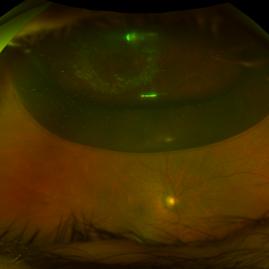
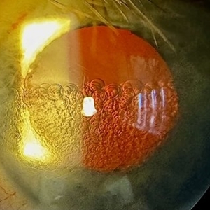
 Giant Retinal Tear with Bare Choroid
Dec 13 2024 by Thirumalesh Mochi Basavaraj, MD
Intra-operative view of a Pediatric Giant Retinal Tear with a view of the Bare Choroid Superiorly.
Photographer: Thirumalesh Mochi Basavaraj
Imaging device: Leica Proveo 8
Condition/keywords: GIANT RETINAL TEAR, Retinal Detachment
-
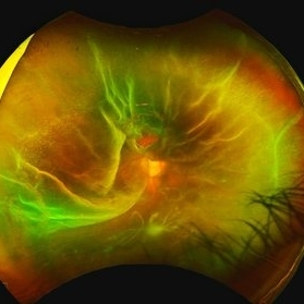
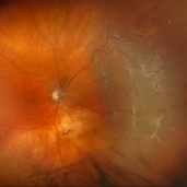
 Intraoperative View of a Giant Retinal Tear
Dec 13 2024 by Thirumalesh Mochi Basavaraj, MD
Intraoperative view of 12 year old child with Giant retinal tear with Retinal detachment.
Photographer: Thirumalesh Mochi Basavaraj
Imaging device: Lumera Proveo 8
Condition/keywords: GIANT RETINAL TEAR, PVR, Retinal Detachment
 Loading…