Search results (13 results)
-
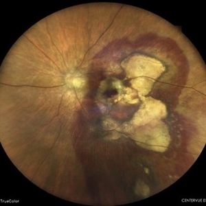
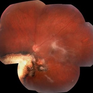
 Chorioretinal Scar
Feb 19 2024 by Sanauddin Samejo , Diploma (Ophthalmic Technician Training Course)
A patient came in to the clinic of Dr Madhav Rao (VR Surgeon).
Photographer: Sanauddin Samejo, Burjeel Hospital, Abu Dhabi, UAE.
Imaging device: Optos Silverstone
Condition/keywords: retinal scar
-
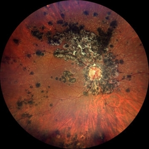
 Subretinal Hemorrhage
Feb 28 2023 by Akansha Sharma
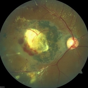
Color fundus photograph of an 84-year old male with subretinal hemorrhage associated with areas of scarring.
Photographer: Dr. Urmil Shah, Dr. Denish Patel, Dr. Akansha Sharma, Bharati Eye Hospital, Ahmedabad, Gujarat
Condition/keywords: choroidal neovascularization (CNV), subretinal hemorrhage
-
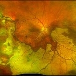
 Tuberculosis-related serpiginous-like choroiditis
Nov 22 2022 by Ricardo Leitão Guerra
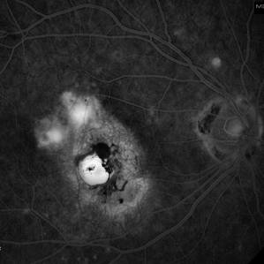
True color BLFI of a 60-year-old male presenting chorioretinal scars from a tuberculosis-related serpiginous-like choroiditis.
Photographer: Ricardo Leitão Guerra
Imaging device: Zeiss Clarus 700
Condition/keywords: serpiginous choroiditis, tuberculosis
-
 Peripheral CNVM with Extensive Scarring
Oct 12 2019 by John S. King, MD
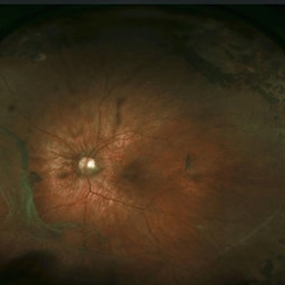
82-year-old white male with an acute loss of vision in the right eye was sent in to rule out a retinal detachment. Vision was 20/350; a dense VH was present, b-scan showed irregular areas of high reflectivity in the periphery that was c/w SRH. Peripherally, a few weeks later, there were areas that could be seen and were c/w peripheral CNVM (old and new). Anti-VEGF was administered. A month later vision was unchanged and patient wanted surgery to remove the VH. Pictured is one week since surgery; large peripheral scars are seen; diffuse areas of SR pigmentation is present; vitreous skirt present; and a few IRHs secondary to DR can be seen. He is currently 20/70 sc.
Photographer: Shelly Blair
Imaging device: Optos CA
Condition/keywords: choroidal neovascular membrane (CNVM), peripheral fundus lesion, vitreous blood
-
 Subretinal Fibrosis (PPCNVM and POHS) OS
Sep 18 2019 by John S. King, MD
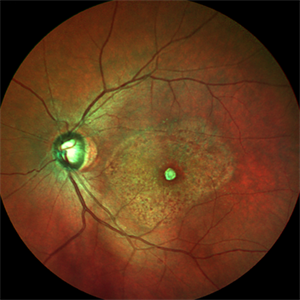
57-year-old white male with history of PPCNVM OS and POHS OU here for a routine visit. History of avastin in 2014, and stable since then. Va OS 20/20. PP scar with macular subretinal fibrosis. No heme or exudates. CR spot supero-nasally.
Photographer: Shelly Blair
Imaging device: Topcon 50
Condition/keywords: choroidal neovascular membrane (CNVM), ocular histoplasmosis syndrome (OHS), peripapillary choroidal neovascularization (PPCNVM), presumed ocular histoplasmosis syndrome (POHS)
-
 Coats' Disease Color Fundus Photograph
Apr 19 2019 by Ahmad B. Tarabishy, MD
57-year-old man referred for macular hemorrhage. Examination reveals a large macular scar along with vascular sheathing, vascular irregularity, and extensive exudation. Fluorescein angiogram revealed findings of lightbulb vascular aneurysms and capillary non-perfusion.
Imaging device: Zeiss Cirrus Photo
Condition/keywords: Coats' disease
-
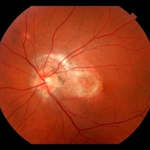
 Macular Scar
Jan 22 2018 by Jason Griffith
44-year-old male with history of CR scarring secondary to histoplasmosis.
Photographer: Jason Griffith, Tennessee Retina, Nashville TN
Imaging device: Topcon TRC 50EX
Condition/keywords: macular scar
-
 Macular Scar Due to Cysticercosis Fundus Photo
Aug 8 2017 by Manuel A Paez-Escamilla, MD, FICO
Fundus photograph of a 69-year-old patient with a long history of eye inflammation and progressive decrease in vision. Multiple trips to Asia and eating undercooked pork.
Photographer: Mark Erickson CRA, COT. The Macula Center. Clearwater, Florida
Condition/keywords: cysticercosis, uveitis
-
 Stickler Syndrome
Dec 8 2016 by Aleksandra V. Rachitskaya, MD, FASRS
Optos wide-field fundus image of a patient with Stickler Syndrome and COL2A1 gene mutation. Patient has perviously undergone prophylactic laser. Lattice, vitreous veils, and laser scars are seen.
Photographer: Anne Pinter, Cole Eye Institute, Cleveland Clinic
Condition/keywords: Stickler Syndrome, vitreoretinal degeneration
-
 Toxoplasmosis Associated Epiretinal Membrane
Oct 27 2016 by Gabriel Costa Andrade, PhD
Fundus photograph of a 26-year-old woman with a chorioretinal scar due to toxoplasmosis and secondary epiretinal membrane.
Photographer: Gabriel Andrade, Federal University of São Paulo, São Paulo, Brazil
Condition/keywords: epiretinal membrane (ERM), toxoplasmosis
-
 Chronical Submacular Hemorrhage in the Setting of Neovascular AMD
Mar 23 2015 by Rita Couceiro, MD, MS
An 80-year-old male, with a history of hypertension and high cholesterol, complained of acute and painless vision loss in his left eye (OS) in the previous 5 months. On observation best corrected visual acuity in OS was hand motion. A dense vitreous opacity in OS precluded fundus examination. Ocular ultrasound revealed vitreous hemorrhage and thickening of the macular area. The patient was submitted to pars plana vitrectomy, which disclosed a large submacular hemorrhage with chronical features and disciform scarring in the setting of neovascular AMD.
Imaging device: Intraoperative fundus photograph
Condition/keywords: neovascular age-related macular degeneration (AMD), submacular hemorrhage, wet age-related macular degeneration (wet AMD)
-
 Scar From IOFB
Nov 26 2014 by Jared Watson
28-year-old male history of corneal perforation following a grinding wheel explosion with subsequent IOFB identified by CT. Temporal scar with associated striae to the fovea, small heme superotemporal striae. Laser scars temporal to fovea.
Photographer: Jared Watson
Condition/keywords: intraocular foreign body
-
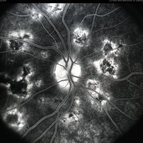 APMPPE, Late Stage, Scar Formation
Mar 4 2013 by Henry J. Kaplan, MD
APMPPE, late stage, scar formation, F/A #2
Condition/keywords: acute posterior multifocal placoid pigment epitheliopathy (APMPPE), white dot syndrome
 Loading…