 Initializing download.
-
 By John S. King, MD
Retina Associates, PA
Co-author(s): Rickey Medlock, MD - Uploaded on Oct 12, 2019.
- Last modified by Caroline Bozell on Feb 28, 2020.
- Image of the week
-
Mar 1, 2020
View all images of the week - Rating
- Appears in
- Miscellaneous
- Condition/keywords
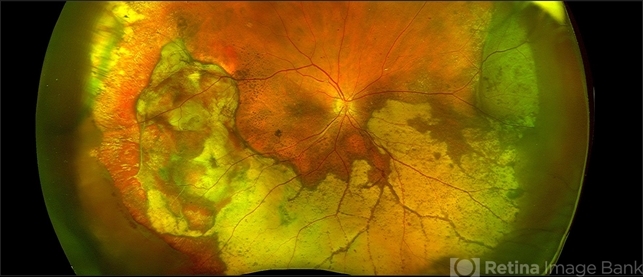
- choroidal neovascular membrane (CNVM), peripheral fundus lesion, vitreous blood
- Photographer
- Shelly Blair
- Imaging device
-
Fundus camera
Optos CA - Description
- 82-year-old white male with an acute loss of vision in the right eye was sent in to rule out a retinal detachment. Vision was 20/350; a dense VH was present, b-scan showed irregular areas of high reflectivity in the periphery that was c/w SRH. Peripherally, a few weeks later, there were areas that could be seen and were c/w peripheral CNVM (old and new). Anti-VEGF was administered. A month later vision was unchanged and patient wanted surgery to remove the VH. Pictured is one week since surgery; large peripheral scars are seen; diffuse areas of SR pigmentation is present; vitreous skirt present; and a few IRHs secondary to DR can be seen. He is currently 20/70 sc.


---thumb.jpg/image-square;max$79,0.ImageHandler "Peripheral Fundus Lesion")


