Search results (834 results)
-
---thumb.jpg/image-square;max$300,300.ImageHandler) Presumed Ocular Histoplasmosis Syndrome
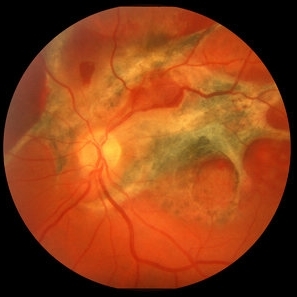
Presumed Ocular Histoplasmosis Syndrome
Feb 26 2013 by Henry J. Kaplan, MD
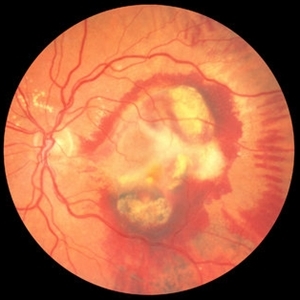
Color fundus photograph of the right eye of a patient with POHS shows typical punched out scars and peripapillary atrophy.
Condition/keywords: presumed ocular histoplasmosis syndrome (POHS)
-
---thumb.JPG/image-square;max$300,300.ImageHandler) Disciform Scar
Disciform Scar
Jul 13 2013 by Jason S. Calhoun
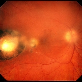
Poor central vision in the left eye due to macular degeneration. Disciform scar.
Photographer: Jason S. Calhoun, Department of Ophthalmology, Mayo Clinic Jacksonville, Florida
Imaging device: TOPCON TRC 50-EX
Condition/keywords: disciform scar, macular degeneration
-
 Ocular Toxocariasis slide 1
Oct 22 2012 by Ronald C. Gentile, MD
40-year-old man from South America was referred for a peripheral retinal scar in his left eye. He had a history of conjunctivitis as a child with exposure to multiple pets (cats and dogs). Fundus photo revealed a peripheral scarred sub-retinal granuloma located superior nasal with a retinal fold and traction extending to the optic nerve.
Photographer: The New York Eye & Ear Infirmary Department of Medical Imaging
Condition/keywords: toxocariasis
-
 Disciform Scar
Jul 13 2013 by Jason S. Calhoun
Chorioretinal scar inferior temporal in the right eye of a middle aged patient.
Photographer: Jason S. Calhoun, Department of Ophthalmology, Mayo Clinic Jacksonville, Florida
Condition/keywords: chorioretinal scar
-
---thumb.jpg/image-square;max$300,300.ImageHandler) Multifocal Choroiditis and Panuveitis Syndrome
Multifocal Choroiditis and Panuveitis Syndrome
Feb 26 2013 by Henry J. Kaplan, MD
Multifocal choroiditis, left eye: multiple punched out scar formations in the posterior pole.
Condition/keywords: multifocal choroiditis, panuveitis
-
 ARMD with Disciform Scar
Oct 16 2012 by Jeffrey G. Gross, MD, FASRS
ARMD with disciform scar, RPE contracture, and subretinal hemorrhage, CF.
Condition/keywords: disciform scar, retinal pigment epithelium (RPE) contracture, subretinal hemorrhage
-
 Toxoplasma Retinochoroiditis
Feb 25 2013 by Henry J. Kaplan, MD
Toxoplasmosis, right eye: reactivation of congenital toxoplasmosis as an active retinitis lesion with overlying vitritis adjacent to an old scar.
Condition/keywords: toxoplasmosis chorioretinitis, toxoplasmosis reactivation
-
---thumb.jpg/image-square;max$300,300.ImageHandler) Multifocal Choroiditis
Multifocal Choroiditis
Feb 26 2013 by Henry J. Kaplan, MD
Multifocal choroiditis, MFC, inactive scars in the periphery.
Condition/keywords: multifocal choroiditis
-
 Ocular Toxoplasmosis Scar, Fluorescein Angiogram
Aug 23 2012 by Gerardo Garcia-Aguirre, MD
Fluorescein angiogram showing a large hypofluorescent round lesion with well-defined borders, where the fluorescence of the choroidal vessels is observed.
Photographer: Noemí Hernández, Asociación para Evitar la Ceguera en México
Imaging device: Zeiss FF4
Condition/keywords: toxoplasmosis
-
 Ocular Toxocariasis slide 1
Oct 22 2012 by Ronald C. Gentile, MD
8-year-old boy with a history of puppy exposure failed his school screening in the right eye. Fundus examination revealed a old scarred granuloma involving the macula. Serum testing for anti-Toxocara antibodies were positive.
Photographer: The New York Eye & Ear Infirmary Department of Medical Imaging
Condition/keywords: scarred granuloma, toxocariasis
-
 Scleral Buckle and Cryo Color
Dec 29 2012 by Barbara Parolini, MD
Panoramic fundus photograph of a 55-year-old man after episcleral sugary for retinal detachment. An encircling scleral buckle and a superotemporal cryotherapy scar are visible.
Photographer: Barbara Parolini, MD
Imaging device: Daytona
Condition/keywords: scleral buckle
-
 Secondary Choroidal Neovascularization Due to Toxoplasmosis
Feb 25 2013 by Henry J. Kaplan, MD
Left eye: secondary choroidal neovascularization and subretinal hemorrhage in a patient with old macular scar of toxoplasma.
Condition/keywords: choroidal neovascularization (CNV), toxoplasmosis, toxoplasmosis chorioretinitis
-
 Macular Scar
Apr 14 2014 by Dipankar Barua, M.Sc
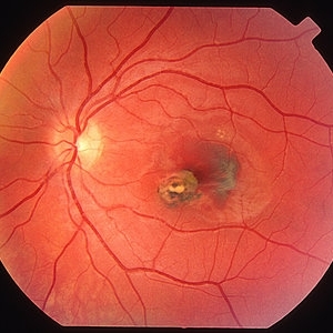
Female patient, 25-years-old. On examination, her vision of the right eye is 6/60 and left eye is 6/6. It seems to be a case of macular scar.
Photographer: Dipankar Barua
Imaging device: Topcon TRC 50 DX (IA)
Condition/keywords: macular scar
-
 Traumatic Chorioretinal Scarring
Oct 15 2012 by Jeffrey G. Gross, MD, FASRS
Traumatic chorioretinal scarring, with less hemorrhage, 1 month later.
Condition/keywords: chorioretinal scar
-
 Atrophic Scar
Oct 16 2012 by Ratimir Lazic, MD, PhD
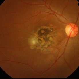
Color fundus image of a 76-year-old female. In this color image the atrophic scar in large macular area and mild periphery can be seen. BCVA on that eye is CF on 1m.
Photographer: Marko Lukic, MD
Imaging device: Zeis Visucam Lite 2
Condition/keywords: atrophic scar, retinal pigment epithelium
-
 Toxoplasma chorioretinitis 2
Jan 11 2013 by Alex P. Hunyor, MD
Toxoplasmosis 2 - recurrent toxoplasma chorioretinitis at the margin of previous scar.
Condition/keywords: ocular toxoplasmosis, toxoplasmosis, toxoplasmosis retinitis
-
 Toxoplasma chorioretinitis 1
Jan 11 2013 by Alex P. Hunyor, MD
Toxoplasmosis 1 - chorioretinal scar from previous toxoplasma chorioretinitis. See image 2 - recurrent todo adjacent to this scar
Condition/keywords: inactive toxoplasmosis, ocular toxoplasmosis, toxoplasmosis, toxoplasmosis retinitis
-
 Chorioretinal Scar
Apr 1 2016 by Nichole Lewis
Chorioretinal scar.
Photographer: Nichole Lewis - Pennsylvania Retina Specialists, Camp Hill, PA
Condition/keywords: chorioretinal scar
-
---thumb.jpg/image-square;max$300,300.ImageHandler) Presumed Ocular Histoplasmosis Syndrome
Presumed Ocular Histoplasmosis Syndrome
Feb 26 2013 by Henry J. Kaplan, MD
POHS; left eye: large punched out pigmented scars and peripapillary atrophy.
Condition/keywords: presumed ocular histoplasmosis syndrome (POHS)
-
---thumb.jpg/image-square;max$300,300.ImageHandler) APMPPE Late Stage Scar Formation
APMPPE Late Stage Scar Formation
Feb 27 2013 by Henry J. Kaplan, MD
APMPPE late stage, multiple scar formation, left eye #2.
Condition/keywords: acute posterior multifocal placoid pigment epitheliopathy (APMPPE), late stage, white dot syndrome
-
---thumb.jpg/image-square;max$300,300.ImageHandler) Toxo Macular Scar
Toxo Macular Scar
Oct 15 2013 by Sjakon G Tahija, MD
Fundus photograph of a 22-year-old woman with a congenital choreoretinal scar from toxoplasma in the left eye. Vision in the right eye is 0.05. The right eye is NLP.
Condition/keywords: toxoplasmosis
-
 Ocular Toxocariasis slide 3
Oct 22 2012 by Ronald C. Gentile, MD
The sub-retinal scarred granuloma was white in color and elevated. It had pigment speckling around it. Serum testing was positive for past exposure to Toxocara canis.
Photographer: The New York Eye & Ear Infirmary Department of Medical Imaging
Condition/keywords: toxocariasis
-
---thumb.jpg/image-square;max$300,300.ImageHandler) Multifocal Choroiditis & Panuveitis Syndrome
Multifocal Choroiditis & Panuveitis Syndrome
Feb 26 2013 by Henry J. Kaplan, MD
Multifocal choroiditis, MFC, old scars.
Condition/keywords: multifocal choroiditis, panuveitis
-
 Choroidal Hemangioma
Oct 20 2012 by Hyung-Woo Kwak, MD
Fundus, ICG, and OCT examination showed a typical chorioretinal scar lying concentric to the optic disc. Typical choroidal rupture was performed after intravitreal gas injection under the diagnosis of submacular hemorrhage caused by trauma, after the absorption of submacular hemorrhage
Condition/keywords: chorioretinal scar, choroidal rupture, submacular hemorrhage
-
---thumb.jpg/image-square;max$300,300.ImageHandler) Acute Posterior Multifocal Placoid Pigment Epitheliopathy Late Stage Scar Formation
Acute Posterior Multifocal Placoid Pigment Epitheliopathy Late Stage Scar Formation
Feb 27 2013 by Henry J. Kaplan, MD
APMPPE late stage scar formation. Right Eye Multiple scar formations occurs in some of the patients #1
Condition/keywords: acute posterior multifocal placoid pigment epitheliopathy (APMPPE), late stage, white dot syndrome
 Loading…