File number: 27312
Comments
-
 Suber S. Huang, MD, MBA, FASRS (October 13 2017)
Great case!
-
 Manuel A Paez-Escamilla, MD, FICO (August 25 2017)
Thank you for your comment. Yes, this patient had a CT of the head performed where intracerebral parenchymal calcifications could be seen, the only way to accurately diagnose active cysticercosis would be with either a biopsy or visualization of the parasite. Eosinophilia was present on CBC which agrees with a parasitic infection, ELISA was not performed because its only positive in 50% of cases.
Patient history, clinical examination and presumptive imaging all agreed with Ocular cysticercosis. To our knowledge the patient hasnt had any neurological symptoms (I.E convulsions) even though ocular cysticercosis should be considered neurocysticercosis. - Suber S. Huang, MD, MBA, FASRS (August 25 2017)
Great picture. Please additional clinical details of workup. Parasitic disease was implied, is there laboratory evidence for this?
Sign in to comment.
 Initializing download.
-
 By Manuel A Paez-Escamilla, MD, FICO
Palmetto Retina Center
Co-author(s): Dana M. Deupree M.D. FACS. The Macula Center. Clearwater, Florida - Uploaded on Aug 8, 2017.
- Last modified by Chayal Patel on Jan 14, 2018.
- Image of the week
-
Jan 21, 2018
View all images of the week - Rating
- Appears in
- 8-Aug-2017
- Condition/keywords
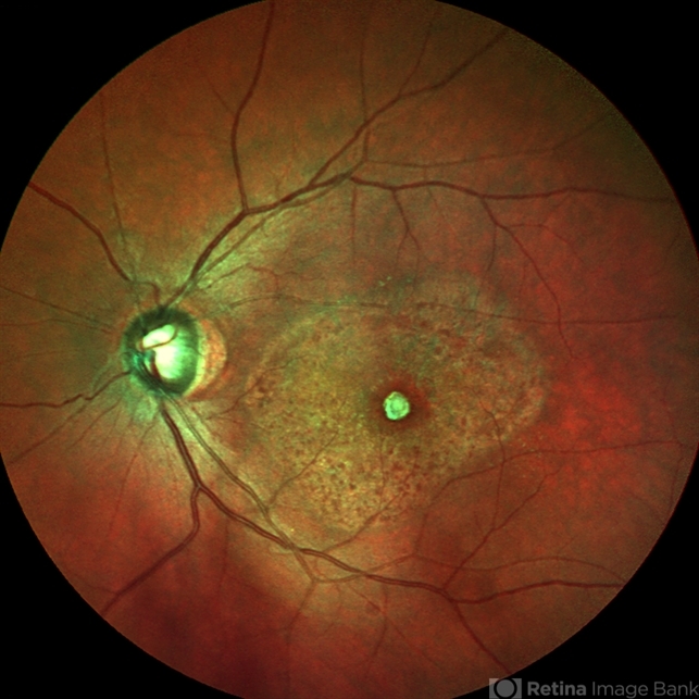
- uveitis, cysticercosis
- Photographer
- Mark Erickson CRA, COT. The Macula Center. Clearwater, Florida
- Imaging device
- Fundus camera
- Description
- Fundus photograph of a 69-year-old patient with a long history of eye inflammation and progressive decrease in vision. Multiple trips to Asia and eating undercooked pork.


---thumb.jpg/image-square;max$79,0.ImageHandler "Cysticerosis")


