Search results (834 results)
-
 Cracking the Angioid Streaks Mystery: Multimodal Mayhem
Nov 26 2025 by SHRADDHA RAJ SHRIVASTAVA
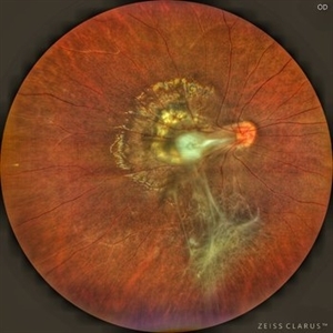
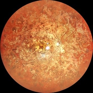
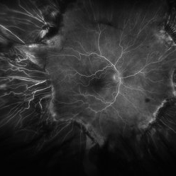
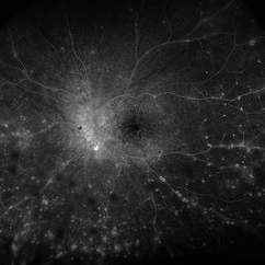
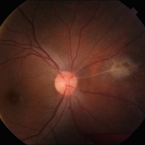
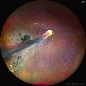
Multimodal imaging of right eye fundus showing Angioid Streaks with scarred CNVM. Color fundus photo shows hyperpigmented irregular lines emanating from the disc in a radiating fashion. Surrounding the angioid streaks and at the posterior pole, we can see numerous dot-like hypopigmented deposits and a disciform scar at macula. G-FAF images better reveal more extensive hypoautofluorescent streaks than are apparent on standard fundus photo. Characteristic “Para-streak phenomenon” of focal hyperautofluorescent spots are seen along the margins of the dark angioid streaks, corresponding to the areas of pigment clumping seen clinically. The para-streak pigment clumps are better delineated on the novel Retro-imaging method, appearing as raised bumps surrounding the angioid streaks.
Photographer: Dr. Shraddha Raj Shrivastava
Imaging device: Nidek Mirante SLO/OCT (Confocal scanning/Spectral domain OCT)
Condition/keywords: Angioid Streaks, Bruch's membrane, disciform scar, fundus autofluorescence (FAF), multimodal imaging, retro mode
-
 Retinitis Pigmentosa: Now available in its Pericentral edition
Nov 7 2025 by SHRADDHA RAJ SHRIVASTAVA
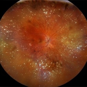
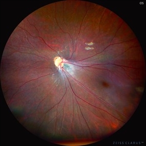
Right eye fundus photo of a 50 year old patient, diagnosed with bilateral Pericentral variant of Retinitis Pigmentosa. True to the subtype, the pigmentation is closer to fixation. There are bony spicules like pigmentary changes and RPE atrophy seen around the macula and disc (posterior pole), just adjacent to the arcades, while the peripheral fundus appears unaffected. The macula shows severe macular atrophy and scarring. Similar changes were observed in the left eye.
Photographer: Dr. Shraddha Raj Shrivastava
Imaging device: Nidek Mirante SLO/OCT (Confocal scanning/Spectral domain OCT)
Condition/keywords: pericentral retinitis pigmentosa, retinitis pigmentosa (RP) dystrophy, Rod cone dystrophy, RP variant
-
 The Retinal Tempest: Toxocara's Trail
Aug 31 2025 by Giriraj Vibhute
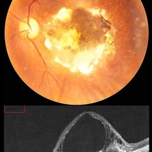
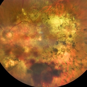
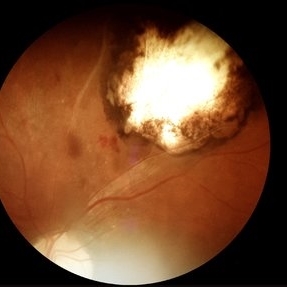
In this striking image, a central white granuloma spirals outward from the optic disc, surrounded by fibrous traction bands and scarring—the telltale markings of intraocular toxocara lesion. The retina is ravaged with proliferative vitreoretinal membranes and peripheral pigmentary changes, starkly illustrating the chronic inflammation and vision-threatening complications caused by Toxocara canis.
Photographer: Dr Giriraj Vibhute, MM Joshi eye institute, Hubli, India.
Condition/keywords: dragged disc, fibrous proliferation, Toxocara, toxocara canis
-
 Healed AZOOR- Multiple White Dot Syndrome
Aug 29 2025 by Aditya S Kelkar, MS, FRCS, FASRS,FRCOphth
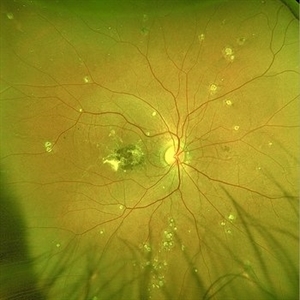
Fundus photograph of a 58 year old woman with multiple, well-defined, punched-out chorioretinal scars scattered throughout the posterior pole and mid-periphery. The macular area shows a large, confluent, yellowish-white scar involving the fovea.
Photographer: Dr. Muskan Mangal
Imaging device: Optos Daytona
Condition/keywords: multifocal choroiditis, multiple evanescent white dot syndrome (MEWDS), punctate inner choroidopathy (PIC)
-
 Macular Mount Everest
Aug 8 2025 by Anand Temkar
A 75 yrs old male came with the chief complains of DOV in LE since past 20 yrs. His BCVA in RE was 6/9 and in LE, it was CF 1 meter. His IOP was 13 mm of Hg in RE and 15 mm of Hg in LE. Patient is a k/c/o DM type 2 since past 20 yrs and is on regular medication. Patient is a k/c/o solitary kidney. Patient gives h/o ( LE ) Intravitreal injection Avastin 3 times 13 yrs ago i/c/o CNVM. In the LE color photo we can see the scarred CNVM along with altered foveal contour. LE OCT also shows cystic spaces with large elevation and scarring.
Photographer: Dr.Anand Temkar- Vasan Eye Hospital, Tiruchirapalli
Condition/keywords: CNVM, macular edema, scarred cnvm
-
 Pseudoxanthoma Elasticum
Aug 7 2025 by Alind Murkhe
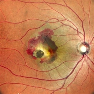
Fundus photograph of a 42 year-old male with pseudoxanthoma elasticum showing Angiod streak, scarred CNVM, Comet tails lesion.
Photographer: Dr Alind Murkhe, Nandadeep Eye Hospital, Sangli, Maharashtra, India
Condition/keywords: Angiod streaks in Pseudoxanthoma elasticum, CNVM
-
 Retinal Aneurysms
Aug 6 2025 by Korey Starkey
54 year-old patient presents with scattered peripheral aneurysms with exudates. FA was performed showing peripheral nonperfusion and aneurysms. Treated patient with PRP and focal laser to aneurysms and continued observation.
Photographer: Kore Starkey
Imaging device: Optos
Condition/keywords: aneurysm, branch retinal vein occlusion (BRVO), chorioretinal scar, circinate ring, exudates, fundus photography, lesion, Optos, retinal aneurysms
-
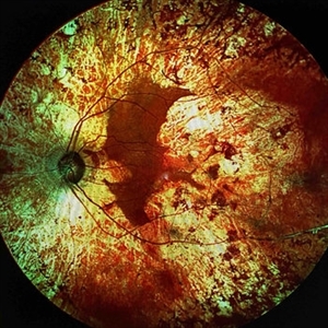
 Ghost Map Retina
Aug 4 2025 by Malvika Singh
Fundus photograph of a 50 year old male showing extensive chorioretinal scarring.
Photographer: Dr Malvika Singh, Retina Foundation, Ahmedabad, India
Imaging device: Mirante SLO/OCT
Condition/keywords: healed choroiditis
-
 Chronic RD with Retinal Dialysis
Jul 23 2025 by Virginia Gebhart
64 year old female with chronic retinal detachment from head trauma 41 years ago. Peripheral scarring from 6:00 to 11:00 with area of subretinal fluid inferotemporally, well demarcated with subretinal bands. Retinal dialysis inferotemporal from 7:00 to 9:00. No surgical repair needed or recommended at this time.
Photographer: Virginia Gebhart, Retina Consultants of Carolina
Imaging device: Optos California
Condition/keywords: chronic retinal detachment, demarcation, RD, Retinal Detachment, retinal dialysis, subretinal bands
-
 Fluorescein Angiogram of ROP With Cryo Scarring
Jul 7 2025 by Jenn Geelan
FA photo of a 34 year old male with prior stage 3 ROP with history of 360 degree cryotherapy.
Photographer: Jenn Geelan, Retina-Vitreous Surgeons of CNY
Imaging device: Optos California
Condition/keywords: cryotheraphy scar, fluorescein angiogram (FA), fundus photograph, retinopathy of prematurity (ROP), ROP, tilted disc
-
 VKH Syndrome
Jun 12 2025 by Virginia Gebhart
22 year old male with VKH Syndrome. Pt has been experiencing severe headaches, distorted vision, hearing loss, weakness, and a large white patch of hair. Significant cell in AC and vitreous, multiple punched-out CR scars in periphery. Referred to rheumatology for possible immunomodulatory treatment
Photographer: Virginia Gebhart, Retina Consultants of Carolina
Imaging device: Optos California
Condition/keywords: montage, multifocal choroiditis, panuveitis, Vogt-Koyanagi-Harada
-
 VKH Syndrome
Jun 12 2025 by Virginia Gebhart
Fluorescein angiogram of 22 year old male with VKH syndrome. Significant cell in AC and vitreous, multiple punched-out CR scars in periphery, mild vascular leakage. Pt referred to rheumatology for immunomodulatory treatment.
Photographer: Virginia Gebhart, Retina Consultants of Carolina
Imaging device: Optos California
Condition/keywords: FA, fluorescein angiogram (FA), multifocal choroiditis, panuveitis, VKH, Vogt-Koyanagi-Harada
-
 Franceschetti's Sign
Jun 5 2025 by César Adrián Gómez Valdivia, MD
Franceschetti's sign found in a 22 year-old female patient diagnosed with ocular toxoplasmosis. These bands typically link an old scar to the optic disc, indicative of previous inflammation. Findings were unilateral.
Photographer: @eyemissu2
Imaging device: TOPCON TRC-50DX
Condition/keywords: chorio, Franceschetti's Sign, toxoplasmosis
-
 When the Macula Decides to Bleed... Artistically (Case of Macular Scar with Subretinal Bleed)
Jun 2 2025 by rohan jain
A case of 42 years old male. Color photograph showing macular scar with subretinal bleed.
Photographer: Dr. ROHAN JAIN
Imaging device: mirante
Condition/keywords: CNVM, macular scar, scar, subretinal hemorrhage, subretinal blood
-
 PCV
PCV
May 20 2025 by LUBNA AHMAD
Fundus image of a 66 year old male with huge branching vascular network, fresh hemorrhage and scarred PCV lesion.
Photographer: Sana Zamani
Imaging device: zeiss clarus 500
Condition/keywords: branching vascular network (BVN), polypoidal choroidal vasculopathy (PCV)
-
 Chorioretinal Macula Scar (Macula View)
May 12 2025 by Briana Hernandez
Zoomed in Macular View of Chorioretinal Macular Scar in 9-year-old female patient.
Photographer: Briana Hernandez, Hilton Head Retina Insitute
Imaging device: Optos
Condition/keywords: chorioretinal scar
-
 Chorioretinal Macula Scar (Ultrawide View)
May 12 2025 by Briana Hernandez
Ultra wide Optos image of Chorioretinal Macular Scar in 9-year-old female patient.
Photographer: Briana Hernandez, Hilton Head Retina Institute
Imaging device: Optos
Condition/keywords: chorioretinal scar, macular scar, ultra-wide field imaging
-
 Retinocoroiditis Inactiva Por Toxoplasmosis
Apr 28 2025 by Paulina Araujo
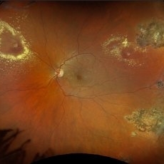
Fundus photography demonstrates a 2-disc-diameter chorioretinal scar in the superior temporal arcade, consistent with inactive toxoplasmic retinochoroiditis. The lesion exhibits pigmented borders and central atrophy, with adjacent splinter hemorrhages and vascular sheathing. No vitreous inflammation or active satellite lesions are present.
Photographer: Paulina D.Araujo Martínez, Asociación para Evitar la Ceguera en México I.A.P., Hospital Dr Luis Sánchez Bulnes.
Condition/keywords: toxoplasmosis chorioretinitis
-
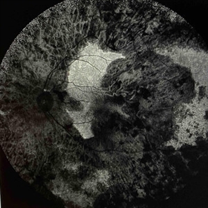 Extensive Chorioretinal Scarring With Partial Macular Sparing
Apr 22 2025 by Maxwell J Wingelaar, MD
Fundus autofluorescence of extensive chorioretinal scarring in the left eye.
Photographer: Killian Roberts
Imaging device: Heidelberg Spectralis AF
Condition/keywords: chorioretinal atrophy, chorioretinal inflammations
-
 Extensive Chorioretinal Scarring with Partial Macular Sparring
Apr 22 2025 by Maxwell J Wingelaar, MD
A multicolor photo showing chorioretinal scarring with partial macular sparing in the left eye.
Photographer: Killian Roberts
Imaging device: Heidelberg Spectralis Multicolor Photo
Condition/keywords: chorioretinal atrophy, chorioretinal inflammations
-
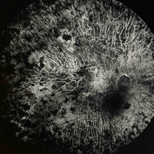 Extensive Chorioretinal Scarring in the Right Eye
Apr 22 2025 by Maxwell J Wingelaar, MD
Fundus autofluorescence of Extensive chorioretinal scarring in the right eye.
Photographer: Killian Roberts
Imaging device: Heidelberg Spectralis AF
Condition/keywords: chorioretinal atrophy, chorioretinal inflammations
-
 Extensive Chorioretinal Scarring in the Right Eye
Apr 22 2025 by Maxwell J Wingelaar, MD
A multicolor photo showing chorioretinal scarring with macular involvement in the right eye
Photographer: Killian Roberts
Imaging device: Heidelberg Spectralis Multicolor Photo
Condition/keywords: chorioretinal atrophy, chorioretinal inflammations
-
 Comets in the Eye (Retinopathy of Prematurity)
Apr 8 2025 by KANWALJEET HARJOT MADAN, M.S. (Ophthalmology); FAICO (Vitreous - Retina)
This is the fundus picture of right eye (RE) of a 4 years female child presented with outward deviation of right eye. Her parents also complained of diminution of vision in both eyes. On examination, her best corrected vision in RE was hand movements close to face and was 20/80 in LE. Posterior segment exam revealed presence of macular scar in RE and presence of dry retinal fold with dragging of retinal vessels. LE fundus revealed presence of nasal drag of optic disc. Parents gave history of untreated ROP as an infant. Retinopathy of Prematurity (ROP) is a Vaso proliferative disorder of Retina occurring in premature infants. Advances in neonatal care and ROP treatment has led these babies to live longer with this disease.
Photographer: Dr. Kanwaljeet Harjot Madan, Thind Eye Hospital, Jalandhar City (Punjab) INDIA.
Imaging device: Zeiss Fundus Camera
Condition/keywords: Retinopathy of Prematurity, Vaso proliferative disorder
-
 Comets in the Eye (Retinopathy of Prematurity)
Apr 8 2025 by KANWALJEET HARJOT MADAN, M.S. (Ophthalmology); FAICO (Vitreous - Retina)
This is the fundus picture of right eye (RE) of a 4 years female child presented with outward deviation of right eye. Her parents also complained of diminution of vision in both eyes. On examination, her best corrected vision in RE was hand movements close to face and was 20/80 in LE. Posterior segment exam revealed presence of macular scar in RE and presence of dry retinal fold with dragging of retinal vessels. LE fundus revealed presence of nasal drag of optic disc. Parents gave history of untreated ROP as an infant. Retinopathy of Prematurity (ROP) is a Vaso proliferative disorder of Retina occurring in premature infants. Advances in neonatal care and ROP treatment has led these babies to live longer with this disease.
Photographer: Dr. Kanwaljeet Harjot Madan, Thind Eye Hospital, Jalandhar City (Punjab) INDIA.
Imaging device: Zeiss Fundus Camera
Condition/keywords: Retinopathy of Prematurity
-
 Angioid Streaks
Mar 18 2025 by T. P . VIGNESH, MBBS,MS
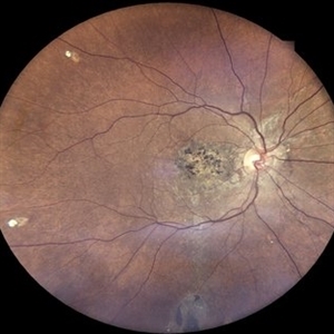
Fundus photograph of a 42-year-old woman with Angioid Streaks and scarred CNVM.
Photographer: Sivanath
Imaging device: EIDON
Condition/keywords: Angioid Streaks
 Loading…