Search results (834 results)
-
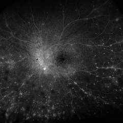
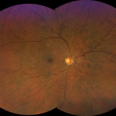
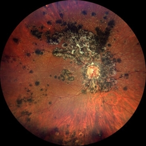 Tuberculosis-related serpiginous-like choroiditis
Nov 22 2022 by Ricardo Leitão Guerra
True color BLFI of a 60-year-old male presenting chorioretinal scars from a tuberculosis-related serpiginous-like choroiditis.
Photographer: Ricardo Leitão Guerra
Imaging device: Zeiss Clarus 700
Condition/keywords: serpiginous choroiditis, tuberculosis
-
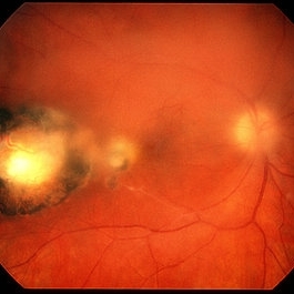
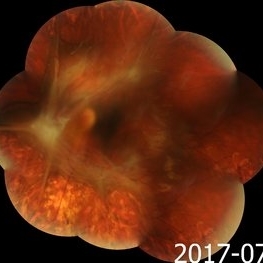
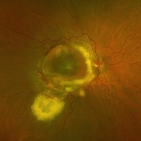
 Disciform Scar
Aug 18 2020 by Aditya S Kelkar, MS, FRCS, FASRS,FRCOphth
Left eye fundus photograph of 75-year-old male, showing large disciform scar post subretinal bleeding secondary to idiopathic polypoidal choroidal vasculopathy
Photographer: Dr.Mounika Bolisetty
Imaging device: CLARUS 500
Condition/keywords: disciform scar, idiopathic polypoidal choroidal vasculopathy
-
---thumb.jpg/image-square;max$300,300.ImageHandler) APMPPE Late Stage Scar Formation
APMPPE Late Stage Scar Formation
Feb 27 2013 by Henry J. Kaplan, MD
APMPPE late stage scar formation. F/A hypofluorescence in the lesions area is due to masking effect of pigments . #1
Condition/keywords: acute posterior multifocal placoid pigment epitheliopathy (APMPPE), late stage, white dot syndrome
-
 Chorioretinitis with Overlying Vitreous Stranding/Vitritis
Mar 23 2023 by Isaac Agranoff
Fundus photograph of a 37-year-old woman presenting with chorioretinitis with overlying vitreous stranding/vitritis that has remained unchanged for multiple years. Patient presented with irritation and blurred vision and her vision was 20/40 OD. The OCT revealed evidence of low-grade inflammation and the recommend treatment was anti-inflammatory eye drops at this time and to obtain second opinion with another physician in the office.
Photographer: Isaac Agranoff, Technician
Imaging device: Optos California
Condition/keywords: chorioretinal scar, chorioretinitis, inflammation, Optos, ultra-wide field imaging, vitritis
-
 Congenital Toxoplasmosis
Feb 2 2021 by Niloofar Piri, MD
43-year-old female with large oval chorioretinal scar in posterior pole with heavy RPE hyperplasia and history of hydrocephalus s/p VP shunt since birth. Findings are consistent with congenital toxoplasmosis.
Condition/keywords: congenital toxoplasmosis
-
 Peripheral CNVM with Extensive Scarring
Oct 12 2019 by John S. King, MD
82-year-old white male with an acute loss of vision in the right eye was sent in to rule out a retinal detachment. Vision was 20/350; a dense VH was present, b-scan showed irregular areas of high reflectivity in the periphery that was c/w SRH. Peripherally, a few weeks later, there were areas that could be seen and were c/w peripheral CNVM (old and new). Anti-VEGF was administered. A month later vision was unchanged and patient wanted surgery to remove the VH. Pictured is one week since surgery; large peripheral scars are seen; diffuse areas of SR pigmentation is present; vitreous skirt present; and a few IRHs secondary to DR can be seen. He is currently 20/70 sc.
Photographer: Shelly Blair
Imaging device: Optos CA
Condition/keywords: choroidal neovascular membrane (CNVM), peripheral fundus lesion, vitreous blood
-
 Retinocoroiditis Inactiva Por Toxoplasmosis
Apr 28 2025 by Paulina Araujo
Fundus photography demonstrates a 2-disc-diameter chorioretinal scar in the superior temporal arcade, consistent with inactive toxoplasmic retinochoroiditis. The lesion exhibits pigmented borders and central atrophy, with adjacent splinter hemorrhages and vascular sheathing. No vitreous inflammation or active satellite lesions are present.
Photographer: Paulina D.Araujo Martínez, Asociación para Evitar la Ceguera en México I.A.P., Hospital Dr Luis Sánchez Bulnes.
Condition/keywords: toxoplasmosis chorioretinitis
-
 Retinopathy of Prematurity
Aug 26 2021 by Stefanie Palmer
Patient has neovascular ridge temporal with elevation of vessels above the PRP scars. The image was obtained with the flying baby technique.
Photographer: Stefanie Palmer, CRA
Condition/keywords: retinopathy of prematurity (ROP)
-
 Retinopathy of Prematurity
Aug 26 2021 by Stefanie Palmer
Patient has neovascular ridge temporal with elevation of vessels above the PRP scars. The image was obtained with the flying baby technique.
Photographer: Stefanie Palmer, CRA
Condition/keywords: retinopathy of prematurity (ROP)
-
 Toxoplasma Retinochoroiditis
Feb 25 2013 by Henry J. Kaplan, MD
Toxoplasmosis, right eye: reactivation of congenital toxoplasmosis as an active retinitis lesion with overlying vitritis adjacent to an old scar.
Condition/keywords: toxoplasmosis chorioretinitis, toxoplasmosis reactivation
-
 VKH Syndrome
Jun 12 2025 by Virginia Gebhart
Fluorescein angiogram of 22 year old male with VKH syndrome. Significant cell in AC and vitreous, multiple punched-out CR scars in periphery, mild vascular leakage. Pt referred to rheumatology for immunomodulatory treatment.
Photographer: Virginia Gebhart, Retina Consultants of Carolina
Imaging device: Optos California
Condition/keywords: FA, fluorescein angiogram (FA), multifocal choroiditis, panuveitis, VKH, Vogt-Koyanagi-Harada
-
---thumb.jpg/image-square;max$300,300.ImageHandler) Acute Toxoplasmosis
Acute Toxoplasmosis
Aug 14 2013 by From the Collections of Thomas M. Aaberg, MD and Thomas M. Aaberg Jr., MD
Focal retinitis with scar.
Condition/keywords: acute toxoplasmosis, focal retinitis
-
 Advanced Proliferative Diabetic Retinopathy
Nov 4 2017 by Hamid Ahmadieh, MD
Merged color fundus photograph of the left eye of a 30-year-old woman with type1 diabetes since childhood. Note laser scars, severe fibrous proliferation, traction RD and macular dragging.
Photographer: Shabnam Poureh, Negah Eye Center, Tehran, Iran
Condition/keywords: color fundus photograph, diabetes, fibrous proliferation, proliferative diabetic retinopathy (PDR), severe traction
-
---thumb.jpg/image-square;max$300,300.ImageHandler) Age Related Macular Degeneration - Geographic Atrophy
Age Related Macular Degeneration - Geographic Atrophy
May 3 2013 by Suber S. Huang, MD, MBA, FASRS
Geographic Atrophy.
Imaging device: Retina Diseases Imaging Analysis Reading Center
Condition/keywords: advanced geographic atrophy, atrophic scar, atrophic spot, geographic atrophy, macula lesion, pigment epithelial atrophy
-
 BDUMP
BDUMP
Dec 11 2018 by John S. King, MD
67-year-old white female with normal vision four months ago, consulted for dry AMD. She reported that vision in the left eye had worsened over the last two months and had progressively gotten worse. Denied history of cancer, or her primary eye doctor ever mentioning choroidal nevi. Va cc was 20/30 OD and 20/100 OS. No RAPD. IOP 9-10 OU. Anterior segment had some stellate like pigmented dusting of the endothlium, a/c was quiet, 2+NSC OU. Vitreous quiet; multiple, flat, pigmented choroidal lesions varying in size was seen the in fundus. Area in the temporal macula extending up to the superior arcade in the left eye that was suspicious for a mass; it did have a "giraffe like" pattern on one of the early FA pics; the OCT in this area showed thickening of the choroid without a definite mass lesion, and overlying thickening of the RPE, or exudative like scar, with SRF directly above. Consulted with Dr. Matt Wilson, who confirmed diagnosis, and had patient evaluated by oncology, who diagnosed non-small cell lung cancer.
Photographer: Stacey Coleman
Imaging device: Topcon
Condition/keywords: bilateral diffuse uveal melanocytic proliferation (BDUMP)
-
 Childhood Acquired Ocular Toxoplasmosis
Sep 13 2023 by Deepak Bhojwani, MS
Fundus image of a 16 year old boy diaagnosed with Ocular Toxoplasmosis since the age of 10 years showing the classic toxo chorioretinitis scar on the posterior pole. Luckily the scar is loacted juxtatemporal to fovea on OCT and so the boy has good vision of 20/30.
Photographer: DR DEEPAK BHOJWANI
Imaging device: OPTCAL COHERENCE TOMOGRAPHY
Condition/keywords: posterior uveitis, toxo chorioretinitis
-
 Choroidopathy
May 27 2020 by Jamin S. Brown, MD
Fluorescein angiography image of 28 year old female with focal chorioretinal inflammation, macular or paramacular OS. chorioretinal scar OS.
Photographer: Jeffrey Barker, Retina-Vitreous Surgeons of CNY
Condition/keywords: choroidopathy
-
 Chronical Submacular Hemorrhage in the Setting of Neovascular AMD
Mar 23 2015 by Rita Couceiro, MD, MS
An 80-year-old male, with a history of hypertension and high cholesterol, complained of acute and painless vision loss in his left eye (OS) in the previous 5 months. On observation best corrected visual acuity in OS was hand motion. A dense vitreous opacity in OS precluded fundus examination. Ocular ultrasound revealed vitreous hemorrhage and thickening of the macular area. The patient was submitted to pars plana vitrectomy, which disclosed a large submacular hemorrhage with chronical features and disciform scarring in the setting of neovascular AMD.
Imaging device: Intraoperative fundus photograph
Condition/keywords: neovascular age-related macular degeneration (AMD), submacular hemorrhage, wet age-related macular degeneration (wet AMD)
-
 Coat's Disease
Jan 14 2025 by Kimberly Wakester
Fundus photographs of an 7-year-old boy with Coat's Disease in the right eye. There is subfoveal lipid end scarring in the macula and "light bulb" type telangiectasias temporally noted on exam and shown in Optos color photos. FA findings show anastomoses, capillary dropout, and "light bulb" type telangiectasias temporally with mild late leakage. Patient will be monitored at this time and have repeat imaging in 4 months.
Photographer: Kimberly Wakester, COA
Imaging device: Optos California
Condition/keywords: Coat's disease
-
 Congenital Hypertrophy of RPE: "Bear Tracks"
Aug 5 2021 by Niloofar Piri, MD
Ultrawide field fundus photograph of a 79-year-old patient who was incidentally found to have extensive bear track lesions in both eyes. Left eye was treated for NVG in the past and bear tracks were only visible temporal to the macula where there was no laser scars. He was referred to be seen by gastroenterologist and have a colonoscopy given high association with FAP and Gardner's syndrome.
Photographer: Jacob Grodsky, MD, St. Louis University
Condition/keywords: bear tracks, congenital hypertrophy of the retinal pigment epithelium (CHRPE)
-
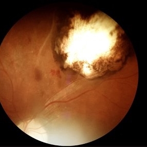
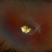
 DISCIFORM SCAR AND RETINAL PIGMENT EPITHELIUM (RPE) DETACHMENT IN A CASE OF IDIOPATHIC POLYPOIDAL CHOROIDAL VASCULOPATHY (IPCV)
Oct 21 2023 by Aditya S Kelkar, MS, FRCS, FASRS,FRCOphth
Right eye fundus photograph of a 83 year old female demonstrating Disciform Scar And Retinal Pigment Epithelium (RPE) Detachment In A Case Of Idiopathic Polypoidal Choroidal Vasculopathy (IPCV).
Photographer: DR APURVA MUKADAM
Imaging device: OPTOS DAYTONA
Condition/keywords: disciform scar
-
 Disseminated Chorioretinitis With Unknown Etiology
Apr 5 2018 by Kim Barrett
Ultra-wide field fluorescein angiogram of a 31-year-old female with intermittent pain in her left eye. Her condition has been managed in Liberia until recently when she moved to the United States. She suffers from multiple modalities including central retinal artery occlusion, posterior synechiae of the iris, interstitial keratitis, disseminated chorioretinitis, as well as HIV. An infectious cause is high on the differential in light of her HIV status. DDx: hypertensive crisis, an embolism (? IV drug use), coagulopathy, trauma, infectious. Blood work was normal. Her current vision is 20/30 right eye and 20/400 left eye.
Photographer: Kim Barrett, COA
Imaging device: Optos
Condition/keywords: central retinal artery occlusion (CRAO), chorioretinal scar, ciliary artery sparring, disseminated chorioretinitis, HIV, left eye, optic atrophy, staining
-
 Ehlers-Danlos Syndrome
Apr 22 2021 by Harita Shah
Fundus photograph of a 37-year-old male, known case of Ehlers-Danlos Syndrome, having left eye CNVM scar with angioid streaks.
Photographer: Harita Shah, Banker's Retina Clinic & Laser Centre
Imaging device: Topcon TRC 50DX
Condition/keywords: Ehlers-Danlos syndrome
-
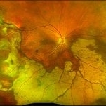
 Extensive Chorioretinal Scarring With Partial Macular Sparing
Apr 22 2025 by Maxwell J Wingelaar, MD
Fundus autofluorescence of extensive chorioretinal scarring in the left eye.
Photographer: Killian Roberts
Imaging device: Heidelberg Spectralis AF
Condition/keywords: chorioretinal atrophy, chorioretinal inflammations
-
 Fibrotic granuloma vs. Pseudoduplication of the Optic Disc
Nov 29 2023 by Virginia Gebhart
74 year-old female with presumed fibrotic granuloma. Previously diagnosed as pseudoduplication of the optic disc by general ophthalmologist. OCT showed elevation in the RPE, more consistent with granuloma. Pt has been aware for many years, asymptomatic. Will observe.
Photographer: Virginia Gebhart
Imaging device: Topcon 50DX
Condition/keywords: fibrotic scar, granuloma, Pseudoduplication of optic disc
 Loading…