Search results (24 results)
-
 Repaired Retinal Detachment with Scleral Buckle
Mar 25 2025 by Kimberly Wakester
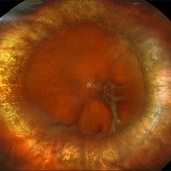
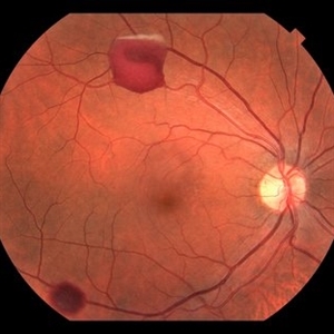
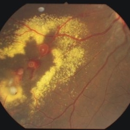
Optomap RGB montage of an 64-year-old woman with a repaired retinal detachment with scleral buckle in the right eye. There is nasal and inferior pre-retinal membranes with traction. PPV was recommended but patient defers to proceed with sx at this time. Will continue to follow patient closely for worsening traction. Patient was educated on how to monitor their peripheral vision and was advised to report any changes immediately.
Photographer: Kimberly Wakester, COA, OCT-C
Imaging device: Optos California
Condition/keywords: pre-retinal membrane with traction, repaired RD, scleral buckle
-
 Pericentral Retinitis Pigmentosa
Sep 6 2024 by Mauricio Bayram-Suverza, MD
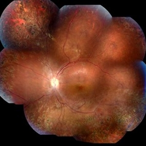
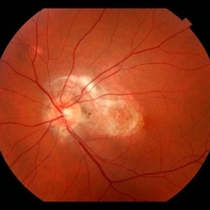
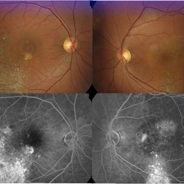
A 65-year-old male patient reports experiencing bilateral blind spots that have gradually intensified over time. Genetic testing was unrevealing. The fundus autofluorescence image shows a hypoautofluorescent ring in the posterior pole, especially nasal to the nerve and along arcades.
Photographer: Mauricio Bayram-Suverza, Casey Eye Institute, OHSU.
Imaging device: Optos California
Condition/keywords: fundus autofluorescence (FAF), inherited retinal disease, nyctalopia, retinal dystrophy, retinitis pigmentosa
-
 Optic Nerve Melanocytoma
Apr 3 2023 by Gustavo Aguirre Suarez
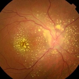
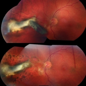
Fundus photograph of a 36-year-old female with a lesion dependent on the optic nerve head with subretinal extension, elevated, about 1.5 disc diameters, dark brown to black in color, involving more than three quarters of the neuroretinal ring towards the inferonasal area.
Photographer: Dr. Gustavo Aguirre-Suarez
Imaging device: Zeiss Visucam 500
Condition/keywords: melanocytic lesion, Melanocytoma
-
 Methotrexate Bubble following Intravitreal Injection for PVR
Sep 21 2022 by Zach Seim
Ultra-widefield fundus photograph of an 81 year old female with a Methotrexate bubble following an Intravitreal Injection for Proliferative Vitreoretinopathy. Patient has been presenting to the office for two week interval Methotrexate injections in her left eye. The image was taken prior to her eighth injection which revealed a residual Methotrexate bubble in her inferior retinal image. This patient was seeing "lots" of floaters, as well as having visual acuity of cc20/400 cc20/200 PH.
Photographer: Zach Seim
Imaging device: OPTOS California
Condition/keywords: bubble, fundus photograph, fundus photography, intravitreal injection, left eye, methotrexate, nasal retina, Optos, proliferative vitreoretinopathy (PVR), pseudocolor, ultra-wide field imaging
-
 Retinoschisis
Mar 28 2021 by JEFFERSON R SOUSA, Tecg.º (Biomedical Systems Technology)
A 14-year-old male patient was admitted for visual evaluation. Visual acuity s/c in the right eye and 20/80 in the left eye. According to family members, he reported low vision since childhood. He had already undergone treatment with photocoagulation in another service to which he had a diagnostic hypothesis of Coats' disease. Laboratory tests were requested (HIV, TOXO, TOXOCARIASIS, ECA, VDRL, PPD). In the evaluation it was observed important exudation in the posterior pole, some vascular irregularities in the right eye. In the left eye, there is retinoschisis affecting the entire posterior pole and the region nasal to the optic disc, macula with a characteristic aspect of a cartwheel. Well exemplified by OCT-A (Structrure Deep: IPL - 25, OPL - 25).
Photographer: JEFFERSON R SOUSA - Study Center and Ophthalmological Research Dr. Andre M V Gomes, Institute Dr. Suel Abujamra São Paulo-Brazil
Imaging device: Topcon TRC-50 DX, Imaginet 4.0, angle de 50 graus. Flash 50w-s
Condition/keywords: Coats' disease, retinoschisis
-
 Rhegmatogenous Retinal Detachment
Mar 3 2021 by Patrik Rajs
A 51-year-old female patient presented with inferior nasal scotoma and 5/10 vision in the right eye due to a retinal detachment with a giant retinal horseshoe tear.
Photographer: Patrik Rajs, EYE CLINIC of Jan Evangelista Purkyne University and Masaryk Hospital, Czech Republic, Ústí nad Labem
Imaging device: Clarus 700
Condition/keywords: giant retinal tear
-
 Morning Glory Disc Anomaly
Nov 11 2020 by Yoshihiro Yonekawa, MD, FASRS
Color fundus photograph of a young boy with morning glory disc anomaly. Notice the concavity surrounding the enlarged disc, radial vasculature, and nasally dragged macula. MRI was negative for moyamoya disease, a known association.
Photographer: Alicia Thresher, Mid Atlantic Retina
Imaging device: Topcon
Condition/keywords: disc coloboma, Morning Glory Syndrome, pediatric retina
-
 CMV Retinitis with Frosted Branch Angiitis
Sep 23 2020 by Nimesh A. Patel, MD, FASRS
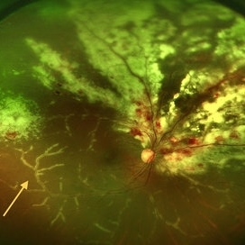
Fundus photo showing peri-vascular inflammation of both arteries and veins with translucent exudation (yellow arrow). Superior nasally, there is classic retinal whitening with retinal hemorrhages superior. This patient was found to have a low CD4 count and a diagnosis of AIDS was made.
Condition/keywords: cytomegalovirus (CMV), HIV, uveitis
-
 Blunt Ocular Trauma Due to Firework Injury
Jun 9 2020 by Brittany Rota
Ultra- widefield pseudocolor image of an 18-year-old male with blunt ocular trauma in the right eye due to a firework injury. The patient presented with commotio retinae (sclopteria), an acute vitreous hemorrhage, choroidal rupture, and a subretinal hemorrhage. The referring physician performed surgery on the lateral rectus muscle which was macerated but not severed, and several orbital fibrous foreign bodies were removed from the posterior orbit. The globe was intact. There is no evidence of retinal tear in the region of sclopetaria; however, there is complete necrosis of the temporal peripheral choroid and retina. The vitreous hemorrhage was slowly clearing on his exam 6-9-2020. The patient is developing subretinal fibrosis. The physician is concerned about the choroidal rupture that is visible through the submacular hemorrhage. There is one rupture that appears to course directly under the fovea. The physician states that if this is the case, his vision most likely will be 20/200 or worse. His vision was hand motion in all fields except nasally, which he was unable to see hand motion at his visit on 6-9-2020.
Photographer: Brittany Rota
Imaging device: Optos California
Condition/keywords: blunt trauma, choroidal rupture, commotio retinae, fibrosis, firework injury, fundus photograph, hand motion, necrotizing retina, Optos, pseudocolor, subretinal hemorrhage, vitreous hemorrhage
-
 Retinal Arteriovenous Malformation
Jun 6 2020 by Albert Li, MD, FASRS
Montaged infrared retinal imaging of a 37-year-old asymptomatic man with a grade II arteriovenous malformation (AVM) in the nasal mid-periphery. The presentation of the AVM can be classified with three categories. Grade 1 AVMs are characterized by an abnormal capillary plexus between the major communicating vessels. Grade 2 AVMs are defined by the direct arteriovenous communication without the interposition of arterioles or capillaries. Grade 3 AVMs are characterized by widespread, large caliber anastomosing vessels that are associated with decreased visual acuity and intracranial AVMs. Because retinal AVMs are mostly asymptomatic and non-progressive, further testing may not be indicated unless there are concomitant neurological signs and symptoms or if there is a strong clinical suspicion of a grade 3 retinal AVM. Observation was recommended for the patient in this image. On his most recent follow-up at four months, the patient remained asymptomatic with a stable appearance of the lesion.
Imaging device: Heidelberg Spectralis
Condition/keywords: arteriovenous anastomosis, arteriovenous malformation
-
 CRVO with Secondary CLRAO
May 28 2020 by Richard M Martindale, MD
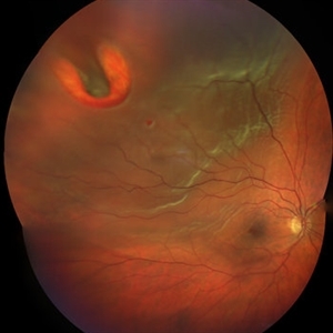
Non-ischemic CRVO (VA 20/30) with secondary CLRAO (nasal macular pallor) in a hypertensive 69yo female. Pathophysiologically, the cilioretinal artery occlusion occurs secondary to elevation in the hydrostatic pressure in the retinal venous system relative to the choroidal perfusion pressure (which supplies the cilioretinal artery).
Photographer: Retina Consultants of Alabama, P. C.
Imaging device: Optos
Condition/keywords: cilioretinal artery occlusion, non-ischemic central retinal vein occlusion (CRVO)
-
 Surfboard to OS - Choroidal Rupture, Retinal and Choroidal Avulsion, Retinal Detachment
May 20 2020 by Theodore Leng, MD, MS, FASRS
33-year-old male who sustained a surfboard to the left orbit with orbital fractures, choroidal rupture, nasally and retinal and choroidal avulsion nasally. He also had a horseshoe retinal tear superotemorally and a resultant rhegmatogenous retinal detachment.
Condition/keywords: choroidal avulsion, choroidal rupture, retinal avulsion, retinal detachment with retinal defect
-
 A Motor Vehicle Accident Causing Valsalva Retinopathy OD, While Racing A Side By Side 4 Wheel Off-Road Vehicle
Apr 29 2020 by John S. King, MD
43-year-old white male who was injured while racing a side by side 4-wheel off-road vehicle (see Video: https://imagebank.asrs.org/file/53854/sxs-crash-during-a-race-causing-valsalva-retinopathy-od). He presented about three weeks after the injury. He was being seen by his local eye doctor who wanted an evaluation for the retinal heme and scotoma. His main complaint was a central/parcentral scotoma described as a greyish area in vision. Va 20/50 OD, nomotensive, no APD (by technician), anterior segment u/r; see picture for the fundus exam - of note there are superficial/preretinal heme, with layering of the heme superiorly, and small superficial heme at nasal edge of the optic disc; in the parafoveal region nasally there is some mottling of the RPE that may indicate an area of prior commotio retinae (also possible to have TON), which may account for his scotoma. Really bad accident (video), and amazingly, he had no LOC or injuries other than the right retina. Helmet and racing harness seat belt were used.
Photographer: Asli Ahmed
Imaging device: Topcon 50
Condition/keywords: valsalva retinopathy
-
 Subretinal Fibrosis (PPCNVM and POHS) OS
Sep 18 2019 by John S. King, MD
57-year-old white male with history of PPCNVM OS and POHS OU here for a routine visit. History of avastin in 2014, and stable since then. Va OS 20/20. PP scar with macular subretinal fibrosis. No heme or exudates. CR spot supero-nasally.
Photographer: Shelly Blair
Imaging device: Topcon 50
Condition/keywords: choroidal neovascular membrane (CNVM), ocular histoplasmosis syndrome (OHS), peripapillary choroidal neovascularization (PPCNVM), presumed ocular histoplasmosis syndrome (POHS)
-
 Uveitis Posterior
Jul 19 2019 by JEFFERSON R SOUSA, Tecg.º (Biomedical Systems Technology)
A 23-year-old male patient attended the clinic with low vision of the right eye. In the evaluation it presented important fundoscopical alterations like retinal exudations in the posterior pole and nasal retina, aspects of macular star. It was proven that it was a posterior uveitis.
Photographer: JEFFERSON R SOUSA - Study Center and Ophthalmological Research Dr. Andre M V Gomes, Institute Dr. Suel Abujamra São Paulo-Brazil
Imaging device: Topcon TRC-50 DX, Imaginet 4.0, angle de 50 graus. Flash 50w-s
Condition/keywords: uveitis
-
 Laser Induced BRAO in IRVAN Syndrome
May 3 2019 by Deependra Vikram Singh, MD FASRS
Fundus photograph of a 26-year-old man with IRVAN syndrome referred for vitreous surgery in OS for secondary rhegmatogenous retinal detachment. OD has received laser photocoagulation for capillary nonperfusion areas and retinal artery macroaneurysm associated with retinal vasculitis. Fundus photograph of OD shows laser induced nasal BRAO. Case re-emphasizes why laser for macroaneurysm should be avoided in cases with IRVAN.
Photographer: Deependra V Singh, Eye-Q Superspecialty Eye Hospitals. Gurugram, India
Imaging device: Zeiss Visucam 500
Condition/keywords: arteriolar macroaneurysm, branch retinal artery occlusion (BRAO), laser photocoagulation
-
 Weiss Ring
Jan 9 2019 by John S. King, MD
77-year-old white male with ERM and PVD OD; sheet of vitreous with weiss ring in the nasal mid-vitreous cavity.
Photographer: Macey Highfill, RN
Imaging device: Topcon 50
Condition/keywords: posterior vitreous detachment, Weiss ring
-
 Bilateral Lebers Miliary Aneurysm in a Female
Sep 5 2017 by Ogugua Ndubuisi Okonkwo, MD, FRCS (Edin), FASRS
Fundus photograph of the active left eye of a 26-year-old female with bilateral LMA. Shows severe exudation in the nasal retina by leaking aneurysms.
Condition/keywords: aneurysm
-
 Combined Hamartoma
Oct 5 2016 by Guruprasad S. Ayachit, MBBS,MS
Fundus photograph of a 9-year-old boy with an ill-defined lesion extending from nasal to the disc going on to include the papillomacular bundle; 14X10 mm in greatest dimensions. There is a thick epiretinal membrane causing distortion and straightening of temporal vascular arcade.
Photographer: Shravan Masurkar, M M Joshi Eye Institute, Hubli
Imaging device: Topcon TRC50DX
Condition/keywords: combined hamartoma
-
 Amelanotic Melanoma
Oct 5 2015 by Scott C. Oliver, MD
58-year-old female with inferonasal choroidal tumor detected on routine eye exam.
Photographer: William Yates, UCHealth Eye Center
Imaging device: Topcon TRC-50DX
Condition/keywords: melanoma
-
 IOFB Combined
Mar 12 2015 by Ahmad B. Tarabishy, MD
A 26-year-old gentleman presented with a metallic intraocular foreign body embedded in the nasal retina (above). Post-operative appearance two weeks after vitrectomy, foreign body removal, endolaser, and gas (below).
Photographer: Jessica Armbruster
Imaging device: Topcon TRC-50EX
Condition/keywords: encapsulated intraocular foreign body, non metallic retained intraocular foreign body (RIOFB), penetrating trauma
-
 Chronic Central Serous Chorioretinopathy (CSCR)
Nov 15 2014 by Rita Couceiro, MD, MS
53-year-old black male, with no relevant prior medical history, complained of bilateral blurry vision for the previous 16 years. On examination, visual acuity was 20/50 on the right eye (OD) and 20/100 on the left eye (OS). Anterior segment evaluation was unremarkable. Fundoscopy revealed pigmentary changes near the macular area in both eyes, with a mottling configuration, suggesting chronic CSCR. Fluorescein angiography showed an ink-blot pattern, with leakage superior to the fovea in OD and nasal to the fovea in OS.
Photographer: Telma Gala - Hospital de Santa Maria, Lisbon, Portugal
Condition/keywords: chronic central serous chorioretinopathy (CSCR)
-
 Optic Nerve Coloboma With 2 Pits, Nasal and Temporal Color
Nov 21 2013 by Alexandre Durao Alves Pereira, MD
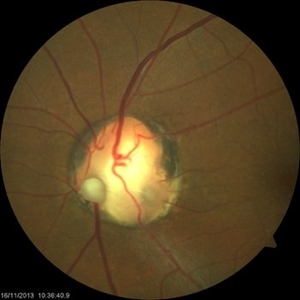
Fundus photograph, color, red free, blue lite and FAF of a optic nerve coloboma with 2 pits, one nasal and other temporal.
Photographer: Alexandre Pereira
Imaging device: Visucam 300
Condition/keywords: color photo, optic nerve coloboma
-
 Rhegmatogenous Retinal Detachment
Aug 23 2012 by Gabriela Lopezcarasa Hernandez, MD
30-year-old male with floaters and inferonasal scotoma.
Photographer: Gabriela Lopezcarasa Hernandez, Hospital Angeles Lomas
Imaging device: ZEISS FF4
Condition/keywords: floaters, inferonasal scotoma
 Loading…