Search results (458 results)
-
---thumb.JPG/image-square;max$300,300.ImageHandler) Weiss Ring (Floater)
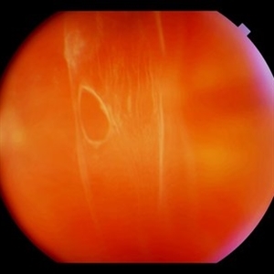
Weiss Ring (Floater)
Jul 10 2013 by Jason S. Calhoun
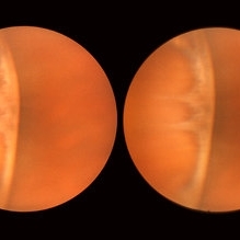
Patient comes in complaining of a floater towards the nasal aspect of her vision. Fundus photograph with anterior shot, shows a weiss ring pulled off from the optic nerve.
Photographer: Jason S. Calhoun, Department of Ophthalmology, Mayo Clinic Jacksonville, Florida
Condition/keywords: floaters, Weiss ring
-
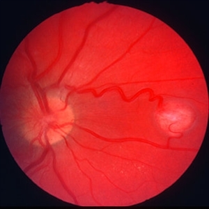
 Severe NPDR
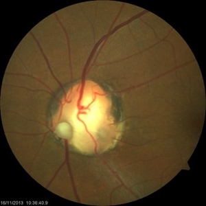
Mar 29 2013 by Henry J. Kaplan, MD
Severe NPDR , IRMA visible inferonasally.
Condition/keywords: nonproliferative diabetic retinopathy
-
 Normal Nasal Ora Serrata
Nov 9 2012 by Norman Byer
This shows the normal nasal ora serrata. Note the dentate processes which divide the nasal ora into prominent bays and teeth
Condition/keywords: dentate processes, normal nasal ora serrata, ora bay, ora teeth
-
---thumb.JPG/image-square;max$300,300.ImageHandler) Weiss Ring (Floater)
Weiss Ring (Floater)
Jul 10 2013 by Jason S. Calhoun
Patient comes in complaining of a floater towards the nasal aspect of her vision. Fundus photograph with anterior shot, shows a weiss ring pulled off from the optic nerve.
Photographer: Jason S. Calhoun, Department of Ophthalmology, Mayo Clinic Jacksonville, Florida
Condition/keywords: floaters, Weiss ring
-
 Normal Nasal Ora Serrata
Nov 9 2012 by Norman Byer
This is the normal nasal ora serrata showing a prominent meridional fold. Such folds are most commonly seen at the lower part of the upper nasal quadrant, and are present in 26% of the population. They are a normal developmental variation and are often bilateral.
Condition/keywords: meridional fold, normal developmental variation, normal nasal ora serrata, upper nasal quadrant
-
 Wegener's Granulomatosis / Nasal Necrosis
Feb 24 2015 by David Callanan, MD
Wegener's granulomatosis / nasal necrosis.
Condition/keywords: Wegener's granulomatosis
-
 Uveitis Posterior
Jul 19 2019 by JEFFERSON R SOUSA, Tecg.º (Biomedical Systems Technology)
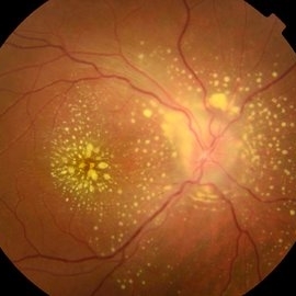
A 23-year-old male patient attended the clinic with low vision of the right eye. In the evaluation it presented important fundoscopical alterations like retinal exudations in the posterior pole and nasal retina, aspects of macular star. It was proven that it was a posterior uveitis.
Photographer: JEFFERSON R SOUSA - Study Center and Ophthalmological Research Dr. Andre M V Gomes, Institute Dr. Suel Abujamra São Paulo-Brazil
Imaging device: Topcon TRC-50 DX, Imaginet 4.0, angle de 50 graus. Flash 50w-s
Condition/keywords: uveitis
-
 Ocular Toxocariasis slide 1
Oct 22 2012 by Ronald C. Gentile, MD
40-year-old man from South America was referred for a peripheral retinal scar in his left eye. He had a history of conjunctivitis as a child with exposure to multiple pets (cats and dogs). Fundus photo revealed a peripheral scarred sub-retinal granuloma located superior nasal with a retinal fold and traction extending to the optic nerve.
Photographer: The New York Eye & Ear Infirmary Department of Medical Imaging
Condition/keywords: toxocariasis
-
 Myelinated Nerve Fiber Layer
Oct 8 2012 by Jeffrey G. Gross, MD, FASRS
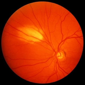
Myelinated nerve fiber layer superonasal retina.
Condition/keywords: myelinated nerve fibers, superonasal retina
-
 Enclosed Ora Bay On The Temporal Side
Nov 9 2012 by Norman Byer
This is a developmental abnormality in a 59-year-old man. It is an enclosed ora bay on the temporal side, an isolated island of normal pars plana epithelium. It is important not to confuse this entity with a retinal break. It has smooth, sloping borders not a sharp, thin, visible retinal edge as a retinal break would have. The border looks exactly like that of the ora serrata, and the grayish pigmented base has the same appearance as the normal pars plana.
Condition/keywords: developmental abnormality, enclosed ora bay, grayish pigmented base, horizontal nasal meridian, pars plana epithelium, smooth sloping borders, temporal retina
-
 Coloboma
Mar 29 2013 by Henry J. Kaplan, MD
Optic disc and inferonasal choroidal coloboma in the same patient #2.
Condition/keywords: coloboma, coloboma of choroid, coloboma of optic disc
-
 Weiss Ring
Jan 9 2019 by John S. King, MD
77-year-old white male with ERM and PVD OD; sheet of vitreous with weiss ring in the nasal mid-vitreous cavity.
Photographer: Macey Highfill, RN
Imaging device: Topcon 50
Condition/keywords: posterior vitreous detachment, Weiss ring
-
 Pigment Epithelial Detachment late FA with small occult CNV
Jul 6 2012 by Tarek S. Hassan, MD, FASRS
72-year-old man with VA loss and metamorphopsia of 2 months duration. PED found, testing done to rule out CNV. Very suspicious for CNV in superonasal fovea/parafovea.
Condition/keywords: choroidal neovascularization (CNV), pigment epithelial detachment (PED)
-
 Optic Nerve Coloboma With 2 Pits, Nasal and Temporal Color
Nov 21 2013 by Alexandre Durao Alves Pereira, MD
Fundus photograph, color, red free, blue lite and FAF of a optic nerve coloboma with 2 pits, one nasal and other temporal.
Photographer: Alexandre Pereira
Imaging device: Visucam 300
Condition/keywords: color photo, optic nerve coloboma
-
 Rhegmatogenous Retinal Detachment
Aug 23 2012 by Gabriela Lopezcarasa Hernandez, MD
30-year-old male with floaters and inferonasal scotoma.
Photographer: Gabriela Lopezcarasa Hernandez, Hospital Angeles Lomas
Imaging device: ZEISS FF4
Condition/keywords: floaters, inferonasal scotoma
-
 Cotton Wool Spot
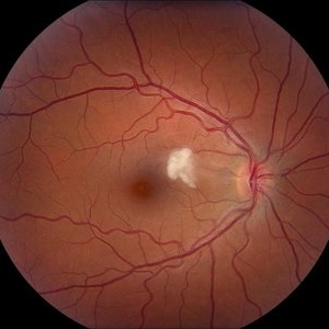
Jul 10 2013 by Jason S. Calhoun
Fundus photograph shows a young male with a single cotton wool spot just nasal to the macula in the right eye.
Photographer: Jason S. Calhoun, Department of Ophthalmology, Mayo Clinic Jacksonville, Florida
Condition/keywords: cotton wool spots, hypertension
-
 Exposed Scleral Buckle, with Exposed Suture, Infection - Infero Nasal View, Upgaze
Feb 4 2013 by James B. Soque, CRA, OCT-C, COA, FOPS
External Photograph of a 66-year-old WM with Hx of SBOD in 2009, graft attempt failed, infection resulted. Scheduled for removal of SBOD.
Photographer: James Soque CRA COA
Imaging device: External Photo, Topcon TRC 50 DX, MERGE software
Condition/keywords: scleral buckle, suture exposed
-
 Inferonasal Branch Retinal Vein Occlusion
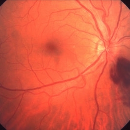
Aug 23 2012 by Gerardo Garcia-Aguirre, MD
Fundus of a 55-year-old male showing intraretinal hemorrhages in the inferonasal quadrant.
Photographer: Noemí Hernández, Asociación para Evitar la Ceguera en México
Condition/keywords: branch retinal vein occlusion (BRVO), intraretinal hemorrhage
-
---thumb.jpg/image-square;max$300,300.ImageHandler) Congenital RPE Hypertrophy
Congenital RPE Hypertrophy
Aug 8 2013 by From the Collections of Thomas M. Aaberg, MD and Thomas M. Aaberg Jr., MD
Well - demarcated CHRPE inferonasal to the optic disc of right eye.
Condition/keywords: retinal pigment epithelium (RPE) hypertrophy
-
 Dialysis of Retina in Upper Nasal Quadrant
Nov 9 2012 by Norman Byer
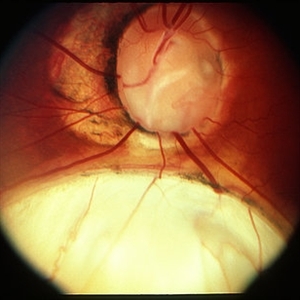
This 20-year-old wrestler sustained a sharp powerful blow to his right eye from his opponent’s thumb. One hour later he saw hundreds of black specs in his vision and was found to have this dialysis of his retina in the upper nasal quadrant. Note the triangular piece of retina in the center that remained attached to the ora serrata causing the retinal flap to resemble a man’s flared shirt collar.
Condition/keywords: retinal dialysis, retinal tear, upper nasal quadrant
-
 Retinal capillary hemangioma
Jan 11 2013 by Alex P. Hunyor, MD
Retinal capillary haemangioma nasal to optic disc, right eye.
Condition/keywords: retinal capillary hemangioma, Von Hippel-Lindau
-
 Chronic Retinal Detachment: Features Slide 2
Oct 22 2012 by Ronald C. Gentile, MD
Chronic retinal detachments can be associated with demarcation lines (tidemarks), subretinal bands or sheets, and retinal cysts. Fundus photo of a chronic retinal detachment reveals a branching subretinal band superior nasal to the macula with a portion extending to the inferior margin of the optic disc.
Photographer: The New York Eye & Ear Infirmary Department of Medical Imaging
Condition/keywords: chronic retinal detachment, subretinal bands
-
 Exposed Scleral Buckle, with Infection - Infero Nasal View
Feb 4 2013 by James B. Soque, CRA, OCT-C, COA, FOPS
External photograph of a 66-year-old WM with Hx of SBOD in 2009, graft attempt failed, infection resulted. Scheduled for removal of SBOD.
Photographer: James Soque CRA COA
Imaging device: External Photo, Topcon TRC 50 DX, MERGE software
Condition/keywords: scleral buckle, suture exposed
-
---thumb.JPG/image-square;max$300,300.ImageHandler) Myelinated Nerve Fiber Layer
Myelinated Nerve Fiber Layer
Jul 12 2013 by Jason S. Calhoun
Myelinated nerve fiber layer surrounding the nasal aspect of the optic nerve in the left eye.
Photographer: Jason S. Calhoun, Department of Ophthalmology, Mayo Clinic Jacksonville, Florida
Condition/keywords: myelinated nerve fibers
-
 Congenital Hypertrophy of the Retinal Pigment Epithelium (CHRPE)
Jul 14 2013 by Jason S. Calhoun
Fundus photo shows congenital hypertrophy of the retinal pigment epithelium (CHRPE), superior, nasally in the left eye.
Photographer: Jason S. Calhoun, Department of Ophthalmology, Mayo Clinic Jacksonville, Florida
Imaging device: TOPCON TRC 50-EX
Condition/keywords: congenital hypertrophy of the retinal pigment epithelium (CHRPE)
 Loading…