Search results (458 results)
-
 Macular Coloboma
Jun 5 2025 by César Adrián Gómez Valdivia, MD
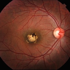
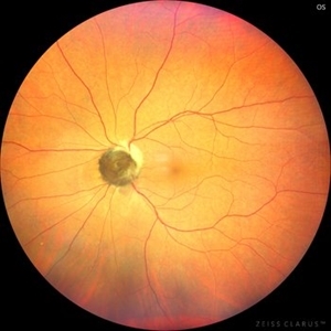
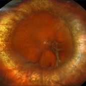
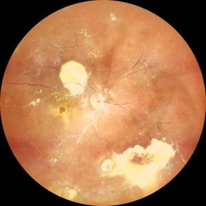
Macular Coloboma found in a 28 year-old male patient, visual acuity was 20/60. Resulting due to fusion failure of the optic fissure, colobomas are commonly found in the infero-nasal quadrant. If the retina is involved, it is reduced to glial tissue with no underlying RPE or choroid. This appears as an area of whitening often with pigment deposition at the junction of the coloboma and normal retina. Findings were bilateral.
Photographer: @eyemissu2
Imaging device: TOPCON TRC-50DX
Condition/keywords: coloboma
-
 Myelinated Nerve Fibers
Jun 4 2025 by Paulina Araujo
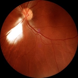
The 55-degree central fundus photograph of the left eye reveals myelination of the nerve fiber layer along the inferior nasal arcade.
Photographer: Paulina D.Araujo Martínez, Asociación para Evitar la Ceguera en México I.A.P., Hospital Dr Luis Sánchez Bulnes.
Condition/keywords: myelinated nerve fibers
-
 Choroidal Rupture
Jun 4 2025 by Paulina Araujo
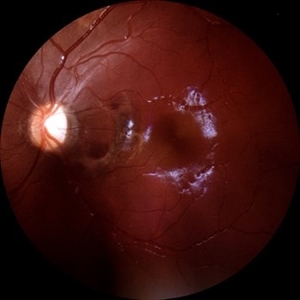
The 55-degree central fundus photograph of the left eye reveals a choroidal rupture in the nasal parafoveal area secondary to blunt ocular trauma.
Photographer: Paulina D.Araujo Martínez, Asociación para Evitar la Ceguera en México I.A.P., Hospital Dr Luis Sánchez Bulnes.
Condition/keywords: choroidal rupture
-
 Aggressive ROP
Jun 3 2025 by Anjana Mirajkar, MS Ophthalmology
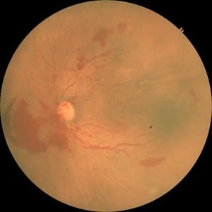
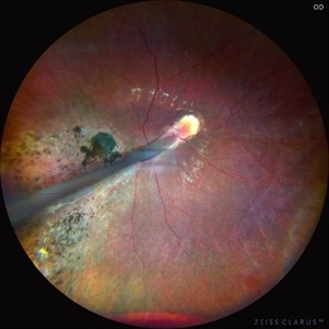
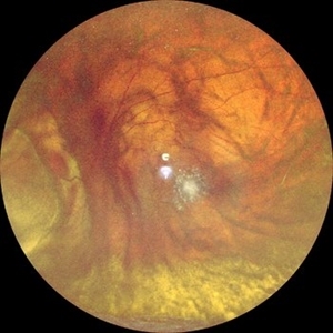
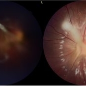
Fundus photograph of OS of a premature baby of GA 29+2, Birth weight of 1325gms and Post menstrual age of 34+2, showing tortuosity and dilatation of vessels with looping in Zone 1 posterior with large pre retinal bleed nasal to disc suggestive of A-ROP.
Photographer: Vishnu Gaikwad- Optometrist H.V .Desai eye hospital, Pune
Imaging device: Retcam
Condition/keywords: aggressive posterior retinopathy of prematurity (APROP)
-
 Optic Nerve Melanocytoma
May 4 2025 by KANWALJEET HARJOT MADAN, M.S. (Ophthalmology); FAICO (Vitreous - Retina)
This is a fundus picture of a young 42-year male who visited for a routine eye exam. His BCVA was 20/20 in both eyes. Anterior segment examination was normal. His left eye showed grey-black pigmentation at the infero-nasal margin of the optic disc. Fundus of the right eye was normal. The patient was diagnosed to have optic disc melanocytoma on multimodal imaging and was advised regular follow-up. Optic nerve melanocytoma is typically a benign tumor made up of melanocytes and melanin. It can grow, but rarely transforms into a malignancy. Patients with Optic Nerve Melanocytoma should be periodically examined for evidence of growth, loss of visual field and optic nerve compression.
Photographer: Dr. Kanwaljeet Harjot Madan, Thind Eye Hospital, Jalandhar City (Punjab) INDIA.
Imaging device: Zeiss Fundus Camera
Condition/keywords: melanocytoma, melanoma, optic nerve
-
 Giant Retinal Tear with Multiple Retinal Breaks
Apr 21 2025 by Hrishikesh Naik, MS
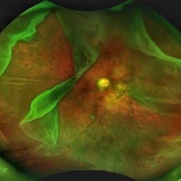
A 28 year old high myope with retinal detachment associated with a supero-temporal giant retinal tear in addition to multiple peripheral horseshoe tears and an additional supero-nasal retinal tear.
Photographer: Hrishikesh Naik
Imaging device: Optos Daytona
Condition/keywords: giant retinal tear, High Myopia, horseshoe tear, retinal break, retinal detachment
-
 Comets in the Eye (Retinopathy of Prematurity)
Apr 8 2025 by KANWALJEET HARJOT MADAN, M.S. (Ophthalmology); FAICO (Vitreous - Retina)
This is the fundus picture of right eye (RE) of a 4 years female child presented with outward deviation of right eye. Her parents also complained of diminution of vision in both eyes. On examination, her best corrected vision in RE was hand movements close to face and was 20/80 in LE. Posterior segment exam revealed presence of macular scar in RE and presence of dry retinal fold with dragging of retinal vessels. LE fundus revealed presence of nasal drag of optic disc. Parents gave history of untreated ROP as an infant. Retinopathy of Prematurity (ROP) is a Vaso proliferative disorder of Retina occurring in premature infants. Advances in neonatal care and ROP treatment has led these babies to live longer with this disease.
Photographer: Dr. Kanwaljeet Harjot Madan, Thind Eye Hospital, Jalandhar City (Punjab) INDIA.
Imaging device: Zeiss Fundus Camera
Condition/keywords: Retinopathy of Prematurity, Vaso proliferative disorder
-
 Comets in the Eye (Retinopathy of Prematurity)
Apr 8 2025 by KANWALJEET HARJOT MADAN, M.S. (Ophthalmology); FAICO (Vitreous - Retina)
This is the fundus picture of right eye (RE) of a 4 years female child presented with outward deviation of right eye. Her parents also complained of diminution of vision in both eyes. On examination, her best corrected vision in RE was hand movements close to face and was 20/80 in LE. Posterior segment exam revealed presence of macular scar in RE and presence of dry retinal fold with dragging of retinal vessels. LE fundus revealed presence of nasal drag of optic disc. Parents gave history of untreated ROP as an infant. Retinopathy of Prematurity (ROP) is a Vaso proliferative disorder of Retina occurring in premature infants. Advances in neonatal care and ROP treatment has led these babies to live longer with this disease.
Photographer: Dr. Kanwaljeet Harjot Madan, Thind Eye Hospital, Jalandhar City (Punjab) INDIA.
Imaging device: Zeiss Fundus Camera
Condition/keywords: Retinopathy of Prematurity
-
 Repaired Retinal Detachment with Scleral Buckle
Mar 25 2025 by Kimberly Wakester
Optomap RGB montage of an 64-year-old woman with a repaired retinal detachment with scleral buckle in the right eye. There is nasal and inferior pre-retinal membranes with traction. PPV was recommended but patient defers to proceed with sx at this time. Will continue to follow patient closely for worsening traction. Patient was educated on how to monitor their peripheral vision and was advised to report any changes immediately.
Photographer: Kimberly Wakester, COA, OCT-C
Imaging device: Optos California
Condition/keywords: pre-retinal membrane with traction, repaired RD, scleral buckle
-
 Hosreshoe Tears on Posterior Pole
Mar 22 2025 by Deepak Bhojwani, MS
A fundus image of an asymptomatic 64 year old male with large horseshoe shaped breaks in inferonasal quadrant on posterior pole, an unusual location for retinal breaks.
Photographer: DR DEEPAK BHOJWANI
Condition/keywords: horseshoe tear, posterior pole break, retinal break
-
 Choroidal Melanoma Masquerading as PEHCR
Mar 3 2025 by Tejaswita Verma
A 65 year old diabetic male presented with large nasal retinal mass giving the appearance of organized dehaemoglobinized subretinal hemorrhage with breakthrough vitreous hemorrhage , with 6/6P vision. Enucleation specimen showed histopathology confirmed choroidal melanoma.
Photographer: DR. TEJASWITA VERMA
Imaging device: MIRANTE
Condition/keywords: vitreous hemorrhage
-
 S/P Vitreo Retinal Surgery
Feb 27 2025 by Angela Rico
Patient referred to our office for Evaluation and Treatment of re- detached retina following previous repair of RD. 10% gas Bubble- Macular detachment- PVR Temporal Star fold super-temporal - Multiple irregular Tears infero nasal
Photographer: Angela Rico M.D.
Imaging device: California Optos
Condition/keywords: PVR, retinal detachment
-
 Choroidal Melanoma Masquerading as Subretinal Hemorrhage With Breakthrough VH
Jan 23 2025 by Tejaswita Verma
A 65 year old diabetic male presented with large nasal retinal mass giving the appearance of organized dehaemoglobinized subretinal hemorrhage with breakthrough vitreous hemorrhage , with 6/6P vision. Enucleation specimen showed histopathology confirmed choroidal melanoma.
Photographer: DR. TEJASWITA VERMA
Imaging device: MIRANTE
-
 Sectoral Ocular Melanocytosis
Jan 17 2025 by Virginia Gebhart
67 year old female with congenital sectoral ocular melanocytosis. Pigmentation on nasal sclera and nasal iris of right eye, as well as deep pigmentation nasally of fundus. Will continue close observation
Photographer: Virginia Gebhart
Imaging device: Topcon 50DX/Samsung Galaxy
Condition/keywords: choroidal melanocytosis, heterochromia, ocular melanocytosis, Oculodermal Melanocytosis
-
 Sub ILM Dehaemoglobinised Hemorrhage With Retinal Detachment in Vitrectomised Eye
Jan 16 2025 by Anand Temkar
A 39 yrs old male was referred to us with this presentation after a month of his first vitrectomy surgery done for VH e/w. His serum homocysteine was raised but MRI brain was within normal limits. We can see the sub ILM dehaemoglobinised hemorrhage (supero-temporal to macula) and retinal detachment (inferiorly and nasally).
Photographer: Dr.Anand Temkar- Retina Foundation, Ahmedabad
Imaging device: Mirante
Condition/keywords: dehemoglobinized hemorrhage, Retinal Detachment, SUB ILM hemorrhage
-
 Sub ILM Dehaemoglobinised Hemorrhage With Retinal Detachment
Jan 16 2025 by Anand Temkar
A 39 year old male was referred to us with this presentation after a month of his first vitrectomy surgery done for VH e/w. His serum homocysteine was raised but MRI brain was within normal limits. We can see the sub ILM dehaemoglobinised hemorrhage (supero-temporal to macula) and Retinal detachment (inferiorly and nasally).
Photographer: Dr.Anand Temkar- Retina Foundation, Ahmedabad
Imaging device: Mirante
Condition/keywords: dehemoglobinized hemorrhage, Retinal Detachment, SUB ILM hemorrhage
-
 Venolymphatic Mass With Disc Edema
Dec 5 2024 by Tejaswita Verma
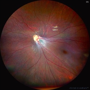
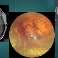
Fundus picture of a 26 year old male who presented with right eye abaxial proptosis, MRI confirmed venolymphatic mass inferomedial in location located near the optic disc with disc edema , nasal elevation ,retinal folds. Vision was 6/18 . He was planned for intralesional bleomycin injection.
Photographer: DR. TEJASWITA VERMA
Imaging device: MIRANTE
Condition/keywords: disc edema, intraorbital mass, proptosis
-
 Venolymphatic Mass with Retinal Folds
Nov 25 2024 by Tejaswita Verma
Fundus picture of a 26 year old male who presented with right eye abaxial proptosis, MRI confirmed venolymphatic mass inferomedial in location located near the optic disc with disc edema , nasal elevation ,retinal folds. Vision was 6/18 . He was planned for intralesional bleomycin injection.
Photographer: DR. TEJASWITA VERMA
Imaging device: MIRANTE
Condition/keywords: disc edema, intraorbital mass, proptosis, retinal folds
-
 Coats Disease
Sep 29 2024 by Tejaswita Verma
Fundus photo of the RE of a 14 y/o female ,nil premorbid presented with reduced vision in the RE ,diagnosed incidentally on ophthalmological examination elsewhere .Vision was finger counting 3 meters in the RE . Fundus picture reveals macular scar , subretinal and intraretinal exudation ,with scattered hemorrhages esp. in STQ, sclerosed vessels in superior, superonasal quadrant ,nasal, inferonasal quadrant, CR scars inferiorly, Telengiectatic vessels S/O Coat's disease. She was advised RE anti VEGF x1 + laser PRP + PST kenacort under GA with guarded prognosis.
Photographer: DR. TEJASWITA VERMA
Imaging device: MIRANTE
Condition/keywords: Coats' disease
-
 Look With Your Heart
Sep 20 2024 by Virginia Gebhart
FA of 65 year old male with exudative AMD superior to a chorioretinal defect in the nasal macula. FA shows classic CNV with late leakage. Treated with IVA, will consider PDT if no improvement.
Photographer: Virginia Gebhart, Retina Consultants of Carolina
Imaging device: Optos California
Condition/keywords: choroidal neovascularization (CNV), exudative age-related macular degeneration, FA early phase
-
 Pericentral Retinitis Pigmentosa
Sep 6 2024 by Mauricio Bayram-Suverza, MD
A 65-year-old male patient reports experiencing bilateral blind spots that have gradually intensified over time. Genetic testing was unrevealing. The fundus autofluorescence image shows a hypoautofluorescent ring in the posterior pole, especially nasal to the nerve and along arcades.
Photographer: Mauricio Bayram-Suverza, Casey Eye Institute, OHSU.
Imaging device: Optos California
Condition/keywords: fundus autofluorescence (FAF), inherited retinal disease, nyctalopia, retinal dystrophy, retinitis pigmentosa
-
 Subhyaloid Hemorrhage
Jul 31 2024 by Arthi Mohankumar , MS,MRCS ED, FICO,FAICO
A 35 year old male presented with complaints of seeing a black spot in left eye for past one day after working out in the gym the previous day. He has history of uncontrolled diabetes and hypertension. Fundus exam of the left eye revealed a sub hyaloid hemorrhage nasal to the disc with minimal background Diabetic and hypertensive changes. His baseline CBG was 200 mg/dl and BP was 170/100 He was suggested observation initially considering the nasal location. But patient found the scotoma very disturbing and eventually underwent yag hyaloidotomy
Photographer: Arthi Mohankumar
Condition/keywords: Sub hyaloid haemorrhage, valsalva retinopathy
-
 Posterior-PFV
Jul 27 2024 by Gokcen Deniz Gulpinar Ikiz
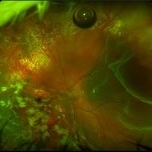
7 Year old girl presented with blurred vision on the left eye, with intermittent esotopia. She had been followed conservatively for intermittent esotropia on the left eye, recently advised for patching of the right eye. The vision is 1.0 on the right eye and 0.4 (Snellen) on the left eye. Anterior segment is natural bilaterally, except 20 PD esotropia on the left eye, with alternation and fixation. Refraction was +0.25 +0.25 x180 and +1.00-1.50 x60 on the right and left eyes respectively. Dilated fundus examination was natural on the right eye. However, there was a fibrotic stalk originating from the optic nerve head extending to the vitreous, terminating in the middle of the vitreous cavity, in a spider web configuration. Which also causes nasal dragging of the macula, leading to partial shallow detachment of the fovea nasally. Vitrectomy is advised for the left eye, with lens preserving approach, to preserve the current functional potential and the anatomy of the globe in long term.
Photographer: Gokcen Deniz Gulpinar Ikiz, Special Eye Clinic
Condition/keywords: amblyopia, posterior PFV, vitrectomy
-
 Macula Off Retinal Detachment
Jun 25 2024 by Zach Seim
Optos Fundus photo of a 47 year old female with a Macula Off Retinal Detachment right eye, presenting with loss of nasal visual field. Patient's vision at presentation was DCC 20/100-1. Patient was counseled and decided to proceed with surgery.
Photographer: Zach Seim
Imaging device: OPTOS California
Condition/keywords: macula off retinal detachment, Optos, OPTOS CALIFORNIA, right eye
-
 Fluorescein Angiography Montage
Jun 21 2024 by BENITO VERGARA, MD
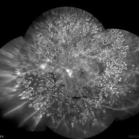
Montage of an angiography with fluorescein from the left eye of a 32 year-old male with diabetic retinopathy previously treated with panretinal photocoagulation, that shows leakage at optic nerve and upper nasal arcade.
Photographer: Benito Vergara, Asociación Para Evitar la Ceguera en México.
Imaging device: Zeiss Clarus 700
Condition/keywords: Angiography Montage, angiography with fluorescein, diabetic retinopathy, FA montage, fluorescein angiogram (FA), peripheral scars
 Loading…