 Initializing download.
-
 By Albert Li, MD, FASRS
Vitreoretinal Consultants of New York
Co-author(s): David Na - Uploaded on Jun 6, 2020.
- Last modified by Caroline Bozell on Nov 20, 2020.
- Image of the week
-
Nov 22, 2020
View all images of the week - Rating
- Appears in
- 6-Jun-2020
- Condition/keywords
- arteriovenous anastomosis, arteriovenous malformation
- Imaging device
-
Scanning laser ophthalmoscope
Heidelberg Spectralis - Description
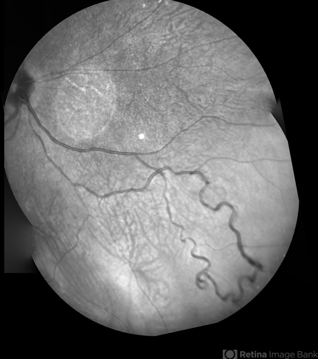
- Montaged infrared retinal imaging of a 37-year-old asymptomatic man with a grade II arteriovenous malformation (AVM) in the nasal mid-periphery. The presentation of the AVM can be classified with three categories. Grade 1 AVMs are characterized by an abnormal capillary plexus between the major communicating vessels. Grade 2 AVMs are defined by the direct arteriovenous communication without the interposition of arterioles or capillaries. Grade 3 AVMs are characterized by widespread, large caliber anastomosing vessels that are associated with decreased visual acuity and intracranial AVMs. Because retinal AVMs are mostly asymptomatic and non-progressive, further testing may not be indicated unless there are concomitant neurological signs and symptoms or if there is a strong clinical suspicion of a grade 3 retinal AVM. Observation was recommended for the patient in this image. On his most recent follow-up at four months, the patient remained asymptomatic with a stable appearance of the lesion.


")


