Search results (458 results)
-
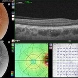
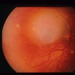
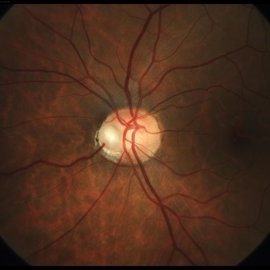 Nasal Optic Disc Pit
May 3 2022 by Bernardo Araújo
Asymptomatic patient. 41-year-old woman.
Photographer: Bernardo Araújo, Retina Clinic, São Paulo, SP, Brazil.
Condition/keywords: nasal, optic disc
-
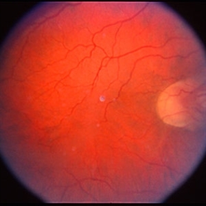
 Candida Endophthalmitis
Apr 1 2019 by Gary R. Cook, MD, FACS
Nasal view of left eye of a 41-year-old white female prostitute and IV drug user with endogenous Candida endophthalmitis OS; V.A.= H.M.
Imaging device: Topcon VT-50
Condition/keywords: candida endophthalmitis, endogenous endophthalmitis
-
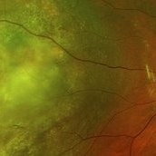 Nasal Choroidal Mass
Jan 7 2018 by John S. King, MD
Mets vs Melanoma
Imaging device: Optos
Condition/keywords: choroidal mass
-
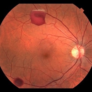
 Optic Nerve Malformation
Feb 19 2013 by From the Collections of Thomas M. Aaberg, MD and Thomas M. Aaberg Jr., MD
Nasal ectasia, no history.
Condition/keywords: color photo, optic nerve malformation
-
 Optic Nerve Malformation
Feb 19 2013 by From the Collections of Thomas M. Aaberg, MD and Thomas M. Aaberg Jr., MD
Nasal ectasia, no history.
Condition/keywords: color photo, optic nerve malformation
-
 Optic Nerve Malformation
Feb 19 2013 by From the Collections of Thomas M. Aaberg, MD and Thomas M. Aaberg Jr., MD
Nasal ectasia and inferior conus; OD 20/25; OS 20/70.
Condition/keywords: conus, ectasia, optic nerve malformation
-
 Optic Nerve Malformation
Feb 19 2013 by From the Collections of Thomas M. Aaberg, MD and Thomas M. Aaberg Jr., MD
Nasal ectasia and inferior conus; OD 20/25; OS 20/70.
Condition/keywords: conus, ectasia, optic nerve malformation
-
 Optic Nerve Malformation
Feb 19 2013 by From the Collections of Thomas M. Aaberg, MD and Thomas M. Aaberg Jr., MD
Nasal ectasia and inferior conus; OD 20/25; OS 20/70.
Condition/keywords: conus, ectasia, optic nerve malformation
-
 Optic Nerve Malformation
Feb 19 2013 by From the Collections of Thomas M. Aaberg, MD and Thomas M. Aaberg Jr., MD
Nasal fundus ectasia; VF loss.
Condition/keywords: ectasia, optic nerve malformation
-
 Optic Nerve Malformation
Feb 19 2013 by From the Collections of Thomas M. Aaberg, MD and Thomas M. Aaberg Jr., MD
Nasal fundus ectasia; VF loss.
Condition/keywords: ectasia, optic nerve malformation
-
 Optic Nerve Malformation
Feb 19 2013 by From the Collections of Thomas M. Aaberg, MD and Thomas M. Aaberg Jr., MD
Nasal fundus ectasia; VF loss.
Condition/keywords: ectasia, optic nerve malformation
-
 Situs-Inversus-Left-eye
Mar 29 2023 by Nizamuddin HM Shaik, MD, FRCS
Situs inversus of the optic disc is a rare, usually bilateral, congenital embryological abnormality associated with high myopia, optic disc coloboma or tilted optic disc. Our patient, 24 years old lady without these conditions presented with bilateral situs inversus. Her BCVA OD 0.4 and OS 0.5. It is characterized by emergence of the retinal vessels in an anomalous direction with dysversion of the optic disc.
Photographer: Mahmoud A Abdelmaguid
Condition/keywords: Nasalization of temporal retinal vessels
-
 Situs-Inversus-OD
Mar 29 2023 by Nizamuddin HM Shaik, MD, FRCS
Situs inversus of the optic disc is a rare, usually bilateral, congenital embryological abnormality associated with high myopia, optic disc coloboma or tilted optic disc. Our patient, 24 years old lady without these conditions presented with bilateral situs inversus. Her BCVA OD 0.4 and OS 0.5. It is characterized by emergence of the retinal vessels in an anomalous direction with dysversion of the optic disc.
Photographer: Mahmoud A Abdelmaguid
Condition/keywords: Nasalization of temporal vessels
-
 Stage 4A Retinopathy of Prematurity
Nov 7 2013 by Maria Ana Martinez-Castellanos, MD
Nasal area of the left eye of a baby with ROP stage 4A.
Photographer: Maria A. Martinez-Castellanos. Asociacion para Evitar la Ceguera en Mexico
Imaging device: RetCam II
Condition/keywords: retinopathy of prematurity stage 4a, tractional retinal detachment
-
 24 Hours Post Scleral Wound Closure+ Scleral Buckle+25 g Vitrectomy+Silicon Oil
Jan 23 2015 by Carlos Quezada-Ruiz, MD, FASRS
24 hours post op fundus photograph of a 43-year-old man who had perforating injury to the right eye with a small piece of plastic while he was hammering. OD LP, subconjunctival hemorrhage, clear cornea, hyphema, irido and ciclodyalisis as well as a luxated lens with traumatic cataract and a dense vitreous hemorrhage. B-US showed rhegmatogenous retinal detachment with a tear and a big inferior hemorrhagic choroidal detachment. 360 peritomy revealed 2-entry scleral wounds were found in zone II (M V and M VI) and closure was performed. 25 G PPV was performed with the infusion canal placed in the AC through the limbus. Lensectomy and removal of a dense recent vitreous hemorrhage revealed a white detached retina with an exit wound through the temporal inferior segment of the optic nerve with a nasal GRT and sub retinal hemorrhage as well as temporal inferior choroidal, PVD was induced and PFOs helped stabilizing the retina while vitrectomy and sub-retinal hemorrhage was removed through the GRT. Fluid air exchange was made and 360 endolaser over the buckle indentation was done and silicon oil was used as endotamponade. This picture was taken 24 hrs after the surgery.
Photographer: Lilibeth Rodriguez, Instituto de la Visión. Torreon, Mexico.
Condition/keywords: central retinal artery occlusion (CRAO), giant retinal tear, trauma
-
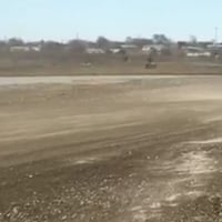
 A Motor Vehicle Accident Causing Valsalva Retinopathy OD, While Racing A Side By Side 4 Wheel Off-Road Vehicle
Apr 29 2020 by John S. King, MD
43-year-old white male who was injured while racing a side by side 4-wheel off-road vehicle (see Video: https://imagebank.asrs.org/file/53854/sxs-crash-during-a-race-causing-valsalva-retinopathy-od). He presented about three weeks after the injury. He was being seen by his local eye doctor who wanted an evaluation for the retinal heme and scotoma. His main complaint was a central/parcentral scotoma described as a greyish area in vision. Va 20/50 OD, nomotensive, no APD (by technician), anterior segment u/r; see picture for the fundus exam - of note there are superficial/preretinal heme, with layering of the heme superiorly, and small superficial heme at nasal edge of the optic disc; in the parafoveal region nasally there is some mottling of the RPE that may indicate an area of prior commotio retinae (also possible to have TON), which may account for his scotoma. Really bad accident (video), and amazingly, he had no LOC or injuries other than the right retina. Helmet and racing harness seat belt were used.
Photographer: Asli Ahmed
Imaging device: Topcon 50
Condition/keywords: valsalva retinopathy
-
 A Motor Vehicle Accident Causing Valsalva Retinopathy OD, While Racing A Side By Side 4 Wheel Off-Road Vehicle
May 5 2020 by John S. King, MD
A 43-year-old white male who was injured while racing his side by side 4 wheel off-road vehicle (this is a video he showed me on his phone). He presented about three weeks after the injury. He was being seen by his local eye doctor who wanted an evaluation for the retinal heme and scotoma. His main complaint was a central/parcentral scotoma described as a greyish area in vision. Va 20/50 OD, nomotensive, no APD (by technician), anterior segment u/r; see {https://imagebank.asrs.org/file/53828/sxs-crash-during-a-race-causing-valsalva-retinopathy-od} for the fundus exam - of note there are superficial/preretinal heme, with layering of the heme superiorly; in the parafoveal region nasally there is some mottling of the RPE that may indicate an area of prior commotio retinae (also possible to have TON), which may account for his scotoma. Really bad accident, and amazingly, he had no LOC or injuries other than the right retina. Helmet and racing harness seat belt were used.
Condition/keywords: motor vehicle accident, trauma, valsalva retinopathy
-
 Aborted Arteriolitis
Feb 15 2013 by From the Collections of Thomas M. Aaberg, MD and Thomas M. Aaberg Jr., MD
Fundus photograph showing activated toxoplasma retinochoroiditis with active retinal whitening adjacent to a hyperpigmented scar in the superonasal mid-periophery.
Condition/keywords: ocular toxoplasmosis
-
 Active Vasculitis with Proliferative Retinopathy
Jan 30 2021 by Raja Rami P Reddy, MD FRCS FASRS
25-year-old boy with unilateral recent onset visual loss. Fundus shows areas of active vasculitis nasally and large neovascular complexes temporally and on the disc and early fibrous membrane formation. Fellow eye fundus is normal. Further investigations suggested tubercular etiology
Photographer: Raja Rami Reddy P
Imaging device: fundus camera
Condition/keywords: proliferative retinopathy, tuberculosis, vasculitis
-
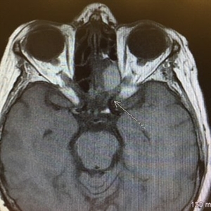 Acute Compressive Optic Neuropathy
Jun 1 2019 by John S. King, MD
84-year-old white female with acute loss of vision in the left eye one day ago was sent here after going to the ED per primary eye provider. She described vision loss as a grey curtain that became total darkness. She had left sided temporal tenderness and some left sided neck pain. In the ED the cardiac work-up was u/r, the ESR and CRP were normal, and the CTH showed some non-specific opacification in the L ethmoid sinus. Acuity was HM OS with RAPD, normal EOMs, no proptosis or ptosis, posteriorly no SVPs were noted; the optic discs were pink and flat; no emboli or retinal whitening present; some bear tracks located nasally (see photo). She was referred to Dr. Doyle, who ordered an MRI, which showed a large mucocele with bony erosion into the left orbit, along with some ON enhancement possibly from compression (see images). She was operated that night and later recovered to 20/40 in that eye with a residual, inferior arcuate scotoma.
Condition/keywords: bear tracks, optic neuropathy
-
---thumb.jpg/image-square;max$300,300.ImageHandler) Acute Histoplasmosis Choroiditis In Immunocompetent Boy
Acute Histoplasmosis Choroiditis In Immunocompetent Boy
Oct 4 2013 by Maurice F. Rabb
On examination both brothers had visual acuities of 20/20 in each eye. The younger brother had a single distinct creamy-white 500 micron lesion located in the choroid temporal to the macula of the right eye. Fundus photographs of the older brother are shown. The right eye had at least five whitish lesions that appeared to be located deep to the retina. There were two lesions present superotemporal to the fovea, one immediately inferior to the fovea, one inferior to the optic nerve head, and one superonasally. In the left eye there was a single choroiditis lesion immediately nasal to the optic nerve head as well as some questionable lesions approximately one disc diameter inferior to the optic nerve head.
Condition/keywords: acute histoplasmosis choroiditis in immunocompetent boy
-
 Acute Histoplasmosis Choroiditis In Immunocompetent Boy
Oct 4 2013 by Maurice F. Rabb
On examination both brothers had visual acuities of 20/20 in each eye. The younger brother had a single distinct creamy-white 500 micron lesion located in the choroid temporal to the macula of the right eye. Fundus photographs of the older brother are shown. The right eye had at least five whitish lesions that appeared to be located deep to the retina. There were two lesions present superotemporal to the fovea, one immediately inferior to the fovea, one inferior to the optic nerve head, and one superonasally. In the left eye there was a single choroiditis lesion immediately nasal to the optic nerve head as well as some questionable lesions approximately one disc diameter inferior to the optic nerve head.
Condition/keywords: acute histoplasmosis choroiditis in immunocompetent boy
-
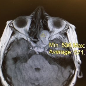 Acute Optic Neuropathy Due to Large Mucocele
Jun 1 2019 by John S. King, MD
84-year-old white female with acute loss of vision in the left eye one day ago was sent here after going to the ED per primary eye provider. She described vision loss as a grey curtain that became total darkness. She had left sided temporal tenderness and some left sided neck pain. In the ED the cardiac work-up was u/r, the ESR and CRP were normal, and the CTH showed some non-specific opacification in the L ethmoid sinus. Acuity was HM OS with RAPD, normal EOMs, no proptosis or ptosis, posteriorly no SVPs were noted; the optic discs were pink and flat; no emboli or retinal whitening present; some bear tracks located nasally (see photo). She was referred to Dr. Doyle, who ordered an MRI, which showed a large mucocele with bony erosion into the left orbit, along with some ON enhancement possibly from compression (see Images). She was operated that night and later recovered to 20/40 in that eye with a residual, inferior arcuate scotoma.
Condition/keywords: bear tracks, optic neuropathy
-
 Acute Optic Neuropathy Due to Large Mucocele (Incidental Bear Tracks)
Jun 1 2019 by John S. King, MD
84-year-old white female with acute loss of vision in the left eye one day ago was sent here after going to the ED per primary eye provider. She described vision loss as a grey curtain that became total darkness. She had left sided temporal tenderness and some left sided neck pain. In the ED the cardiac work-up was u/r, the ESR and CRP were normal, and the CTH showed some non-specific opacification in the L ethmoid sinus. Acuity was HM OS with RAPD, normal EOMs, no proptosis or ptosis, posteriorly no SVPs were noted; the optic discs were pink and flat; no emboli or retinal whitening present; some bear tracks located nasally (see photo). She was referred to Dr. Doyle, who ordered an MRI, which showed a large mucocele with bony erosion into the left orbit, along with some ON enhancement possibly from compression (see images). She was operated that night and later recovered to 20/40 in that eye with a residual, inferior arcuate scotoma.
Photographer: Karin Aletter
Imaging device: Topcon 50
Condition/keywords: bear tracks, optic neuropathy
-
 Acute Partial Vitreous Separation
Dec 10 2012 by Yale L. Fisher, MD
This is a partial vitreous separation that demonstrates a mildly reflective curvilinear shape of the partially separated vitreous face. The vitreous is attached inferonasally and inferotemporally, but detached and freely mobile at 6 o'clock.
Condition/keywords: video
 Loading…