Search results (32 results)
-
 Who Stole My Blood Supply?
Jan 25 2025 by Muna Bhende, MD
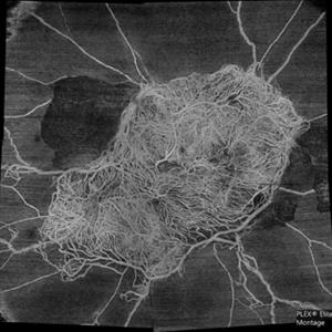
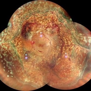
Superficial capillary plexus slab montage image of a young female diabetic with florid proliferation . There is no flow in the capillaries anterior to the tangle of new vessels indicating severe retinal ischemia.
Photographer: Mohanapriya L , Medical Research Foundation, Sankara Nethralaya, Chennai, India
Imaging device: PLEX elite 9000
Condition/keywords: florid type PDR, OCTA
-
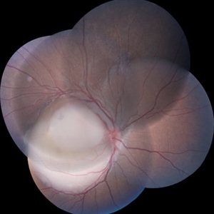
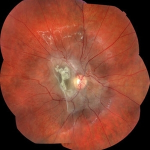
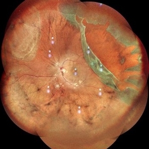 Giant Retinal Tear
Oct 11 2024 by Anjana Mirajkar, MS Ophthalmology
Fundus photograph montage of LE showing a giant retinal extending from 12 to 4 o clock.
Photographer: Dr. Anjana Mirajkar -Retina Foundation, Ahmedabad
Imaging device: Mirante-Nidek
Condition/keywords: GIANT RETINAL TEAR
-
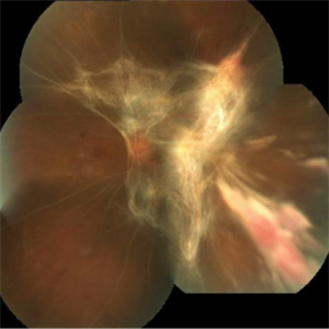
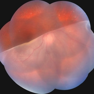 Giant Retinal Tear
Jul 15 2024 by Arthi Mohankumar , MS,MRCS ED, FICO,FAICO
Fundus montage of a 15 year old boy with Marfans syndrome who presented with defective vision in the right eye.
Photographer: Arthi Mohankumar
Condition/keywords: giant retinal tear, Retinal detachment
-
 High risk Proliferative Diabetic Retinopathy treated with Pan Retinal Photocoagulation
Nov 5 2022 by Somnath Chakraborty, MD
A Fundus Photo Montage of 43 year old Asian Male with Type 2 Diabetes Mellitus since 7 years who presented with sudden onset diminition of vision in his Left eye. BCVA OS was 20/200. He was diagnosed to have Pre retinal bleed due to Proliferative Diabetic Retinopathy and was treated with Pan Retinal Photocoagulation. This image shows a large neo-cascular frond at the disc and superior to it with Pre-retinal bleed and Fresh laser marks along
Photographer: Pulak Roy
Condition/keywords: diabetic blindness, diabetic retinopathy vitrectomy study (DRVS), fresh laser burns, laser photocoagulation, preretinal hemorrhage, proliferative diabetic retinopathy (PDR)
-
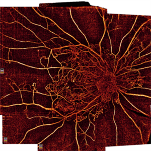 Proliferative Diabetic Retinopathy
Oct 16 2021 by Timur Shaimov
32 y.o. female with Type 1 Diabetes with no glucose compensation for several years. A manual montage of several 8x8 mm OCT angiograms were obtained for this Widefield OCTA image.
Photographer: Timur Shaimov
Imaging device: RTVue xR Avanti
Condition/keywords: OCT Angiography, proliferative diabetic retinopathy (PDR)
-
 RD Montage
Jul 3 2021 by Somnath Chakraborty, MD
Fundus photo montage of the left eye of a 56-year-old male showing subtotal retinal detachment with macular involvement and a large circumlinear tear extending from 1 o' clock to 3 o' clock hours.
Photographer: Pulak Roy
Condition/keywords: acute retinal detachment, retinal detachment of the macula, retinal tear, retinal tear with detachment
-
 Proliferative Diabetic Retinopathy with Choroidal Effusion Status Post PRP
Dec 15 2020 by Manish Nagpal, MD, FRCS (UK), FASRS
A 17-year-old juvenile diabetic patient came to us with extensive neovascular proliferations and PRP done a week back and had developed 360 degree choroidal effusion as seen in this wide field montage image
Photographer: Sham Talati, Retina Fellow , Retina Foundation, Ahmedabad, India
Imaging device: Mirante CSLO
Condition/keywords: choroidal effusion, diabetic retinopathy, proliferative diabetic retinopathy (PDR)
-
 Retinal Arteriovenous Malformation
Jun 6 2020 by Albert Li, MD, FASRS
Montaged infrared retinal imaging of a 37-year-old asymptomatic man with a grade II arteriovenous malformation (AVM) in the nasal mid-periphery. The presentation of the AVM can be classified with three categories. Grade 1 AVMs are characterized by an abnormal capillary plexus between the major communicating vessels. Grade 2 AVMs are defined by the direct arteriovenous communication without the interposition of arterioles or capillaries. Grade 3 AVMs are characterized by widespread, large caliber anastomosing vessels that are associated with decreased visual acuity and intracranial AVMs. Because retinal AVMs are mostly asymptomatic and non-progressive, further testing may not be indicated unless there are concomitant neurological signs and symptoms or if there is a strong clinical suspicion of a grade 3 retinal AVM. Observation was recommended for the patient in this image. On his most recent follow-up at four months, the patient remained asymptomatic with a stable appearance of the lesion.
Imaging device: Heidelberg Spectralis
Condition/keywords: arteriovenous anastomosis, arteriovenous malformation
-
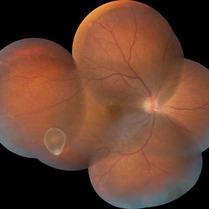
 Retinal Tear
Apr 30 2020 by Giselle DeOliveira
Fundus photograph montage of 32-year-old male with retinal tear after repair.
Photographer: Giselle DeOliveira, University of Miami, Bascom Palmer Eye Institute
Imaging device: Topcon
Condition/keywords: retinal tear
-
 Diabetic Macular TRD
Jan 10 2020 by Somnath Chakraborty, MD
Fundus Montage image of the left eye of a 48-year-old type 2 diabetic with post PRP macular extensive tractional retinal detachment involving macula.
Photographer: Pulak Roy
Condition/keywords: diabetic retinopathy, proliferative diabetic retinopathy (PDR), tractional retinal detachment, vitrectomy, vitreomacular surgery
-
 Retinoblastoma Group C
Dec 29 2019 by Vishal Agrawal, MD, FRCS,FACS,FASRS
Fundus montage of a 3-year-old boy with mass lesion involving macula and abutting the disc of the right eye.The superior half of the tumor shows preretinal extension.
Photographer: Vishal Agrawal MD
Imaging device: Zeiss
Condition/keywords: retinoblastoma
-
 Coats' Disease With Exudative Retinal Detachment and Retinal Macrocyst
Dec 9 2019 by Sophia El Hamichi, MD
A 3-year-old male with a presentation of a complex Coats' disease in the left eye with exudative retinal detachment, abnormal telangiectatic vasculature, and inferotemporal retinal macrocyst/retinoschisis.
Photographer: Abby Orcutt-Hayes, Murray Ocular Oncology and Retina
Imaging device: RetCam
Condition/keywords: Coats' disease, exudative detachment, montage, retinal macrocyst
-
 Retinoblastoma Stage 5 After One Cycle of Systemic Chemotherapy and Laser Ablation
Sep 17 2019 by Sophia El Hamichi, MD
A 1-year-old patient with stage 5B retinoblastoma, fundus after one cycle of systemic chemotherapy and laser ablation.
Photographer: Abby Orcutt-Hayes, Murray Ocular Oncology and Retina
Condition/keywords: chemoreduction, laser photocoagulation, montage, retinoblastoma, stage 5
-
 Retinal CRAO With Emboli
Jun 27 2019 by Somnath Chakraborty, MD
Left eye fundus photo montage of a 43-year-old male with central retinal artery occlusion with bright yellow multiple retinal (cholesterol) emboli both at the disc and also along multiple retinal arteries.
Photographer: Pulak Roy
Condition/keywords: arterial embolus, central retinal artery occlusion (CRAO), cholesterol embolus
-
 Varicella Zoster Virus Retinitis
Jan 17 2019 by Netan Choudhry, MD, FRCS(C) FASRS
Wide-field montage image of a patient with unilateral varicella-zoster virus retinitis following ingestion of animal feces in Colombia.
Photographer: Carmelina Timboli, Vitreous Retina Macula Specialists of Toronto
Imaging device: Optos California (Optos PLC, Edinburgh, UK)
Condition/keywords: retinitis, varicella zoster virus (VZV)
-
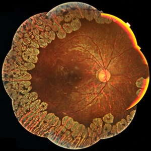 Gyrate Atrophy
Jan 6 2019 by Hashim Ali Khan, OD, FAAO
Montage of Multiple Fundus Photographs from the right eye of a 25-year-old woman with gyrate atrophy.
Photographer: Ahmed Abbass
Imaging device: Topcon TRC-NW8F
Condition/keywords: gyrate atrophy, hereditary retinal dystrophy, retinal dystrophy
-
 Astrocytic Hamartoma
Jan 4 2019 by Netan Choudhry, MD, FRCS(C) FASRS
Fundus photograph montage of a 7-year-old boy with an astrocytic hamartoma involving the macula.
Photographer: John Golding BA, Vitreous Retina Macula Specialists of Toronto
Imaging device: Topcon TRC-50 Dx
Condition/keywords: astrocytic hamartoma
-
 Intraocular Foreign Body
Feb 7 2019 by Somnath Chakraborty, MD
Left eye fundus photo montage of a 45-year-old male showing a large iron foreign body, impacted inferior to the infero-temporal branch vessels with a large patch of surrounding chorio-retinal atrophy, secondary to resolving Commotio retinae
Photographer: Saptarshi Mehta
Condition/keywords: commotio retinae, intraocular foreign body, trauma
-
 Retinochoroidal Coloboma With Aberrant Vasculature
Nov 10 2018 by Chintan D Desai, MBBS, DO, DNB, FICO
Fundus photo montage of a 32-year-old female with a retinochoroidal coloboma Ida Mann classification type 3 with a spring coil shaped aberrant vessel.
Photographer: Kankan Talukdar
Imaging device: Zeiss FF4
Condition/keywords: chorioretinal coloboma
-
 SLE Retinopathy
Jul 10 2018 by Deepak Bhojwani, MS
Colour fundus montage image of a 33-year-old young lady with history of Systemic Lupus Erythematosus of 6 years showing classic SLE retinopathy with multiple cotton wool spots , few haemorrhages and multiple small vessel sheathing s/o SLE vasculitis.
Photographer: Deepak Bhojwani
Condition/keywords: systemic lupus erythematosus (SLE) retinopathy, systemic lupus erythematosus (SLE) vasculitis
-
 Extensive Tractional Retinal Detachment in Proliferative Diabetic Retinopathy
Jun 4 2018 by Diva Kant Misra, MBBS, DO, DNB, MNAMS, FVRS
Montage fundus photograph of a 54-year-old male diabetic patient showing extensive TRD with PDR.
Photographer: DIVA KANT MISRA
Condition/keywords: diabetes, proliferative diabetic retinopathy (PDR), tractional retinal detachment
-
 Multiple Retinal Lesions Secondary to Blunt Trauma
Jun 19 2018 by Somnath Chakraborty, MD
A montage of the right eye of a 15-year-old boy, who was struck by a football. The image shows multiple choroidal ruptures in the macular area, with sub-retinal blood and multiple, large retinal tears temporally. There is also an area of juxtapapillary, pigmentary changes.
Photographer: Saptarshi Mehta, Retina Institute of Bengal
Condition/keywords: blunt trauma, choroidal rupture, giant retinal tear, subretinal hemorrhage
-
 Bear Tracks
Apr 27 2018 by Giselle DeOliveira
Fundus Montage photograph of 13-year-old girl.
Photographer: Giselle DeOliveira, University of Miami , Bascom Palmer Eye Institute
Condition/keywords: bear tracks
-
 Intravitreal Cysticercosis With Full Thickness Macular Hole
Apr 30 2018 by Vishal Agrawal, MD, FRCS,FACS,FASRS
Fundus montage picture of a 40-year-old man presenting with decreased vision in the right eye for the past 2 months. Live intravitreal cysticercosis can be seen lying on the retina. Zooming the image reveals the full thickness macular hole. The scolex invaginates with the light of the camera causing double image of the cyst because of movement .
Photographer: Vishal Agrawal MD,FRCS
Imaging device: Zeiss 524
Condition/keywords: cysticercosis, full thickness macular hole
-
 Post Traumatic Optic Nerve Head Avulsion
Nov 18 2017 by Vishal Agrawal, MD, FRCS,FACS,FASRS
Right eye fundus picture of a 24-year-old male patient who suffered blunt trauma 7 days back with a wooden stick . He presented with NLP vision with a non reacting dilated pupil. Fundus montage picture shows ONH avulsion,CRAO,peripapillary resolving hemorrhages and cicatricial tissue at the edge.
Photographer: Vishal Agrawal, MD, SMS Medical College, Jaipur, India
Imaging device: Zeiss 524
Condition/keywords: avulsion, central retinal artery occlusion (CRAO)
 Loading…