Search results (250 results)
-
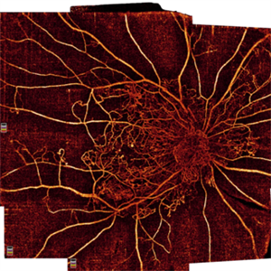
 Wide-Field-OCT-montage
Jan 8 2018 by Netan Choudhry, MD, FRCS(C) FASRS
This is an SD-OCT montage image of a 55 year old male with optic neuropathy representing a wide-field OCT spanning 130 degrees.
Photographer: John Golding, Vitreous Retina Macula Specialists of Toronto
Imaging device: Heidelberg Spectralis OCT system
Condition/keywords: wide angle imaging
-
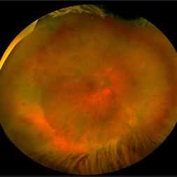
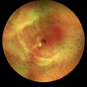
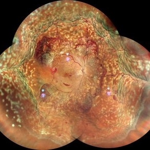 Proliferative Diabetic Retinopathy with Choroidal Effusion Status Post PRP
Dec 15 2020 by Manish Nagpal, MD, FRCS (UK), FASRS
A 17-year-old juvenile diabetic patient came to us with extensive neovascular proliferations and PRP done a week back and had developed 360 degree choroidal effusion as seen in this wide field montage image
Photographer: Sham Talati, Retina Fellow , Retina Foundation, Ahmedabad, India
Imaging device: Mirante CSLO
Condition/keywords: choroidal effusion, diabetic retinopathy, proliferative diabetic retinopathy (PDR)
-
 Retinoblastoma
Apr 27 2018 by Brenda Fallas
2-year-old boy with stage D+ retinoblastoma of the right eye.
Photographer: Brenda Fallas, Bascom Palmer Eye Institute, Miami, FL
Imaging device: RETCAM III 130 degree lens montage
Condition/keywords: tumor, tumor seeding
-
 Post Traumatic Optic Nerve Head Avulsion
Nov 18 2017 by Vishal Agrawal, MD, FRCS,FACS,FASRS
Right eye fundus picture of a 24-year-old male patient who suffered blunt trauma 7 days back with a wooden stick . He presented with NLP vision with a non reacting dilated pupil. Fundus montage picture shows ONH avulsion,CRAO,peripapillary resolving hemorrhages and cicatricial tissue at the edge.
Photographer: Vishal Agrawal, MD, SMS Medical College, Jaipur, India
Imaging device: Zeiss 524
Condition/keywords: avulsion, central retinal artery occlusion (CRAO)
-
 Whole Eye OCT
Jan 4 2019 by Netan Choudhry, MD, FRCS(C) FASRS
Swept-Source OCT montage of a 45-year-old male with Alports disease and posterior subcapsular cataract.
Photographer: John Golding BA, Vitreous Retina Macula Specialists of Toronto
Imaging device: Topcon DRI Triton
Condition/keywords: Alports disease, optical coherence tomography (OCT), swept source
-
 Bear Tracks
Apr 27 2018 by Giselle DeOliveira
Fundus Montage photograph of 13-year-old girl.
Photographer: Giselle DeOliveira, University of Miami , Bascom Palmer Eye Institute
Condition/keywords: bear tracks
-
 Retinal CRAO With Emboli
Jun 27 2019 by Somnath Chakraborty, MD
Left eye fundus photo montage of a 43-year-old male with central retinal artery occlusion with bright yellow multiple retinal (cholesterol) emboli both at the disc and also along multiple retinal arteries.
Photographer: Pulak Roy
Condition/keywords: arterial embolus, central retinal artery occlusion (CRAO), cholesterol embolus
-
 Retinal Tear
Apr 30 2020 by Giselle DeOliveira
Fundus photograph montage of 32-year-old male with retinal tear after repair.
Photographer: Giselle DeOliveira, University of Miami, Bascom Palmer Eye Institute
Imaging device: Topcon
Condition/keywords: retinal tear
-
 Coats' Disease Montage
Feb 5 2021 by Akansha Sharma
Fundus photograph of a 5-year-old male child who presented with unilateral diminution of vision since one month.
Photographer: Dr. Nivesh Gupta, M.S., Retina Foundation, Ahmedabad
Condition/keywords: angiomatosis retinae, Coats' disease, exudative detachment, subretinal exudates
-
 Coats' Disease With Exudative Retinal Detachment and Retinal Macrocyst
Dec 9 2019 by Sophia El Hamichi, MD
A 3-year-old male with a presentation of a complex Coats' disease in the left eye with exudative retinal detachment, abnormal telangiectatic vasculature, and inferotemporal retinal macrocyst/retinoschisis.
Photographer: Abby Orcutt-Hayes, Murray Ocular Oncology and Retina
Imaging device: RetCam
Condition/keywords: Coats' disease, exudative detachment, montage, retinal macrocyst
-
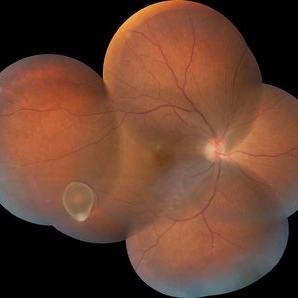
 Giant Retinal Tear
May 27 2020 by Jamin S. Brown, MD
Fundus photo montage of 55-year-old male with retinal detachment and giant retinal tear.
Photographer: Stefanie Palmer CRA, Retina-Vitreous Surgeons of CNY
Condition/keywords: giant retinal tear
-
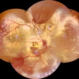
 Proliferative Diabetic Retinopathy
Oct 16 2021 by Timur Shaimov
32 y.o. female with Type 1 Diabetes with no glucose compensation for several years. A manual montage of several 8x8 mm OCT angiograms were obtained for this Widefield OCTA image.
Photographer: Timur Shaimov
Imaging device: RTVue xR Avanti
Condition/keywords: OCT Angiography, proliferative diabetic retinopathy (PDR)
-
 Acute Retinal Necrosis secondary to Herpes Zoster Ophthalmicus
Jan 9 2018 by Olivia Rainey
Ultra-wide field Optos pseudocolor montage of an 40-year-old female presenting with acute retinal necrosis secondary to herpes zoster ophthalmicus affecting her right eye.
Photographer: Olivia Rainey
Imaging device: Optos California
Condition/keywords: acute retinal necrosis, color fundus photograph, Herpes zoster, montage, Optos, ultra-wide field imaging
-
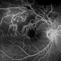 Branch Retinal Vein Occlusion- Fluorescein Angiogram, Montage
Apr 15 2016 by James B. Soque, CRA, OCT-C, COA, FOPS
A fluorescein angiogram of an 80-year-old white female with a superotemporal branch retinal vein occlusion, and retinal edema of the right eye. Currently receiving Lucentis 0.5 injection therapy.
Photographer: James Soque, CRA OCT-C COA, Island Retina, Shirley, NY
Imaging device: Topcon TRC, MERGE Imaging Software V. 11.2.0
Condition/keywords: branch retinal vein occlusion (BRVO), montage, non-perfused branch retinal vein occlusion (BRVO)
-
 Color Montage Picture
Jan 15 2019 by Aniruddha Maiti, MBBS DO DNB FRVS FICO MRCS FACS FASRS FRCOphth
Dilated tortuous vessels with intraretinal and subretinal hemorrhage at fovea.
Photographer: Sangeeta Mohanta
Imaging device: Zeiss FF450 plus IR
Condition/keywords: dilated tortuous vessels, subretinal hemorrhage, Wyburn-Mason
-
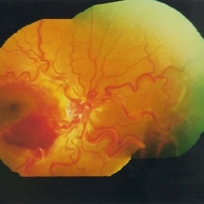
 Diabetic Retinopathy, CSME, Exudates, NVD, Color Fundus Photo, Montage
Mar 18 2015 by James B. Soque, CRA, OCT-C, COA, FOPS
A 58-year-old diabetic male with a longstanding history of diabetic eye disease. Left eye color fundus photo shows extensive CSME, Clinically Significant Macular Edema, with deposits of hard exudates at fixation. There is extensive scattering of hard exudates and sheathing of the vessels.
Photographer: James B Soque, CRA COA
Imaging device: Topcon TRC 50 DX, OIS 5 MP Camera, MERGE software
Condition/keywords: background diabetic retinopathy (BDR), creamy yellow exudates, diabetes, exudates over the posterior pole, neovascularization of the disc (NVD), vessel sheathing
-
 Familial Exudative Vitreoretinopathy
Jan 21 2019 by Netan Choudhry, MD, FRCS(C) FASRS
Widefield montage pseudocolor image of a 35-year-old woman with prior history of FEVR.
Photographer: Carmelina Timboli, Vitreous Retina Macula Specialists of Toronto
Imaging device: Optos California (Optos PLC, Edinburgh, UK)
Condition/keywords: familial exudative vitreoretinopathy (FEVR)
-
 Fluorescein Angiogram of Coats's Disease With Exudative Retinal Detachment
Dec 9 2019 by Sophia El Hamichi, MD
A 3-year-old male presenting a complex Coats' disease of the left eye with exudative retinal detachment, abnormal telangiectatic vasculature with peripheral nonperfusion and leakage.
Photographer: Abby Orcutt-Hayes, Murray Ocular Oncology and Retina
Condition/keywords: Coats' disease, exudative detachment, fluorescein angiogram (FA), montage, retinal macrocyst
-
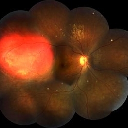 Focal Chroidal Hemangioma
Sep 18 2018 by Somnath Chakraborty, MD
Right eye fundus photo montage of a 17-year-old boy showing a focal choridal hemangioma temporally.
Photographer: Saptarshi Mehta, Retina Institute of Bengal
Condition/keywords: choroidal hemangioma
-
 Giant Retinal Tear
Apr 1 2016 by Nichole Lewis
Giant retinal tear montaged on Anterior Segment due to the Detachment being very bullous.
Photographer: Nichole Lewis - Pennsylvania Retina Specialists, Camp Hill, PA
Condition/keywords: giant retinal tear, retinal tear
-
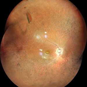 Horseshoe Tear in Retinitis Pigmentosa
Mar 22 2021 by ASRS Staff
Montage of 25-year-old patient, high myopic patient came with complaint of diminution of vision in both eyes and on posterior segment examination of right eye, HST was present along with maculopathy.
Imaging device: Nidek Mirante
Condition/keywords: maculopathy, retinitis pigmentosa
-
 Intravitreal Cysticercosis With Full Thickness Macular Hole
Apr 30 2018 by Vishal Agrawal, MD, FRCS,FACS,FASRS
Fundus montage picture of a 40-year-old man presenting with decreased vision in the right eye for the past 2 months. Live intravitreal cysticercosis can be seen lying on the retina. Zooming the image reveals the full thickness macular hole. The scolex invaginates with the light of the camera causing double image of the cyst because of movement .
Photographer: Vishal Agrawal MD,FRCS
Imaging device: Zeiss 524
Condition/keywords: cysticercosis, full thickness macular hole
-
 Montage OF a Combined Case of CRVO and CRAO
May 15 2014 by Manish Nagpal, MD, FRCS (UK), FASRS
30-year-old anemic lady presented with a acute loss of vision. Her vision was just hand movements in the affected eye and the other eye was normal.
Photographer: pooja barot, Optometrist, Retina Foundation, Ahmedabad
Condition/keywords: central retinal artery occlusion (CRAO), central retinal vein occlusion (CRVO), macular edema
-
 Montage of the Fundus Depicting the IOFB in the Inferotemporal Region
Jun 30 2014 by Manish Nagpal, MD, FRCS (UK), FASRS
A patient came to us with raised intraocular pressure and normal visual acuity. He gave history of injury 2 years back with a iron particle while hammering a nail. On examination foreign body in the infero temporal region stuck in the retina with surrounding hypopigmented scarred area suggestive of resolved oedema and localised reaction. We operated the patient for vitrectomy and removed the IOFB along with laser barrage to the scarred area. The surgery and post operative recovery was uneventful.
Photographer: pooja barot, Optometrist, Retina Foundation, Ahmedabad
Condition/keywords: intraocular foreign body
-
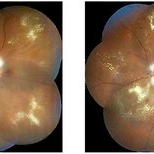 NPDR With Myelinated Nerve Fibers
Nov 5 2018 by Diva Kant Misra, MBBS, DO, DNB, MNAMS, FVRS
Bilateral montage funds photo images of a 56-year-old diabetic patient showing signs of NPDR along with myelinated nerve fibers.
Photographer: Hiteshwar Saikia
Condition/keywords: diabetes, hard exudates, myelinated nerve fibers, nonproliferative diabetic retinopathy
 Loading…