Search results (251 results)
-
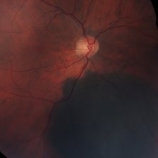
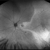
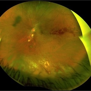
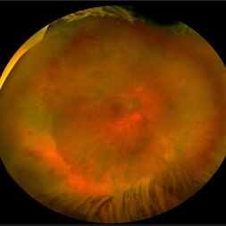 Acute Retinal Necrosis secondary to Herpes Zoster Ophthalmicus
Jan 9 2018 by Olivia Rainey
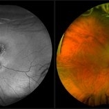
Ultra-wide field Optos pseudocolor montage of an 40-year-old female presenting with acute retinal necrosis secondary to herpes zoster ophthalmicus affecting her right eye.
Photographer: Olivia Rainey
Imaging device: Optos California
Condition/keywords: acute retinal necrosis, color fundus photograph, Herpes zoster, montage, Optos, ultra-wide field imaging
-
 Branch Retinal Vein Occlusion
Sep 11 2018 by Olivia Rainey
Ultra-wide field pseudocolor montage of an 84-year-old female with a branch retinal vein occlusion affecting her left eye. Patient recently had a PPV for a epiretinal membrane in her left eye and shortly after developed an occlusion.
Photographer: Olivia Rainey
Imaging device: Optos
Condition/keywords: branch retinal artery occlusion (BRAO), hemorrhage, left eye, montage, Optos, pseudocolor, ultra-wide field imaging
-
 Branch Retinal Vein Occlusion- Fluorescein Angiogram, Montage
Apr 15 2016 by James B. Soque, CRA, OCT-C, COA, FOPS
A fluorescein angiogram of an 80-year-old white female with a superotemporal branch retinal vein occlusion, and retinal edema of the right eye. Currently receiving Lucentis 0.5 injection therapy.
Photographer: James Soque, CRA OCT-C COA, Island Retina, Shirley, NY
Imaging device: Topcon TRC, MERGE Imaging Software V. 11.2.0
Condition/keywords: branch retinal vein occlusion (BRVO), montage, non-perfused branch retinal vein occlusion (BRVO)
-
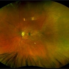
 Central Retinal Vein Occlusion
Jul 13 2018 by Olivia Rainey
Ultra-wide field, pseudocolor montage of a patient presenting with a central retinal vein occlusion, as well as, an inferior chorioretinal scar in their right eye.
Photographer: Olivia Rainey
Imaging device: Optos
Condition/keywords: central retinal vein occlusion (CRVO), chorioretinal scar, montage, Optos, pseudocolor, ultra-wide field imaging
-
 Choroidal Melanoma, Color Fundus Photo
Oct 26 2017 by James B. Soque, CRA, OCT-C, COA, FOPS
71-year-old white male with VA 20/40 OD, c/o shadow in upper right quadrant of visual field. Color Montage of right eye reveals choroidal Melanoma. Patient being evaluated at Sloan Kettering, NYC.
Photographer: James B Soque, CRA, OCT-C, COA, FOPS, Island Retina, Shirley, New York
Imaging device: Topcon TRC 50 DX, with MERGE Winstation 11.2.0
Condition/keywords: color fundus photograph, color photo, fundus autofluorescence (FAF), montage
-
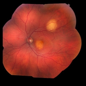 Choroidal Metastasis
Jan 24 2018 by Olivia Rainey
Color fundus montage of a 35-year-old male with choroidal metastasis from the lung. Before the diagnosis was confirmed, the patient had multiple CT scans revealing only pneumonia, with no signs of cancer.
Photographer: Olivia Rainey
Imaging device: Topcon 50DX
Condition/keywords: choroidal metastasis, color fundus photograph, lung cancer metastasis, montage
-
 Coats' Disease
Aug 24 2018 by Kim Barrett
Montage fluorescein angiography of 14-year-old male with Coats' Disease of the left eye. Multiple focal laser treatments. Current uncorrected visual acuity is 20/15-1 OU.
Photographer: Kim Barrett, C.O.A. Retina Specialist of Michigan
Imaging device: Heidelberg Spectralis
Condition/keywords: adolescent, Coats' disease, fluorescein angiogram (FA), Heidelburg Spectralis, laser photocoagulation, left eye, macroaneurysm, montage
-
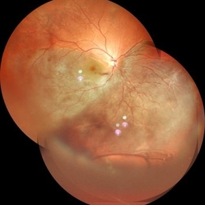
 Coats' Disease With Exudative Retinal Detachment and Retinal Macrocyst
Dec 9 2019 by Sophia El Hamichi, MD
A 3-year-old male with a presentation of a complex Coats' disease in the left eye with exudative retinal detachment, abnormal telangiectatic vasculature, and inferotemporal retinal macrocyst/retinoschisis.
Photographer: Abby Orcutt-Hayes, Murray Ocular Oncology and Retina
Imaging device: RetCam
Condition/keywords: Coats' disease, exudative detachment, montage, retinal macrocyst
-
 Coats's Disease Montage Color Photo
Oct 22 2019 by Sophia El Hamichi, MD
A 7-year-male patient with Coats' disease in the right eye.
Photographer: Abby Orcutt-Hayes, Murray Ocular Oncology and Retina
Condition/keywords: Coats' disease, montage
-
 Combined Hamartoma of the Retina and Retinal Pigment Epithelium
Oct 7 2019 by Sophia El Hamichi, MD
A 7-year-old female followed for combined hamartoma of the retina and the retinal pigment epithelium with amblyopia
Photographer: Abby Orcutt-Hayes, Murray Ocular Oncology and Retina
Condition/keywords: combined hamartoma, montage
-
 Cytomegalovirus Retinitis
Jan 16 2018 by Olivia Rainey
Color fundus montage of an 37-year-old, HIV positive male with CMV retinitis affecting his right eye. Patient's vision was sc20/20-1. He received an intravitreal Ganciclovir injection as well. The referring physcian suspects his condition is secondary to his chemotherapy for large B cell lymphoma or stomach cancer. The patient had not started taking oral Valgancyclovir.
Photographer: Olivia Rainey
Imaging device: Topcon 50dx
Condition/keywords: CMV retinitis, color fundus photograph, cytomegalovirus (CMV), HIV, montage
-
 Dislocated IOL in Vitreous with RD
May 11 2020 by Gayathri Mohan
Color fundus photo montage of a patient showing a dislocated posterior chamber intraocular lens in the vitreous cavity inferiorly along with a sub total retinal detachment.
Photographer: Gayathri Mohan, Retina Foundation
Imaging device: Mirante, Nidek
Condition/keywords: dislocated posterior chamber intraocular lens (PCIOL), montage
-
 Familial Exudative Vitreoretinopathy
Feb 2 2018 by Olivia Rainey
Ultra-wide field montage of a 37-year-old female with familial exudative vitreoretinopathy affecting her left eye. Cryotherapy, laser destruction of retinopathy, and a scleral buckle was performed to stabilize the retina in 2017.
Photographer: Olivia Rainey
Imaging device: Optos
Condition/keywords: familial exudative vitreoretinopathy (FEVR), fibrotic neovascularization, laser scarring, left eye, montage, Optos, scleral buckle, tractional retinal detachment, ultra-wide field imaging
-
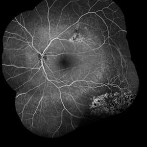
 Fluorescein Angiogram of Coats's Disease With Exudative Retinal Detachment
Dec 9 2019 by Sophia El Hamichi, MD
A 3-year-old male presenting a complex Coats' disease of the left eye with exudative retinal detachment, abnormal telangiectatic vasculature with peripheral nonperfusion and leakage.
Photographer: Abby Orcutt-Hayes, Murray Ocular Oncology and Retina
Condition/keywords: Coats' disease, exudative detachment, fluorescein angiogram (FA), montage, retinal macrocyst
-
 Fluorescein Angiogram of Combined Hamartoma of the Retina and Retinal Pigment Epithelium
Oct 7 2019 by Sophia El Hamichi, MD
A 7-year-old female followed for combined hamartoma of the retina and the retinal pigment epithelium with amblyopia.
Photographer: Abby Orcutt-Hayes, Murray Ocular Oncology and Retina
Condition/keywords: combined hamartoma, fluorescein angiogram (FA), montage
-
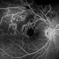
 Hemi Vein Occlusion, Fluorescein Angiogram, Montage
Dec 17 2015 by James B. Soque, CRA, OCT-C, COA, FOPS
74-year-old woman, with recurrent superior hemi vein occlusion, montage image of fluorescein angiogram left eye. Currently receiving Lucentis injections OS.
Photographer: James Soque, CRA COA
Imaging device: Topcon RC 50 DX Fundus Camera with MERGE Winstation Software for Fluorescen Angiography
Condition/keywords: montage, occlusion of retinal vein, superior arcade
-
 Macula Off Retinal Detachment with CNV
Nov 11 2019 by Olivia Rainey
Ultra-wide field pseudocolor photograph of a 42-year-old female with a long-standing, macula-off retinal detachment affecting her left eye. Patient was unaware of vision loss until testing her visual acuity and she denied seeing flashing lights. Patient decided to proceed with scleral buckling. The CNV is potentially secondary the retinal detachment, but may be myopic related or idiopathic. The CNV appears fibrotic and inactive. The patient was warned that this will absolutely limit how much vision she recovers once the retina is reattached.
Photographer: Olivia Rainey
Imaging device: Optos California
Condition/keywords: choroidal neovascularization (CNV), chronic retinal detachment, fundus autofluorescence (FAF), left eye, montage, Optos, retinal detachment of the macula, ultra-wide field imaging
-
 Macula Off Retinal Detachment with CNV
Nov 11 2019 by Olivia Rainey
Ultra-wide field pseudocolor photograph of a 42-year-old female with a long-standing, macula-off retinal detachment affecting her left eye. Patient was unaware of vision loss until testing her visual acuity and she denied seeing flashing lights. Patient decided to proceed with scleral buckling. The CNV is potentially secondary the retinal detachment, but may be myopic related or idiopathic. The CNV appears fibrotic and inactive. The patient was warned that this will absolutely limit how much vision she recovers once the retina is reattached.
Photographer: Olivia Rainey
Imaging device: Optos California
Condition/keywords: choroidal neovascularization (CNV), left eye, montage, Optos, pseudocolor, retinal detachment of the macula, ultra-wide field imaging
-
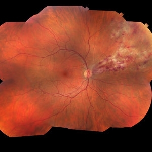
 Malignant Melanoma
Sep 11 2018 by Olivia Rainey
Ultra-wide field autofluorescence and pseudocolor montage of a 57-year-old male s/p I-125 brachytherapy for malignant melanoma affecting his right eye. The patient’s radiation retinopathy has resulted in retinal vascular occlusive disease and optic nerve edema.
Photographer: Olivia Rainey
Imaging device: Optos
Condition/keywords: branch retinal vein occlusion (BRVO), fundus autofluorescence (FAF), I-125 brachytherapy, malignant melanoma, montage, Optos, pseudocolor, radiation retinopathy, ultra-wide field imaging
-
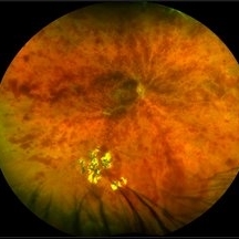
 Montage Photo: Retinopathy of Prematurity Stage 4A Treated with Laser and Bevacizumab Intravitreal Injection
Jan 6 2020 by Sophia El Hamichi, MD
A 41-week-old girl, with a gestational age of 23 weeks and 4 days presenting with stage 4 A of ROP OU. She was treated with laser and intravitreal injections of bevacizumab.
Photographer: Abby Orcutt-Hayes, Murray Ocular Oncology and Retina
Condition/keywords: intravitreal bevacizumab, laser photocoagulation, montage, retinopathy of prematurity stage 4a
-
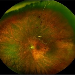
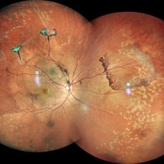 Neovascularization in a Case of Idiopathic Vasculitis
Aug 18 2021 by ASRS Staff
Right eye, Wide field photograph Motage of 37 year-old male, having idiopathic vasculitis status post PRP. Patient's systemic evaluation was done, and everything was within normal limits.
Imaging device: Nidek Mirante
Condition/keywords: Idiopathic vasculitis, montage, neovascularization (NV), pan-retinal photocoagulation (PRP)
-
 Ocular Metastasis of Breast Cancer
Mar 13 2018 by Olivia Rainey
Color fundus montage of a 45-year-old female presenting with ocular metastasis affecting her left eye. She had been treated for pneumonia, had progressive lumbar back pain, and a 29 pound weight loss recently. She reported that she had a breast lump and a mammogram, but had not been provided the results. After she was sent to oncology, it was confirmed that she has widely metastatic breast cancer and tumors throughout the brain. The oncology team felt she could not wait weeks for her chemotherapy to start and consequently decided to do whole brain radiation and treat the affected eye just posterior to the lens.
Photographer: Olivia Rainey
Imaging device: Topcon DX50
Condition/keywords: choroidal metastasis, color fundus photograph, exudative detachment, left eye, lipid exudation, montage
-
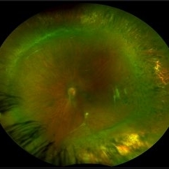
 Ocular Parasitosis
May 22 2016 by Olivia Rainey
Color fundus montage of an 12-year-old boy with ocular parasitosis affecting his left eye. Patient presented with decreased vision and recent travel to Florida. The specimen was lost in the lab and was never recovered.
Photographer: Olivia Rainey
Imaging device: Topcon 50dx
Condition/keywords: color fundus photograph, color photo, intraocular foreign body, left eye, montage, parasite
-
 Penetrating Trauma of an Inadvertent Sub-Tenon's Kenalog Injection
Jan 31 2018 by Olivia Rainey
Ultra-wide field pseudocolor photograph of a 38-year-old female with penetrating trauma after an inadvertent sub-tenon's kenalog injection affecting her left eye. Patient has a large dehemoglobinized vitreous hemorrhage settling inferior near the entry wound. The exit wound has developed chorioretinal scarring and the disruption of several veins near the optic nerve, resulting in a branch retinal vein occlusion.
Photographer: Olivia Rainey
Imaging device: Optos
Condition/keywords: branch retinal vein occlusion (BRVO), chorioretinal scar, color fundus photograph, dehemoglobinized hemorrhage, kenalog, left eye, montage, Optos, penetrating trauma, sub-tenon's, ultra-wide field imaging
-
 Proliferative Diabetic Retinopathy
Mar 1 2021 by Avris Romario Diparaja Siahaan
Fundus photograph (montage photography) of a 57-year-old woman with proliferative diabetic retinopathy in her both eyes.
Photographer: Nanda Lessi Hafni Eka Putri, MD (Ophthalmologist) & Ryan Mishbahuddin (Nurse), Ciawi General Hospital (Rumah Sakit Umum Daerah Ciawi)
Imaging device: DRI OCT Triton Plus
Condition/keywords: fundus photograph, montage, optical coherence tomography (OCT), swept source, wide angle imaging
 Loading…