Search results (250 results)
-
 Retinal Detachment and Lattice Degeneration
Mar 25 2025 by Korey Starkey
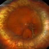
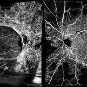
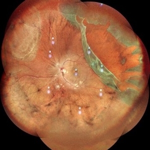
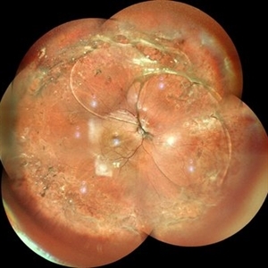
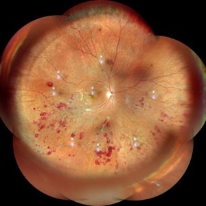
26 year-old patient presented at first visit with rhegmatogenous macula involving retinal detachment of the left eye. Underwent prompt surgical repair. Both eyes also present with lattice degeneration with atrophic holes.
Photographer: Korey Starkey
Condition/keywords: atrophic retinal hole, fundus photography, lattice degeneration, montage photo, Optos, OPTOS CALIFORNIA RGB, retinal detachment, retinal holes, rhegmatogenous retinal detachment, ultra-wide field imaging
-
 Repaired Retinal Detachment with Scleral Buckle
Mar 25 2025 by Kimberly Wakester
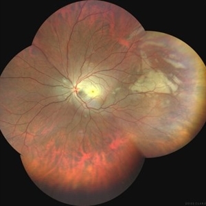
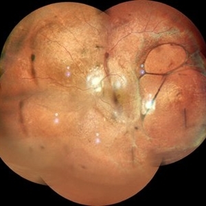
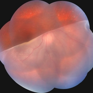
Optomap RGB montage of an 64-year-old woman with a repaired retinal detachment with scleral buckle in the right eye. There is nasal and inferior pre-retinal membranes with traction. PPV was recommended but patient defers to proceed with sx at this time. Will continue to follow patient closely for worsening traction. Patient was educated on how to monitor their peripheral vision and was advised to report any changes immediately.
Photographer: Kimberly Wakester, COA, OCT-C
Imaging device: Optos California
Condition/keywords: pre-retinal membrane with traction, repaired RD, scleral buckle
-
 Central Retinal Artery Occlusion
Mar 21 2025 by T. P . VIGNESH, MBBS,MS
Fundus photo montage of a 29 year old man with Central retinal artery occlusion.
Photographer: Bharathi
Imaging device: Zeiss Clarus
Condition/keywords: central retinal artery occlusion (CRAO)
-
 Branch Retinal Vein Occlusion with Macular Edema
Mar 14 2025 by Drew Mitchell
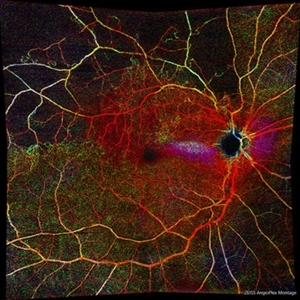
Zeiss Montage Angio 8x8 mm OCT Angiography Retina Depth Encoded Angioplex of a New BRVO in the right eye.
Photographer: Drew Mitchell, OCT-C
Imaging device: Zeiss Cirrus 6000
Condition/keywords: branch retinal vein occlusion (BRVO), macular edema, OCT Angiography
-
 Branch Retinal Vein Occlusion with Macular Edema
Mar 14 2025 by Drew Mitchell
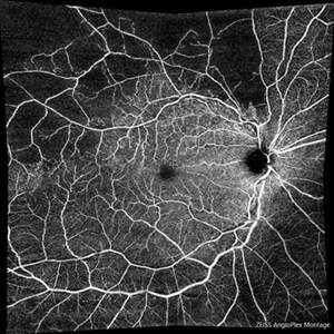
Zeiss Montage Angio 8x8 mm OCT Angiography Superficial Angioplex of a New BRVO in the right eye.
Photographer: Drew Mitchell OCT-C
Imaging device: Zeiss Cirrus 6000
Condition/keywords: branch retinal vein occlusion (BRVO), macular edema, OCT Angiography
-
 Bilateral Proliferative Diabetic Retinopathy OU
Feb 21 2025 by Drew Mitchell
OCT-Angiography 8x8 Montage OU. PDR with active NVE OD. 37 year old male with no visual complaints. Vision is 20/20 in both eyes.
Photographer: Drew Mitchell OCT-C
Imaging device: Zeiss Cirrus 5000
Condition/keywords: CIRRUS 5000 ANGIOPLEX, Diabetes, NVE, OCT Angiography, proliferative diabetic retinopathy (PDR)
-
 Lattice Degeneration With Atrophic Retinal Holes
Jan 30 2025 by Kimberly Wakester
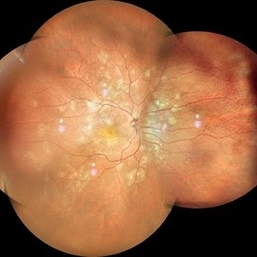
Ultra-wide field montage fundus photograph of a 56-year-old woman with lattice degeneration with atrophic holes statues post laser. Patient also has a small CHRPE temporal to macula and trace ERM that is not visually significant. Will continue follow up care to monitor and treat as needed.
Photographer: Kimberly Wakester, COA
Imaging device: Optos California
Condition/keywords: atrophic retinal hole, CHRPE, epiretinal membrane (ERM), lattice degeneration, montage photo
-
 Who Stole My Blood Supply?
Jan 25 2025 by Muna Bhende, MD
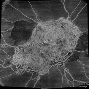
Superficial capillary plexus slab montage image of a young female diabetic with florid proliferation . There is no flow in the capillaries anterior to the tangle of new vessels indicating severe retinal ischemia.
Photographer: Mohanapriya L , Medical Research Foundation, Sankara Nethralaya, Chennai, India
Imaging device: PLEX elite 9000
Condition/keywords: florid type PDR, OCTA
-
 Choroideremia
Jan 23 2025 by Prashant K Bawankule, M.S.
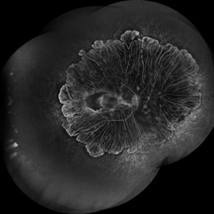
A young male of 25 years, with progressive loss of vision with suspected CNVM. FFA showed 360 degree shutdown with blocked fluorescence in macular region.
Photographer: Prashant Bawankule, Sarakshi Netralaya, Nagpur, Maharashtra , India
Imaging device: Mirante ( by Nidek)
Condition/keywords: Montage of FFA in a case of choroderemia
-
 Retinal Detachment with Multiple OCT Overlays
Jan 7 2025 by Drew Mitchell
Optos 360* Color photo montage with multiple Zeiss Cirrus OCT scan overlays. Retinal Detachment with multiple breaks and a Epiretinal Membrane.
Photographer: Drew Mitchel, OCT-C
Imaging device: Optos California
Condition/keywords: ERM, macular pucker, montage, Optos, OPTOS CALIFORNIA, RD, Retinal Detachment
-
 Uveal Effusion Syndrome
Jan 7 2025 by Drew Mitchell
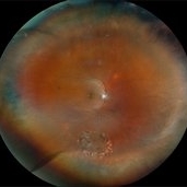
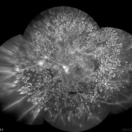
Fundus Autofluorescence Montage of Uveal Effusion Syndrome.
Photographer: Drew Mitchel, OCT-C
Imaging device: Optos California
Condition/keywords: montage, Optos, uveal effusion
-
 Uveal Effusion Syndrome
Jan 7 2025 by Drew Mitchell
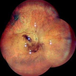
Optos Color Montage of Uveal Effusion Syndrome
Photographer: Drew Mitchell, OCT-C
Imaging device: Optos California
Condition/keywords: color photo, montage, OPTOS, uveal effusion
-
 Giant Retinal Tear
Oct 11 2024 by Anjana Mirajkar, MS Ophthalmology
Fundus photograph montage of LE showing a giant retinal extending from 12 to 4 o clock.
Photographer: Dr. Anjana Mirajkar -Retina Foundation, Ahmedabad
Imaging device: Mirante-Nidek
Condition/keywords: GIANT RETINAL TEAR
-
 X-Linked Juvenile Retinoschisis
Oct 5 2024 by Anand Temkar
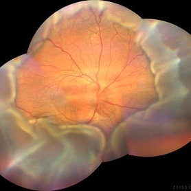
This is a color photo montage of LE of a 15 year old child with X-linked juvenile retinoschisis.
Photographer: Dr.Anand Temkar- Retina Foundation, Ahmedabad
Imaging device: Mirante
Condition/keywords: x-linked retinoschisis (XLRS)
-
 X-Linked Juvenile Retinoschisis
Oct 5 2024 by Anand Temkar
This is a color photo montage of RE of a 15 year old child with X-linked juvenile retinoschisis.
Photographer: Dr.Anand Temkar- Retina Foundation, Ahmedabad
Imaging device: Mirante
Condition/keywords: x-linked retinoschisis (XLRS)
-
 Ischemic BRVO
Aug 26 2024 by Nassim Alejandro Abreu Arbaje, MD
Montage of fluorescein angiography and color fundus photos of a 60 years old male with diabetic retinopathy complicated with extensive retinal neovascularization secondary to and ischemic BRVO.
Photographer: Nassim Abreu, Hospital Dr. Elías Santana
Condition/keywords: BRVO, diabetic retinopathy, Neovascularisation elsewhere (NVE)
-
 Choroidal Detachment
Aug 14 2024 by STEFANY DAVILA
Montage of fundus photography of an elderly male with choroidal detachment 360 degrees after trabeculectomy surgery.
Photographer: Stefany Dávila Avila, Instituto Mexicano de Oftalmología, Querétaro
Imaging device: Zeiss Clarus 700
Condition/keywords: Choroidal, detachment
-
 Membranes Formed Under Silicon Oil and Retina
Jul 18 2024 by Anjana Mirajkar, MS Ophthalmology
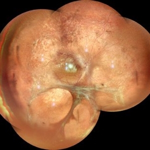
A montage color photo of RE of a 7 year old male with membranes formed between silicon and the retina injected for retinal detachment.
Photographer: Dr. Anjana Mirajkar -Retina Foundation, Ahmedabad
Imaging device: Mirante-Nidek
Condition/keywords: sub-silicon membranes
-
 Giant Retinal Tear
Jul 15 2024 by Arthi Mohankumar , MS,MRCS ED, FICO,FAICO
Fundus montage of a 15 year old boy with Marfans syndrome who presented with defective vision in the right eye.
Photographer: Arthi Mohankumar
Condition/keywords: giant retinal tear, Retinal detachment
-
 Multifocal Choroiditis
Jul 13 2024 by Tejaswita Verma
RE fundus montage of a 34 y/o male showing old and active hypopigmented lesions with macular involvement .He presented with DOV since a month,treated with oral steroids for 15 days elsewhere,with BCVA of CF2mt and positive Mantoux test.
Photographer: DR. TEJASWITA VERMA
Imaging device: MIRANTE
Condition/keywords: multifocal choroiditis
-
 Combined Retinal Artery Macro Aneurysm with Retinal Vein Occlusion
Jun 25 2024 by Aniruddh Soni, DO DNB FLVPEI
Color Fundus and Fluorescein Angiography Montage of a 55 year Lady with Combined Retinal Artery Macro Aneurysm with Retinal Vein Occlusion.
Photographer: Dr Aniruddh Soni, Anupam Eye Hospital, Jaipur, INDIA
Condition/keywords: macroaneurysm, Vein Occlusion
-
 Fluorescein Angiography Montage
Jun 21 2024 by BENITO VERGARA, MD
Montage of an angiography with fluorescein from the left eye of a 32 year-old male with diabetic retinopathy previously treated with panretinal photocoagulation, that shows leakage at optic nerve and upper nasal arcade.
Photographer: Benito Vergara, Asociación Para Evitar la Ceguera en México.
Imaging device: Zeiss Clarus 700
Condition/keywords: Angiography Montage, angiography with fluorescein, diabetic retinopathy, FA montage, fluorescein angiogram (FA), peripheral scars
-
 FFA in Atypical Tubercular Peripheral Occlusive Retinal Vasculitis
Jun 21 2024 by Tejaswita Verma
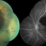
Right eye FFA montage of a 27 year male with peripheral occlusive tubercular vasculitis, showing CNP areas inferiorly and temporally, leakages and blocked fluorescence due to hemorrhages. The patient was advised intravitreal anti-VEGF injection and later sectoral laser once inflammation subsides.
Photographer: DR. TEJASWITA VERMA
Imaging device: MIRANTE
Condition/keywords: obliterative peripheral vasculitis, ocular tuberculosis
-
 Atypical Tubercular Peripheral Occlusive Retinal Vasculitis
Jun 21 2024 by Tejaswita Verma
Fundus montage of the right eye of a 27 year old male with macula threatening occlusive vasculitis showing hemorrhages in inferior, temporal quadrant with vascular sheathing. The patient was Mantoux positive (20 mm induration) and IGRA (TB-GOLD)positive and started on oral steroids. The case was atypical due to no vitritis at presentation which is unusual of tuberculosis. Behcet's disease was ruled out as there was no panuveitis like picture.
Photographer: DR. TEJASWITA VERMA
Imaging device: MIRANTE
Condition/keywords: occlusive vasculitis, ocular tuberculosis
-
 Lasered Horse Shoe Tear with Mild Vitreous Haemorrhage
Jun 12 2024 by Anand Temkar
RE CF Montage of a 55 yrs old male with Lasered HST and Mild VH.
Photographer: Dr.Anand Temkar- Retina Foundation, Ahmedabad
Imaging device: Mirante
Condition/keywords: vitreous hemorrhage
 Loading…