File number: 28403
Comments
-
 Jonathan L. Prenner, MD (December 9 2018)
I'm making it my screensaver!
-
 Suber S. Huang, MD, MBA, FASRS (September 21 2018)
A rare and informative case. Please consider updating pertinent clinical information of age. fellow eye, pertinent and confirmatory laboratory testing. Consider also submitting fellow eye and refining montage. The FA would help document the severity of occlusive vasculopathy. Excellent. Thank you!
-
 Deepak Bhojwani, MS (July 13 2018)
Bingo Hosam Attia...
Your observations and clinical interpretation are too good except the pateint didnt had ophthalmic artery occlusion. I will try to collect all images and angiogram if poosible and upload the clinical case soon... - Hosam Attia, MD (July 12 2018)
Excellent photo!
Thank you for your submission.
May I ask, what is the patient vision (she seems to have optic atrophy as well, ? ophthalmic a. Occlusion), do you have a photo of the other eye, Red free, early & late angiogram frames. it would be nice to include these information as a complete case.
Thank you again - Deepak Bhojwani, MS (July 11 2018)
Thanks James for your kind words.
-
 James B. Soque, CRA, OCT-C, COA, FOPS (July 10 2018)
The rarity of this disease is hallmark in itself. You have done your office quite proudly with your submission! Excellent work sir. Thank you for your submission.
Sign in to comment.
 Initializing download.
-
 By Deepak Bhojwani, MS
- Uploaded on Jul 10, 2018.
- Last modified by Caroline Bozell on Nov 30, 2018.
- Image of the week
-
Dec 2, 2018
View all images of the week - Rating
- Appears in
- Miscellaneous
- Condition/keywords
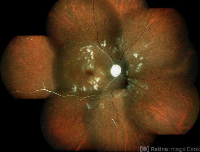
- systemic lupus erythematosus (SLE) vasculitis, systemic lupus erythematosus (SLE) retinopathy
- Photographer
- Deepak Bhojwani
- Imaging device
- Fundus camera
- Description
- Colour fundus montage image of a 33-year-old young lady with history of Systemic Lupus Erythematosus of 6 years showing classic SLE retinopathy with multiple cotton wool spots , few haemorrhages and multiple small vessel sheathing s/o SLE vasculitis.


---thumb.jpg/image-square;max$79,0.ImageHandler "SLE Retinopathy")
---thumb.jpg/image-square;max$79,0.ImageHandler "SLE Vasculitis")
---thumb.jpg/image-square;max$79,0.ImageHandler "SLE Vasculitis")
---thumb.jpg/image-square;max$79,0.ImageHandler "SLE Retinopathy")
---thumb.JPG/image-square;max$79,0.ImageHandler "SLE retinopathy")
---thumb.JPG/image-square;max$79,0.ImageHandler "SLE retinopathy")
---thumb.JPG/image-square;max$79,0.ImageHandler "SLE retinopathy")
---thumb.JPG/image-square;max$79,0.ImageHandler "SLE retinopathy")

