Search results (365 results)
-
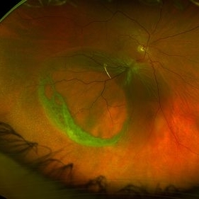
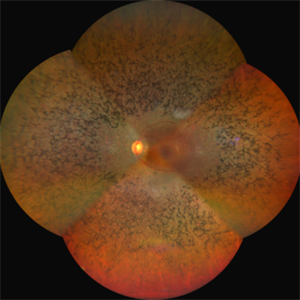
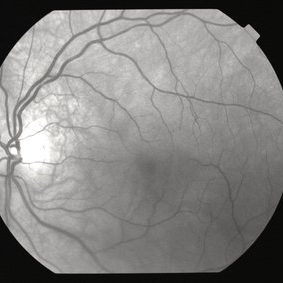
 Colour Photo of a Case of Pigmented Paravenous Chorioretinal Atrophy (PPCRA)
Jun 30 2017 by Manish Nagpal, MD, FRCS (UK), FASRS
This is a rare presentation of a case of PPCRA
Photographer: RAKESH JUNEJA
Condition/keywords: pigmented paravenous chorioretinal atrophy (PPCRA), rare
-
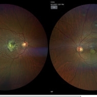
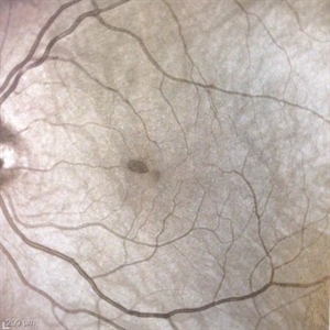
 Colour Photo of a Rare Case of Pigmented Paravenous Chorioretinal Atrophy (PPCRA)
Jun 30 2017 by Manish Nagpal, MD, FRCS (UK), FASRS
A rare case of PPCRA
Photographer: RAKESH JUNEJA
Condition/keywords: pigmented paravenous chorioretinal atrophy (PPCRA), rare
-
 Complex Retinal Detachment with PVR and Starfold
Jun 6 2025 by Jenn Geelan
57 year old male with a Complex Retinoschisis related retinal detachment with PVR and a Posterior Star Fold
Photographer: Jenn Geelan, Retina-Vitreous Surgeons of CNY
Imaging device: Optos California
Condition/keywords: proliferative vitreoretinopathy (PVR), rare, Retinal Detachment, retinoschisis, Starfolds, subretinal fluid
-
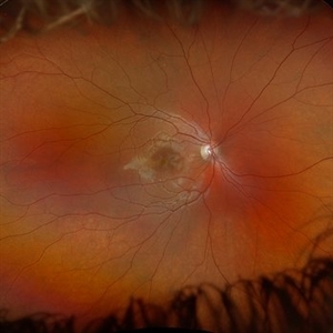
 Macular Coloboma
Jul 17 2024 by Anubhav Chauhan
This is fundus photograph of a 30 year male depicting a Macular coloboma in the right eye. The patient had a sharply defined large, yellowish white, coarsely pigmented, atrophic, round crater like defect at the macula. Spectral domain optical coherence tomography confirmed our diagnosis. The serology testing such as serum IgM, IgG for toxoplasma and cytomegalovirus was negative. His systemic examination was normal.
Photographer: Dr Anubhav Chauhan, Department of Ophthalmology, Shri Lal Bahadur Shastri Government Medical College, Nerchowk, District Mandi, Himachal Pradesh, India
Imaging device: Zeiss
Condition/keywords: macula, rare
-
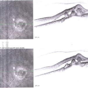
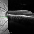
 OCT OD of a Case of Pigmented Paravenous Chorioretinal Atrophy (PPCRA)
Jun 30 2017 by Manish Nagpal, MD, FRCS (UK), FASRS
This is the OCT of a rare case of PPCRA
Photographer: RAKESH JUNEJA
Condition/keywords: pigmented paravenous chorioretinal atrophy (PPCRA), rare
-
 OCT OS of a Case of Pigmented Paravenous Chorioretinal Atrophy (PPCRA)
Jun 30 2017 by Manish Nagpal, MD, FRCS (UK), FASRS
This is the OCT of a rare case of PPCRA.
Photographer: Rakesh Junena
Condition/keywords: pigmented paravenous chorioretinal atrophy (PPCRA), rare
-
 Spiral ERM
Dec 20 2019 by Anfisa Ayalon, MD
OCT of a 60 -year-old man with epiretinal membrane. Note a spiral-like edge.
Photographer: Anfisa Ayalon,MD., Meir Medical Center, Kfar Saba, Israel.
Condition/keywords: epiretinal membrane (ERM), optical coherence tomography (OCT), rare
-
 Choroidal nevus with polypoidal choroidal vasculopathy
Nov 20 2012 by Roy Schwartz, MD
Rare combination of a choroidal nevus complicated by polypoidal choroidal vasculopathy. The lesion is temporal to the fovea.
Photographer: Galit Yair-Pur
Condition/keywords: choroidal nevus, polypoidal choroidal vasculopathy (PCV)
-
 choroidal nevus with polypoidal choroidal vasculopathy
Nov 20 2012 by Roy Schwartz, MD
Rare combination of a choroidal nevus complicated by polypoidal choroidal vasculopathy. The lesion is temporal to the fovea, and leakage of subretinal fluid almost reaching the fovea is demonstrated.
Photographer: Galit Yair-Pur
Condition/keywords: choroidal nevus, polypoidal choroidal vasculopathy (PCV)
-
 choroidal nevus with polypoidal choroidal vasculopathy
Nov 20 2012 by Roy Schwartz, MD
Rare combination of a choroidal nevus complicated by polypoidal choroidal vasculopathy. The lesion is temporal to the fovea, and subretinal fluid almost reaching the fovea is demonstrated.
Photographer: Galit Yair-Pur
Condition/keywords: choroidal nevus, optical coherence tomography (OCT), polypoidal choroidal vasculopathy (PCV)
-
 Rare Bilateral Choroidal Metastasis from Occult Primary Lung Cancer
May 5 2021 by Deependra Vikram Singh, MD FASRS
Fundus photographs and OCT scans of a 73-year-old non-smoker Indian male who presented to our retina clinic in 2013 with blurred vision in left eye for past 2 weeks. BCVA was 20/20 in right eye and 20/40 in left eye. Slit lamp exam was unremarkable for both eyes with no cells in aqueous or anterior vitreous. Fundus examination revealed creamy yellow choroidal lesions in both eyes. Lesion in right eye was one disc diameter (DD) in size and was located close to fovea (Fig-1a). Lesion in the left eye was bigger with a size of 2 DD located superior to fovea (Fig-1b). OCT scan for left eye revealed neurosensory detachment involving fovea (Fig-1c). Fundus fluorescein angiography was inconclusive for right eye and showed late hyper fluorescence the choroidal lesion in left eye. Patient underwent detailed systemic work up for malignancy that revealed primary lung non-small cell carcinoma. He had widespread metastasis affecting liver and brain. Palliative chemotherapy and radiotherapy were initiated 4 weeks after he presented to us. The choroidal lesions show progression on fundus picture and OCT scans done at 4 weeks follow up after initial presentation (Fig – 1d, e, f). The lesions in both eyes show regression at 4 weeks and 12 weeks follow up after initiation of therapy. Unfortunately, patient succumbed at 13 weeks follow up due to disease progression. The case demonstrates rare bilateral choroidal metastasis from primary lung cancer and also highlights that lesions can be asymptomatic till they develop neurosensory detachment as evident from asymptomatic lesion in right eye despite proximity to fovea and symptomatic lesion in left eye with NSD.
Photographer: Deependra Vikram Singh, Eye-Q Superspecialty Eye Hospitals, Gurugram
Imaging device: Topcon
Condition/keywords: choroidal mass, choroidal metastasis
-
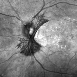 "NVD Flower"
Oct 20 2023 by Daniel Davis, OCT-C
Infrared image of NVD (52F)
Imaging device: Heidelberg Spectralis
Condition/keywords: neovascularization of the disc (NVD)
-
 "Untouched" Intra-ocular Foreign Body - Course Over 7 Years!
Jul 7 2020 by Deependra Vikram Singh, MD FASRS
25-yr-old male presented to our retina clinic in 2007 with history of Hammer and chisel injury to left eye 2 months back. On examination BCVA in left eye was 20/20. Slit-lamp examination revealed iris hole and fundus examination showed an encapsulated metallic intraocular foreign body (IOFB) close to inferior arcade in left eye. Patient was advised Vitreous surgery with IOFB removal. Patient, however did not turn up for Surgery and revisited our clinic after seven years in 2014. On examination his BCVA was 20/20 in left eye and IOFB has reduced in size with brown siderotic deposits seen over IOFB capsule. Examination revealed posterior sub-capsular cataract but no siderotic changes with intraocular pressure (IOP) also being recorded as normal. In view of good visual acuity and no siderotic changes, he was advised regular follow up and ERG. Since most IOFBs would get timely removed by vitreous surgery, this Image capturing the natural course of a metallic IOFB is rare.
Photographer: Deependra Vikram Singh, Eye-Q Hospitals, Gurugram, INDIA
Imaging device: Kowa and Zeiss
Condition/keywords: encapsulated intraocular foreign body
-
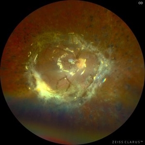 360 Retinotomy in a closed Funnel combined Tractional and rhegmatogenous retinal detachment
Jan 1 2023 by Malek Yassine, MD
This is the results at 6 months of a Bimanual 23 G-PPV with a very extensive and posterior 360 retinotomy for the management of a combined longstanding closed funnel RD, with submacular membranes, intraretinal PVR. Preop VA was a doubtful light perception. Borders of the retinotomy are stable at 6 months under 1300 Cs Silicon oil with some pigmented PVR developping the edges. Macula appears spared. Silicon oil emulsification droplets are well visualized beneath the superior temporal arcade.
Imaging device: Zeiss Clarus 700
Condition/keywords: combined retinal detachment, retinotomy, silicone oil
-
 A Feast for Crows , Retinitis pigmentosa
Sep 22 2022 by wang xiaomei
Fundus photograph of an 55-year-old man with Retinitis Pigmentosa, There is increasing loss of pigment from the pigment epithelium with intraretinal clumping of melanin, appearing most often as coarse clumps in a "bone spicule" configuration, arteriolar narrowing
Photographer: Man, Li, Bao Ji Ophthalmic Hospital
Imaging device: ZEISS CLARUS 500
Condition/keywords: retinitis pigmentosa (RP) dystrophy
-
 A rare case of a 45-year-old male with choroidal neovascular membrane in Familial Dominant Drusen (Doyne Honeycomb Drusen) in both eyes treated with intravitreal injections.
Nov 30 2022 by SHRADDHA ASHOK CHANDORKAR, DNB DO FVRS
A 45-year-old man presented with diminution of vision in both eyes with metamorphopsia, which was painless and gradually progressive in nature. BCVA at presentation were 6/40 and 6/36 for the right and left eye respectively. Anterior segment examination of both eyes was unremarkable. IOP were within normal limits. Fundus examination showed bilateral numerous yellowish white round closely spaced lesions extending radially from the vascular arcades till the periphery associated with an elevated grayish macular choroidal neovascular membrane (CNV) with multiple drusen in the macular area and posterior pole. Impression was Familial Dominant Drusen (Doyne Honeycomb Drusen) associated with CNVM, both eyes. Color fundus photograph and autofluorescence showed Familial Dominant Drusen with CNVM. Subsequently , the patient underwent periodic intravitreal injections of Ranibizumab in both eyes under guarded visual prognosis, for which he tolerated well.
Photographer: NATIONAL INSTITUTE OF OPHTHALMOLOGY, PUNE
Imaging device: ZEISS CLARUS
Condition/keywords: choroidal neovascular membrane (CNVM), Doyne's Honeycomb, FAMILIAL DOMINANT DRUSEN, IMIM (Online Mendelian Inheritance in Man), intravitreal injection, Malattia Leventinese
-
 Acute Macular Neuroretinopathy
Mar 25 2024 by Daniel Davis, OCT-C
18 yo female presenting with hazy vison for 2-3 weeks. VA OD: sc20/20 VA OS: sc20/20 Infrared imaging showed dark gray, petalloid, perifoveal lesions and OCT shows focal signal reduction of the Inner Segment / Outer Segment junction. Elects to observe.
Photographer: Daniel Davis, OCT-C, The Retina Institute, St. Louis
Imaging device: Optos California SWL
Condition/keywords: acute macular neuroretinopathy
-
 Acute Macular Neuroretinopathy
Dec 11 2019 by Lauren Whaley
34-year-old female patient presented with changes in vision after recent upper respiratory infection. Referring doctor originally thought it was a blood pressure issue. She noticed a "C" shape in her vision. Infrared image was captured showing exactly what patient was describing! Doctor confirmed with this image that it was AMN.
Photographer: Lauren R. Whaley, COA
Imaging device: Heidelberg Spectralis
Condition/keywords: 30 degrees, acute macular neuroretinopathy, Heidelburg Spectralis, left eye, macula, near infrared autofluorescence (NIRAF)
-
 Acute Macular Neuroretinopathy
Sep 15 2014 by Thomas A. Ciulla, MD, MBA, FASRS
Color photo. This might be a typical fundus photo, with no definite lesion. However, the infrared photo nicely depicts a typical lesion.
Condition/keywords: acute macular neuroretinopathy, color photo
-
 Acute Macular Neuroretinopathy
Sep 15 2014 by Thomas A. Ciulla, MD, MBA, FASRS
Red-free photo. Similar to the color photo, this red-free photo shows no definite lesion. However, the infrared photo nicely depicts the typical lesion.
Condition/keywords: acute macular neuroretinopathy, red-free
-
 Acute Macular Neuroretinopathy
Aug 13 2014 by Thomas A. Ciulla, MD, MBA, FASRS
Infrared photo. This infrared photo nicely demonstrates a typical small wedge-shaped lesion of acute macular neuroretinopathy. This case highlights the usefulness of the infrared photo for this condition.
Condition/keywords: acute macular neuroretinopathy
-
---thumb.jpg/image-square;max$300,300.ImageHandler) Acute retinal necrosis
Acute retinal necrosis
Feb 15 2013 by From the Collections of Thomas M. Aaberg, MD and Thomas M. Aaberg Jr., MD
Diffuse intraretinal hemorrhages and whitening in the posterior pole consistent with acute retinal necrosis.
Condition/keywords: macular edema, microangiopathy, retinal necrosis, retinal whitening
-
 Amelanotic Melanoma
Aug 12 2025 by César Adrián Gómez Valdivia, MD
This FAF image reveals a hypoautofluorescent mass with areas of dense hyperautofluorescent stippling—a classic pattern suggestive of an amelanotic choroidal melanoma. Amelanotic melanoma is a rare variant of uveal melanoma, accounting for only a minority of cases. Unlike pigmented melanomas, these lesions lack melanin, making them more challenging to detect on conventional color fundus imaging. FAF Characteristics: • Central hypoautofluorescence: due to loss or compression of the RPE • Peripheral hyperautofluorescent speckling: consistent with lipofuscin accumulation or RPE disruption • Often associated with subretinal fluid or orange pigment seen clinically Location: Juxtapapillary, with potential optic nerve involvement—a factor that complicates both diagnosis and
Photographer: @eyemissu2
Imaging device: California ICG OPTOS
Condition/keywords: amelanotic melanoma
-
 Angiographic Storm: Fluorescein Leakage in Retinal Vasculitis
Nov 17 2025 by SHRADDHA RAJ SHRIVASTAVA
This left eye montage fundus fluorescein angiography (FFA) image of a 19 year old male with idiopathic retinal vasculitis, having skip vasculitic lesions predominantly involving retinal veins. There are areas of blocked fluorescence due to intraretinal hemorrhages, the involved veins have filling defects and occlusions, leading to formation of numerous collateral channels. The inflamed vessels also show perivascular fuzzy hyperfluorescent stain due to leakage of dye. We can also see multiple peripheral capillary non perfusion (CNP) areas, with a 'hot disc', suggestive of ongoing inflammation.
Photographer: Dr. Shraddha Raj Shrivastava
Imaging device: Nidek Mirante SLO/OCT (Confocal scanning/Spectral domain OCT)
Condition/keywords: FA late phase leakage, Fundus Fluorescein Angiography, idiopathic retinal vasculitis, optic disc leakage, VASCULITIS
 Loading…