Search results (9 results)
-
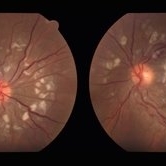
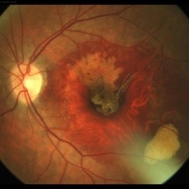 RPE Tear After Anti-VEGF Injection
Mar 17 2021 by RAFAEL REIS PEREIRA, MD
Retinal pigment epithelium (RPE) tear is a rare devastating complication of age-related macular degeneration (AMD). The believed mechanism lies in an adherence of the neovascularization to the undersurface of the RPE creating a contractile force that increases after VEGF therapy and causes the tear.
Photographer: Rafael Reis, Retina Clinic, São Paulo
Condition/keywords: retinal pigment epithelium (RPE) contracture
-
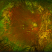
 Retinal Arteriovenous Malformation
Jun 6 2020 by Albert Li, MD, FASRS
Montaged infrared retinal imaging of a 37-year-old asymptomatic man with a grade II arteriovenous malformation (AVM) in the nasal mid-periphery. The presentation of the AVM can be classified with three categories. Grade 1 AVMs are characterized by an abnormal capillary plexus between the major communicating vessels. Grade 2 AVMs are defined by the direct arteriovenous communication without the interposition of arterioles or capillaries. Grade 3 AVMs are characterized by widespread, large caliber anastomosing vessels that are associated with decreased visual acuity and intracranial AVMs. Because retinal AVMs are mostly asymptomatic and non-progressive, further testing may not be indicated unless there are concomitant neurological signs and symptoms or if there is a strong clinical suspicion of a grade 3 retinal AVM. Observation was recommended for the patient in this image. On his most recent follow-up at four months, the patient remained asymptomatic with a stable appearance of the lesion.
Imaging device: Heidelberg Spectralis
Condition/keywords: arteriovenous anastomosis, arteriovenous malformation
-
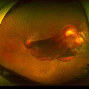
 Submacular PFO
Feb 20 2020 by Kevin J. Blinder, MD, FASRS
This is a 53-year-old gentleman that was referred to us for a second opinion with an inoperable RD with PVR after 3 failed attempts. We performed a PPV, membranectomy, scleral buckling procedure, with silicone oil injection. This case did not require PFO. You can imagine our surprise when we discovered submacular PFO postoperatively. It is very difficult to see the PFO on the Optos. The infrared shows it clearly, with confirmation of the submacular space on the SD-OCT.
Photographer: Jarrod Wehmeier, The Retina Institute; St. Louis, MO
Imaging device: optos
Condition/keywords: submacular perfluorocarbon liquid (PFO)
-
 Acute Macular Neuroretinopathy
Dec 11 2019 by Lauren Whaley
34-year-old female patient presented with changes in vision after recent upper respiratory infection. Referring doctor originally thought it was a blood pressure issue. She noticed a "C" shape in her vision. Infrared image was captured showing exactly what patient was describing! Doctor confirmed with this image that it was AMN.
Photographer: Lauren R. Whaley, COA
Imaging device: Heidelberg Spectralis
Condition/keywords: 30 degrees, acute macular neuroretinopathy, Heidelburg Spectralis, left eye, macula, near infrared autofluorescence (NIRAF)
-
 Ruptured Macroaneurysm
May 22 2019 by Nichole Lewis
FA of a 91-year-old woman with a ruptured macroaneurysm, intraretinal hemorrhage and subretinal hemorrhage. VA 20/400.
Photographer: Nichole Lewis
Condition/keywords: intraretinal hemorrhage, ruptured macroaneurysm, subretinal hemorrhage
-
 Penetrating Trauma with Retinal Detachment
Apr 30 2019 by Olivia Rainey
Ultra-wide field pseudocolor image of a 39-year-old female with penetrating trauma resulting in a retinal detachment with an intraretinal hemorrhage affecting the left eye. Patient was struck with a champagne glass in October of 2018, which lacerated the eyelid and globe. Patient was "seeing red" when she first came to the office and after multiple surgeries she was seeing 20/20 at her last check in April 2019.
Photographer: Olivia Rainey
Imaging device: Optos
Condition/keywords: hemorrhage, left eye, Optos, penetrating trauma, ruptured globe, ultra-wide field imaging
-
 Dengue-Associated-Retinopathy (Anaemic Retinopathy)
Jan 16 2018 by Deepak Bhojwani, MS
22-year-old male with systemic dengue fever and anaemia presenting with roth spots in both eyes (OD>OS). Horizontal raster OCT scans showing intraretinal foveal hameorrhage in right eye.
Photographer: Dr Deepak Bhojwani, Raghudeep Eye Hospital , Ahmedabad
Imaging device: Zeiss- HD- OCT
Condition/keywords: anaemic retinopathy, Roth spots
-
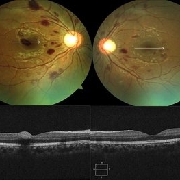
 SLE Retinopathy
Nov 14 2016 by Mitzy E Torres Soriano, MD
25-year-old female patient with systemic lupus erythematosus. Photographs show cotton wool spots, intraretinal hemorrhages and vascular tortuosity. FA demonstrated retinal vasculitis and OCT revealed cystoid macular edema. In this case diagnosis of SLE was made after ocular manifestation.
Photographer: Grupo Laser Vision, Rosario, Argentina
Condition/keywords: cotton wool spots, occlusive retinal vasculitis, occlusive vasculitis, systemic lupus erythematosus, vasculopathy
-
 Foveoschisis secondary to high myopia
Mar 13 2015 by Niloofar Piri, MD
Infrared and HD-OCT of the right eye in a 55-year-old African American female with high myopia (more than -6.00 D), BCVA: 20/25 OU Cartwheel appearance of the fovea in the infrared imaging is visible. HD- OCT demonstartes schisis in different layers of the retina (both NFL and OPL; notice stretching of the Muller cells); VMT is also present . Outer retinal layers are preserved which explains the good vision . She had the same findings in OS.
Photographer: Niloofar Piri, MD
Imaging device: Heidelberg Spectralis
Condition/keywords: high myopia, retinoschisis
 Loading…