Search results (365 results)
-
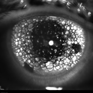 Oil Bubbles
Apr 27 2018 by Mark Lazcano
Infrared photograph of 56-year-old male with retinal detachment oil on lens.
Photographer: Mark Lazcano, University of Miami, Bascom Palmer Eye Institute
Imaging device: Heidelberg Spectralis
Condition/keywords: silicone oil
-
 Pigmented Paravenous Retinochoroidal Atrophy (PPRCA)
Jun 30 2025 by Maria Letícia Costa Holanda
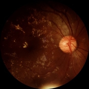
Fundoscopy of a 42-year-old asymptomatic man with pigmented paravenous chorioretinal atrophy. Pigmented paravenous retinochoroidal atrophy (PPRCA) is a rare disorder of unknown etiology. The disease is characterized by pigment accumulation along the distribution of retinal veins. The findings are usually incidental with minimal effect on vision.
Photographer: Guilherme da Cruz Reis, CLINOS Eye Hospital - Feira de Santana (BA),Brazil
Condition/keywords: pigmented paravenous chorioretinal atrophy (PPCRA)
-
 Silicon Oil
Apr 27 2018 by Giselle DeOliveira
Infrared photo of 75-year-old male with retinal detachment.
Photographer: Giselle DeOliveira, University of Miami, Bascom Palmer Eye Institute
Imaging device: Heidelberg Spectralis
Condition/keywords: infrared image, silicone oil
-
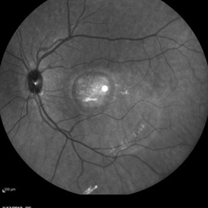
 Acute Macular Neuroretinopathy
Dec 11 2019 by Lauren Whaley
34-year-old female patient presented with changes in vision after recent upper respiratory infection. Referring doctor originally thought it was a blood pressure issue. She noticed a "C" shape in her vision. Infrared image was captured showing exactly what patient was describing! Doctor confirmed with this image that it was AMN.
Photographer: Lauren R. Whaley, COA
Imaging device: Heidelberg Spectralis
Condition/keywords: 30 degrees, acute macular neuroretinopathy, Heidelburg Spectralis, left eye, macula, near infrared autofluorescence (NIRAF)
-
 Cuticular and soft drusen
Jun 14 2021 by Gerardo Garcia-Aguirre, MD
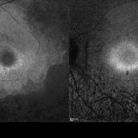
Fundus photograph (left) and Retro mode infrared image (right) of an eye with soft and cuticular drusen. Drusen are highlighted and better visualized with retro mode imaging.
Photographer: Gerardo Garcia-Aguirre
Imaging device: Nidek Mirante
Condition/keywords: drusen, dry age-related macular degeneration (dry AMD)
-
 Siegrist Streaks
Mar 29 2013 by Henry J. Kaplan, MD
Typical Siegrist streaks in hypertensive choridopathy; hyperpigmentations in a linear fashion along choroidal vessels , a rare finding.
Condition/keywords: hypertensive choroidopathy, Siegrist Streaks
-
 Submacular PFO
Feb 20 2020 by Kevin J. Blinder, MD, FASRS
This is a 53-year-old gentleman that was referred to us for a second opinion with an inoperable RD with PVR after 3 failed attempts. We performed a PPV, membranectomy, scleral buckling procedure, with silicone oil injection. This case did not require PFO. You can imagine our surprise when we discovered submacular PFO postoperatively. It is very difficult to see the PFO on the Optos. The infrared shows it clearly, with confirmation of the submacular space on the SD-OCT.
Photographer: Jarrod Wehmeier, The Retina Institute; St. Louis, MO
Imaging device: optos
Condition/keywords: submacular perfluorocarbon liquid (PFO)
-
 Foveoschisis secondary to high myopia
Mar 13 2015 by Niloofar Piri, MD
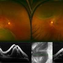
Infrared and HD-OCT of the right eye in a 55-year-old African American female with high myopia (more than -6.00 D), BCVA: 20/25 OU Cartwheel appearance of the fovea in the infrared imaging is visible. HD- OCT demonstartes schisis in different layers of the retina (both NFL and OPL; notice stretching of the Muller cells); VMT is also present . Outer retinal layers are preserved which explains the good vision . She had the same findings in OS.
Photographer: Niloofar Piri, MD
Imaging device: Heidelberg Spectralis
Condition/keywords: high myopia, retinoschisis
-
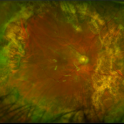
 Pigmented Paravenous Chorioretinal Atrophy (PPCRA)
Jun 27 2025 by Maria Letícia Costa Holanda
Fundoscopy of a 42-year-old asymptomatic man with pigmented paravenous chorioretinal atrophy. Pigmented paravenous retinochoroidal atrophy (PPRCA) is a rare disorder of unknown etiology. The disease is characterized by pigment accumulation along the distribution of retinal veins. The findings are usually incidental with minimal effect on vision.
Photographer: Guilherme da Cruz Reis, CLINOS Eye Hospital - Feira de Santana (BA),Brazil
Condition/keywords: pigmented paravenous chorioretinal atrophy (PPCRA)
-
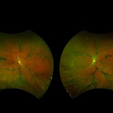
 Pigmented Paravenous Retinochoroidal Atrophy (PPRCA)
Jun 27 2025 by Maria Letícia Costa Holanda
Fundoscopy of a 42-year-old asymptomatic man with pigmented paravenous chorioretinal atrophy. Pigmented paravenous retinochoroidal atrophy (PPRCA) is a rare disorder of unknown etiology. The disease is characterized by pigment accumulation along the distribution of retinal veins. The findings are usually incidental with minimal effect on vision.
Photographer: Guilherme da Cruz Reis, CLINOS Eye Hospital - Feira de Santana (BA),Brazil
Condition/keywords: pigmented paravenous chorioretinal atrophy (PPCRA)
-
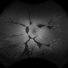
 "NVD Flower"
Oct 20 2023 by Daniel Davis, OCT-C
Infrared image of NVD (52F)
Imaging device: Heidelberg Spectralis
Condition/keywords: neovascularization of the disc (NVD)
-
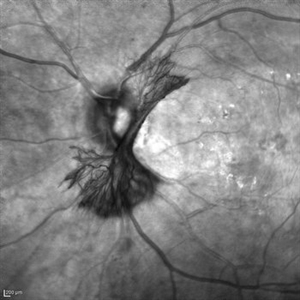
 Best Disease
Mar 9 2013 by Hamid Ahmadieh, MD
Infrared imaging of the left eye of a 49-year-old man with decreased VA due to advanced Best disease.
Photographer: Soodabeh Fooladin, Negah Eye Center, Tehran
Imaging device: Heidelberg Spectralis
Condition/keywords: Best disease, infrared image
-
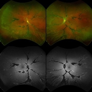
 Case 2 Retinitis Pigmentosa BAF IRAF OD
May 14 2014 by Avris Romario Diparaja Siahaan
Fundus image a 57-year-old man with retinitis pigmentosa on both eyes. These image were taken with blue auto fluorescein mode (BAF) and infrared auto fluorescence (IRAF).
Photographer: Avris Romario Diparaja Siahaan
Imaging device: Heidelberg HRA + OCT Spectralis
Condition/keywords: autofluorescence imaging, fundus photograph, infrared image, retinitis pigmentosa
-
 Case 2 Retinitis Pigmentosa BAF IRAF OS
May 14 2014 by Avris Romario Diparaja Siahaan
Fundus image a 57-year-old man with retinitis pigmentosa on both eyes. These image were taken with blue auto fluorescein mode (BAF) and infrared auto fluorescence (IRAF).
Photographer: Avris Romario Diparaja Siahaan
Imaging device: Heidelberg HRA + OCT Spectralis
Condition/keywords: autofluorescence imaging, fundus photograph, infrared image, retinitis pigmentosa
-
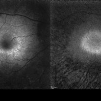
 Central Areolar Choroidal Dystrophy
Jul 7 2015 by Hamid Ahmadieh, MD
Infrared image of both eyes of a 58-year-old man with progressive loss of vision. VA OD is 20/60 and VA OS is 20/400.
Photographer: Soulmaz Shahmohammad, Negah Eye Center, Tehran, Iran
Imaging device: Specteralis
Condition/keywords: central areolar choroidal dystrophy (CACD), infrared image
-
 choroidal nevus with polypoidal choroidal vasculopathy
Nov 20 2012 by Roy Schwartz, MD
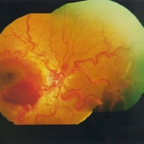
Rare combination of a choroidal nevus complicated by polypoidal choroidal vasculopathy. The lesion is temporal to the fovea, and leakage of subretinal fluid almost reaching the fovea is demonstrated.
Photographer: Galit Yair-Pur
Condition/keywords: choroidal nevus, polypoidal choroidal vasculopathy (PCV)
-
 Color Montage Picture
Jan 15 2019 by Aniruddha Maiti, MBBS DO DNB FRVS FICO MRCS FACS FASRS FRCOphth
Dilated tortuous vessels with intraretinal and subretinal hemorrhage at fovea.
Photographer: Sangeeta Mohanta
Imaging device: Zeiss FF450 plus IR
Condition/keywords: dilated tortuous vessels, subretinal hemorrhage, Wyburn-Mason
-
 Commotio Retinae
Aug 7 2025 by Gabriel Costa Andrade, PhD
Color fundus photograph of a 13-year-old girl who was hit by accidental discharge of gel bullet in her right eye. She presented with retinal whitening with intraretinal hemorrhages in temporal inferior area of the peripheral retina.
Photographer: Gabriel Andrade
Condition/keywords: macula, Retina, Trauma
-
 Congenital Retinal Macrovessel
Oct 13 2023 by Jacob D. Grodsky, MD
41 y/o male who presented with acute onset of blurred vision OD. Visual acuity was 20/200 OD; 20/25 OS. Examination was consistent with congenital retinal macrovessel through the macula with intraretinal hemorrhage as seen in the fundus photo. Intravitreal bevacizumab was injected, and visual acuity improved to 20/40 at 4-week follow-up. MRA head and neck was ordered to rule out other vascular anomalies.
Condition/keywords: congenital retinal macrovessel, RETINAL MACROVESSEL
-
 Diabetic Retinopathy
Jun 4 2025 by Paulina Araujo
The 55-degree central fundus photograph of the right eye demonstrates numerous hard exudates, dot intraretinal hemorrhages, and microaneurysms.
Photographer: Paulina D.Araujo Martínez, Asociación para Evitar la Ceguera en México I.A.P., Hospital Dr Luis Sánchez Bulnes.
Condition/keywords: diabetic retinopathy
-
 Geographic Atrophy - Case 1: Photo 1 of 6
Oct 4 2012 by Gregg T. Kokame, MD, MMM, FASRS
NIRAF (near infrared autofluorescence) Image of patient with Geographic Atrophy
Photographer: Jaclyn Pisano, Retina Consultants of Hawaii
Imaging device: Heidelberg Spectralis
Condition/keywords: autofluorescence imaging, geographic atrophy, near infrared autofluorescence (NIRAF)
-
 Hypertensive Retinopathy Pre
Mar 10 2014 by Dong Yoon Kim, MD
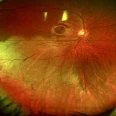
20-year-old women who had severe preeclamsia visited our clinic for decreased visual acuity on her both eyes. Her visual acuity was 20/100 on both eyes. Fundus examination revealed serous retinal detachment on both eyes. OCT examination revealed subretinal and intraretinal fluid.
Photographer: Sun Tae Kim, University of Ulsan, Asan Medical Center
Imaging device: Optos C200 MA scanning laser ophthalmoscope
Condition/keywords: hypertensive retinopathy
-
 Macular Coloboma
Jul 17 2024 by Anubhav Chauhan
This is fundus photograph of a 30 year male depicting a Macular coloboma in the right eye. The patient had a sharply defined large, yellowish white, coarsely pigmented, atrophic, round crater like defect at the macula. Spectral domain optical coherence tomography confirmed our diagnosis. The serology testing such as serum IgM, IgG for toxoplasma and cytomegalovirus was negative. His systemic examination was normal.
Photographer: Dr Anubhav Chauhan, Department of Ophthalmology, Shri Lal Bahadur Shastri Government Medical College, Nerchowk, District Mandi, Himachal Pradesh, India
Imaging device: Zeiss
Condition/keywords: macula, rare
-
 Macular Edema
Jun 4 2025 by Paulina Araujo
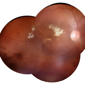
The composite fundus photograph of the right eye demonstrates circinate hard exudates in the thickened macular area, along with flame-shaped intraretinal hemorrhages along the inferior temporal arcade.
Photographer: Paulina D.Araujo Martínez, Asociación para Evitar la Ceguera en México I.A.P., Hospital Dr Luis Sánchez Bulnes.
Condition/keywords: macular edema
-
 Macular fold posterior microphthalmos OS
Apr 24 2022 by Mariam Cernichiaro-Espinosa, MD
Macular OCT of 6-year-old girl with posterior microphthalmos, showing macular fold and one intraretinal cyst OS.
Photographer: Mariam Cernichiaro-Espinosa, Asociación para Evitar la Ceguera, I.A.P. Mexico City, Mexico.
Imaging device: Zeiss Clarus
Condition/keywords: posterior microphthalmos
 Loading…