Search results (365 results)
-
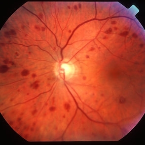
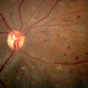
 Siegrist Streaks
Mar 29 2013 by Henry J. Kaplan, MD
Typical Siegrist streaks in hypertensive choridopathy; hyperpigmentations in a linear fashion along choroidal vessels , a rare finding.
Condition/keywords: hypertensive choroidopathy, Siegrist Streaks
-
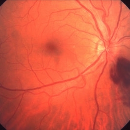
 Ocular ischaemic syndrome colour 1
Jan 11 2013 by Alex P. Hunyor, MD
Ocular ischaemic syndrome, left eye - color image, posterior pole. Note: dilated but not tortuous veins, attenuated arteries, and multiple intraretinal haemorrhages.
Condition/keywords: ocular ischemic syndrome
-
 Lyme Disease
Feb 13 2013 by From the Collections of Thomas M. Aaberg, MD and Thomas M. Aaberg Jr., MD
Papilledema, intra-retinal hemorrhage, periopticneuritis.
Condition/keywords: intraretinal hemorrhage, Lyme disease, periopticneuritis
-
 Intraretinal Microvascular Abnormalities
Oct 12 2012 by Gregg T. Kokame, MD, MMM, FASRS
Intraretinal microvascular abnormalities
Photographer: Jaclyn Pisano, Retina Consultants of Hawaii
Imaging device: Zeiss FF-450 plus
Condition/keywords: fundus photograph, intraretinal microvascular abnormalities, red-free
-
 Venous Loop in Severe Nonproliferative Diabetic Retinopathy
Mar 1 2014 by Homayoun Tabandeh, MD, FASRS
Venous loop, intraretinal microvascular abnormalities, venous beading in patient with severe nonproliferative diabetic retinopathy.
Condition/keywords: diabetic retinopathy, intraretinal microvascular abnormalities, venous loop
-
 Inferonasal Branch Retinal Vein Occlusion
Aug 23 2012 by Gerardo Garcia-Aguirre, MD
Fundus of a 55-year-old male showing intraretinal hemorrhages in the inferonasal quadrant.
Photographer: Noemí Hernández, Asociación para Evitar la Ceguera en México
Condition/keywords: branch retinal vein occlusion (BRVO), intraretinal hemorrhage
-
---thumb.jpg/image-square;max$300,300.ImageHandler) Peripapillary Atrophy
Peripapillary Atrophy
Feb 13 2013 by From the Collections of Thomas M. Aaberg, MD and Thomas M. Aaberg Jr., MD
Papilledema, intra-retinal hemorrhage, periopticneuritis.
Condition/keywords: intraretinal hemorrhage, papilledema, periopticneuritis, peripapillary atrophy
-
 Chronic Macular Hole
Sep 2 2012 by Hyung-Woo Kwak, MD
A large hole with rolled everted edges, adjacent cystoid intraretinal spaces, a shallow rim of subretinal fluids.
Imaging device: Zeiss F450 plus
Condition/keywords: macular hole
-
---thumb.jpg/image-square;max$300,300.ImageHandler) Fibrovascular Proliferation
Fibrovascular Proliferation
Feb 13 2013 by From the Collections of Thomas M. Aaberg, MD and Thomas M. Aaberg Jr., MD
Neovascularization, fibrous proliferation, intraretinal hemorrhage.
Condition/keywords: fibrous proliferation, intraretinal hemorrhage, neovascularization (NV)
-
---thumb.jpg/image-square;max$300,300.ImageHandler) Progressive Outer Retinal Necrosis
Progressive Outer Retinal Necrosis
Feb 15 2013 by From the Collections of Thomas M. Aaberg, MD and Thomas M. Aaberg Jr., MD
Color fundus photograph showing extensive confluent retinal whitening, retinal exudation, intraretinal hemorrhage, and sheathing of retinal vessels consistent with infectious retinitis such as progressive outer retinal necrosis (PORN).
Condition/keywords: occlusive retinitis, retinal necrosis
-
 Cone-Rod Dystrophy
Mar 15 2017 by Hamid Ahmadieh, MD
Infrared and OCT images of the left eye of a 16-year-old boy with decreased visual acuity and color vision deficiency due to cone-rod dystrophy.
Photographer: Abazarnezhad , Negah Eye Center, Tehran, Iran
Imaging device: Spectralis OCT
Condition/keywords: cone dystrophy, infrared image, optical coherence tomography (OCT)
-
---thumb.jpg/image-square;max$300,300.ImageHandler) Peripheral retinal nonperfusion, capillary abnormalities, retinal microaneurysms, and intraretinal hemorrhage
Peripheral retinal nonperfusion, capillary abnormalities, retinal microaneurysms, and intraretinal hemorrhage
Feb 15 2013 by From the Collections of Thomas M. Aaberg, MD and Thomas M. Aaberg Jr., MD
Color fundus photograph showing peripheral retinal nonperfusion, capillary abnormalities, retinal microaneurysms, and intraretinal hemorrhage.
Condition/keywords: peripheral retinal nonperfusion, proliferative retinopathy
-
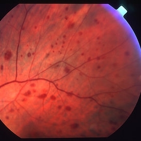 Ocular ischaemic syndrome colour 2
Jan 11 2013 by Alex P. Hunyor, MD
Ocular ischaemic syndrome, left eye - color image, superotemporal midperiphery. Note: dilated but not tortuous veins, attenuated arteries, and multiple intraretinal haemorrhages.
Condition/keywords: ocular ischemic syndrome
-
 PED due to CSCR 3
Sep 2 2012 by Hamid Ahmadieh, MD
Infrared imaging of a 37-year-old man with a serous PED secondary to CSCR.
Photographer: Hamid Ahmadieh, Ophthalmic Research Center, Labbafinejad Medical Center
Imaging device: Heidelberg Spectralis
Condition/keywords: central serous chorioretinopathy (CSCR), pigment epithelial detachment (PED)
-
 SLE Retinopathy
Nov 14 2016 by Mitzy E Torres Soriano, MD
25-year-old female patient with systemic lupus erythematosus. Photographs show cotton wool spots, intraretinal hemorrhages and vascular tortuosity. FA demonstrated retinal vasculitis and OCT revealed cystoid macular edema. In this case diagnosis of SLE was made after ocular manifestation.
Photographer: Grupo Laser Vision, Rosario, Argentina
Condition/keywords: cotton wool spots, occlusive retinal vasculitis, occlusive vasculitis, systemic lupus erythematosus, vasculopathy
-
 Choroidal Melanoma
Feb 2 2018 by Olivia Rainey
Optical coherence tomography with enhanced depth imaging of a 78-year-old female with choroidal melanoma with subretinal fluid affecting her right eye.
Photographer: Olivia Rainey
Imaging device: Heidelberg Spectralis
Condition/keywords: enhanced depth imaging, infrared image, optical coherence tomography (OCT), subretinal fluid, superior retina
-
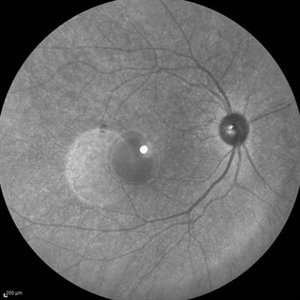
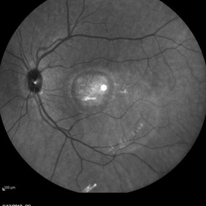
 Geographic Atrophy - Case 1: Photo 3 of 6
Oct 4 2012 by Gregg T. Kokame, MD, MMM, FASRS
Infrared Image of patient with Geographic Atrophy
Photographer: Jaclyn Pisano, Retina Consultants of Hawaii
Imaging device: Heidelberg Spectralis
Condition/keywords: autofluorescence imaging, geographic atrophy
-
 Proliferative Diabetic Retinopathy
Sep 15 2012 by Hamid Ahmadieh, MD
Infrared image of a 30-year-old woman with the history of scatter laser photocoagulation and a preretinal hemorrhage due to active PDR .
Photographer: Hamid Ahmadieh, MD, Ophthalmic Research Center, Labbafinejad Medical Center, Shahid Beheshti University of Medical Sciences
Imaging device: Heidelberg HRA
Condition/keywords: infrared image, preretinal hemorrhage
-
 Intraretinal Microvascular Abnormalities and Venous Beading
Mar 1 2014 by Homayoun Tabandeh, MD, FASRS
Intraretinal microvascular abnormalities and venous beading in a patient with diabetic retinopathy.
Condition/keywords: intraretinal microvascular abnormalities, venous beading
-
 Retinal Detachment
Nov 9 2012 by Norman Byer
This eye of a 25-year-old man has a retinal detachment of about six year’s duration. This photograph shows an intraretinal cyst, which is a secondary result of the longstanding detachment.
Condition/keywords: intraretinal cyst
-
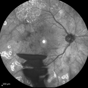
 Geographic Atrophy - Case 1: Photo 1 of 6
Oct 4 2012 by Gregg T. Kokame, MD, MMM, FASRS
NIRAF (near infrared autofluorescence) Image of patient with Geographic Atrophy
Photographer: Jaclyn Pisano, Retina Consultants of Hawaii
Imaging device: Heidelberg Spectralis
Condition/keywords: autofluorescence imaging, geographic atrophy, near infrared autofluorescence (NIRAF)
-
 Geographic Atrophy - Case 1: Photo 4 of 6
Oct 4 2012 by Gregg T. Kokame, MD, MMM, FASRS
NIRAF (near infrared autofluorescence) Image of patient with Geographic Atrophy
Photographer: Jaclyn Pisano, Retina Consultants of Hawaii
Imaging device: Heidelberg Spectralis
Condition/keywords: autofluorescence imaging, geographic atrophy, near infrared autofluorescence (NIRAF)
-
---thumb.jpg/image-square;max$300,300.ImageHandler) CMV with leukemia
CMV with leukemia
Feb 15 2013 by From the Collections of Thomas M. Aaberg, MD and Thomas M. Aaberg Jr., MD
color fundus photograph of a patient with leukemia complicated by CMV retinitis, manifesting as intraretinal hemorrhage, nerve fiber layer infarction, and retinal exudation
Condition/keywords: leukemia
-
 Branch Retinal Vein Occlusion with Macular Edema
Aug 23 2012 by Gerardo Garcia-Aguirre, MD
Fluorescein angiogram composition of the left eye, showing hypofluorescent areas corresponding to intraretinal hemorrhages.
Photographer: Noemí Hernández, Asociación para Evitar la Ceguera en México
Condition/keywords: branch retinal vein occlusion (BRVO), macular edema
-
 Best Disease
Mar 9 2013 by Hamid Ahmadieh, MD
Infrared imaging of the left eye of a 49-year-old man with decreased VA due to advanced Best disease.
Photographer: Soodabeh Fooladin, Negah Eye Center, Tehran
Imaging device: Heidelberg Spectralis
Condition/keywords: Best disease, infrared image
 Loading…