Search results (365 results)
-
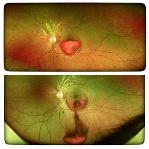
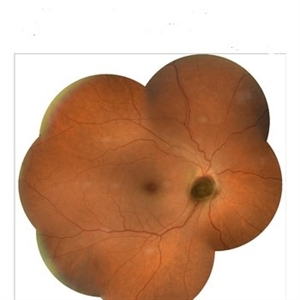
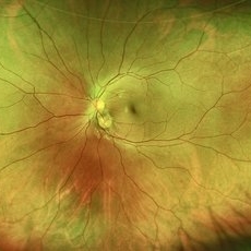
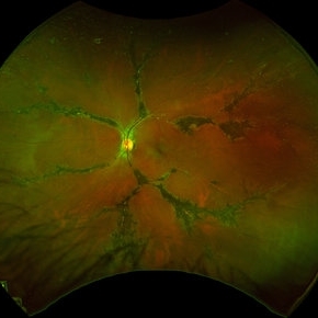
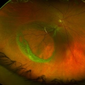
 Leber’s Miliary Aneurysm
Dec 12 2025 by KANWALJEET HARJOT MADAN, M.S. (Ophthalmology); FAICO (Vitreous - Retina)
A 34 year-old male presented with decrease vision in right eye for 3 months. Anterior segment exam was normal. Fundus exam in RE revealed presence of macular edema which was evident on OCT. Multiple retinal vascular aneurysmal dilatations with telangiectasia of the retina blood vessels noted superiorly which was evident on FFA. These aneurysms were multiple, tiny and leaky on FFA. He was diagnosed to have Leber’s miliary aneurysms. It is a rare, typically unilateral eye condition, often seen in young males, characterized by multiple tiny, leaky aneurysms in the retinal blood vessels, leading to deposits of hard exudates and potential vision loss, especially if it affects the macula. It is considered a milder form of Coats' disease.
Photographer: Dr. Kanwaljeet Harjot Madan, Thind Eye Hospital, Jalandhar City (Punjab) INDIA.
Imaging device: Zeiss Fundus Camera
Condition/keywords: FFA, Leber's miliary aneurysm
-
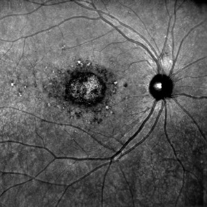
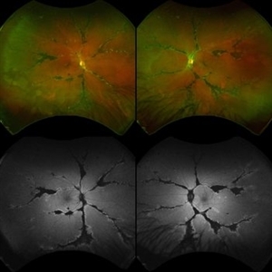
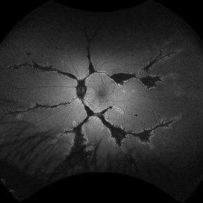
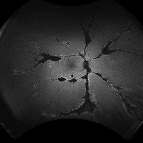
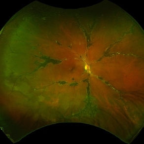
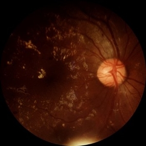
 Angiographic Storm: Fluorescein Leakage in Retinal Vasculitis
Nov 17 2025 by SHRADDHA RAJ SHRIVASTAVA
This left eye montage fundus fluorescein angiography (FFA) image of a 19 year old male with idiopathic retinal vasculitis, having skip vasculitic lesions predominantly involving retinal veins. There are areas of blocked fluorescence due to intraretinal hemorrhages, the involved veins have filling defects and occlusions, leading to formation of numerous collateral channels. The inflamed vessels also show perivascular fuzzy hyperfluorescent stain due to leakage of dye. We can also see multiple peripheral capillary non perfusion (CNP) areas, with a 'hot disc', suggestive of ongoing inflammation.
Photographer: Dr. Shraddha Raj Shrivastava
Imaging device: Nidek Mirante SLO/OCT (Confocal scanning/Spectral domain OCT)
Condition/keywords: FA late phase leakage, Fundus Fluorescein Angiography, idiopathic retinal vasculitis, optic disc leakage, VASCULITIS
-
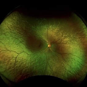
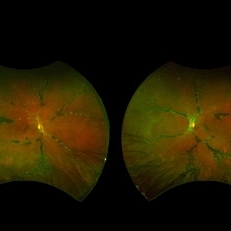
 Unilateral Pigmentary Retinopathy
Nov 9 2025 by Hrishikesh Naik, MS
Montage fundus photographs of a 47 year old female presenting with unilateral vision loss in the left eye. Fundoscopy revealed extensive intraretinal pigment clumps, waxy disc pallor, and marked vessel attenuation in the left eye with a normal fundus in the right. Electroretinography showed unilateral reduction in rod and cone function. Unilateral pigmentary retinopathy, an uncommon variant of retinitis pigmentosa (reported incidence ˜ 5%) presents with RP-like changes in one eye, the fellow eye being completely normal. Proposed causes include lyonization and somatic mosaicism. Conditions which mimic RP should be excluded, and any diagnoses should be supported with electrodiagnostic tests and autofluorescence imaging. Management parallels RP, focusing on cataract and macular complications and long-term follow-up to monitor possible bilateral progression.
Imaging device: Zeiss Visucam 224
Condition/keywords: montage, retinitis pigmentosa, unilateral
-
 Retained PFCL Over the Optic Disc
Oct 14 2025 by rohan jain
Retained PFCL bubble over the optic disc after retinal detachment surgery.
Photographer: Dr. ROHAN JAIN
Imaging device: mirante
Condition/keywords: near infrared autofluorescence (NIRAF), PFCL
-
 Macular Tributary Retinal Venous Occlusion
Sep 7 2025 by Anand Temkar
A 55 yrs old female, k/c/o DM ( type II ) since past 5 yrs ( on medication ). Her vision was 6/6 in RE and 6/24 in her LE. IOP was 16 in both eyes. On examination, RE was WNL, and in LE ( color photo ) we noticed exudates, small hemorrhages, edema and sclerosed vessel ( depicted by black arrow. OCT LE shows altered foveal contour with cystic spaces and intraretinal hyperreflective material ( IRHRM ).
Photographer: Dr.Anand Temkar- Vasan Eye Hospital, Tiruchirappalli
Imaging device: Opticon
Condition/keywords: macular branch retinal vein occlusion (BRVO), venous occlusion
-
 YAG Laser Hyaloidotomy
Aug 31 2025 by Giriraj Vibhute
A 24-year-old young man presented with sudden loss of vision in left eye following history of rigorous coughing. Visual acuity in RE was 6/6, LE was 6/60p. Fundoscopy showed bilateral multiple small intraretinal hemorrhages with LE large premacular subhyaloid hemorrhage just covering the fovea suggestive of bilateral valsalva retinopathy changes. Nd:YAG laser hyaloidotomy was performed to left eye the same day (A250; 2mJ;6 SHOTS). Visual acuity improved to 6/9 immediately following the procedure. After 1 week, the subhyaloid hemorrhage had completely cleared with dispersed intragel hemorrhage in the inferior vitreous cavity with visual acuity of 6/6 in left eye
Photographer: Dr Vani S. MM Joshi eye institute, Hubli
Condition/keywords: valsalva retinopathy, YAG HYALOIDOTOMY
-
 Amelanotic Melanoma
Aug 12 2025 by César Adrián Gómez Valdivia, MD
This FAF image reveals a hypoautofluorescent mass with areas of dense hyperautofluorescent stippling—a classic pattern suggestive of an amelanotic choroidal melanoma. Amelanotic melanoma is a rare variant of uveal melanoma, accounting for only a minority of cases. Unlike pigmented melanomas, these lesions lack melanin, making them more challenging to detect on conventional color fundus imaging. FAF Characteristics: • Central hypoautofluorescence: due to loss or compression of the RPE • Peripheral hyperautofluorescent speckling: consistent with lipofuscin accumulation or RPE disruption • Often associated with subretinal fluid or orange pigment seen clinically Location: Juxtapapillary, with potential optic nerve involvement—a factor that complicates both diagnosis and
Photographer: @eyemissu2
Imaging device: California ICG OPTOS
Condition/keywords: amelanotic melanoma
-
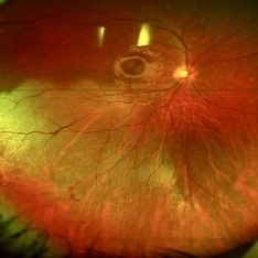
 Oguchi Disease
Aug 12 2025 by Debarun Sharma
A 21 year old female presented with a history of night blindness for the past 16 years when she had difficulty in doing work and navigating places at night. BCVA OU was 6/6. Fundus examination showed the Mizuo-Nakamura phenomenon. ERG was done which showed extinguished rod responses with slightly diminished cone responses. A of Oguchi’s disease was made. The patient was advised for genetic testing and sibling screening. Oguchi’s disease is a rare cause of congenital stationary night blindness with characteristic fundus appearance.
Photographer: Debarun Sharma
Imaging device: Optos
Condition/keywords: Oguchi disease
-
 Commotio Retinae
Aug 7 2025 by Gabriel Costa Andrade, PhD
Color fundus photograph of a 13-year-old girl who was hit by accidental discharge of gel bullet in her right eye. She presented with retinal whitening with intraretinal hemorrhages in temporal inferior area of the peripheral retina.
Photographer: Gabriel Andrade
Condition/keywords: macula, Retina, Trauma
-
 Traction in Progression
Aug 6 2025 by Claudio Brancato, MD
This image captures stage 4A Retinopathy of Prematurity, showing partial retinal detachment sparing the macula. The elevated retina and fibrous ridge indicate tractional forces secondary to extraretinal neovascularization. A striking representation of disease evolution, poised between reversibility and vision loss.
Photographer: Gregorio Lo Giudice, ARNAS Civico Hospital, Palermo, Italy
Imaging device: RETCAM 3 (enhanced via IA)
Condition/keywords: retinopathy of prematurity
-
 Black Swan - Optic Disc Melanocytoma
Aug 5 2025 by SHRADDHA ASHOK CHANDORKAR, DNB DO FVRS
Just like the Black Swan which signifies an event that comes as a surprise, can have a major effect, and is often inappropriately rationalized after the fact with the benefit of hindsight, a 50 yr old presbyopic lady came to OPD with complains of diminution of vision - BCVA being 6/6 N6 in both eyes. Fundus examination revealed a pigmented nodule covering the optic disc .In most cases, fluorescein angiography of a melanocytoma of the optic disk demonstrates hypofluorescence throughout the angiogram. OCT disc showed elevated lesion, OCT macula normal and USG B scan with measurements were done to corroborate the posterior extension and to note increase in size if any on follow ups, perimetry was done to check for any field defects. All tests seemingly within normal limits - Patient was counselled and asked for 6 monthly follow up. Optic Disc Melanocytoma usually unilateral known to be a benign lesion that carries an excellent prognosis, the malignancy of this specific condition is rare 1-2%. The mean age at diagnosis of optic disk melanocytoma is 50 years with a median of 52 and range of 1–91 years. It is possible that melanocytoma is a congenital lesion but may not become clinically apparent until later in life, perhaps due to acquisition of pigment in a previously amelanotic lesion.
Photographer: Dr.Shraddha A. Chandorkar
Imaging device: topcon
Condition/keywords: optic disc melanocytoma
-
 Stars of Stargardt
Aug 4 2025 by Malvika Singh
Infrared fundus photograph of a 22 year old female with Stargardt's disease.
Photographer: Dr Malvika Singh, Retina Foundation, Ahmedabad, India
Imaging device: Mirante SLO/OCT
Condition/keywords: infrared image, Stargardt disease
-
 New Choroidal Melanoma
Jul 16 2025 by Virginia Gebhart
78 year old male with a partially amelanotic dome-shaped lesion with RPE changes, hard exudates, overlying intraretinal fluid and minimal SRF temporally. Exam and ultrasound findings consistent with choroidal melanoma. Pt will be scheduled for brachytherapy pending CT scan results.
Photographer: Virginia Gebhart, Retina Consultants of Carolina
Imaging device: Optos California
Condition/keywords: amelanotic melanoma, choroidal melanoma
-
 Sialidosis
Jul 10 2025 by Jessilla Phou
These are fundus photographs capturing an 18 year old male with Type 1 Sialidosis, a rare inherited lysosomal storage disorder caused by a deficiency in the neuraminidase 1 (Neu1) enzyme. Currently, there are fewer than 1,000 people in the USA who have this disorder. It is characterized by a cherry red spot in the macula which occurs when lipids accumulate in the retinal ganglion cells. This causes the macula to appear red as seen in these fundus images. The patient presented at our office with ataxia, depth perception issues, and slow reaction time. His visual acuity was 20/40, suggestive of early stage Sialidosis.
Photographer: Jessilla Phou
Imaging device: Optos California
Condition/keywords: cherry red spot, fundus photograph, Sialidosis
-
 Pseudoduplication of the Optic Disc
Jul 9 2025 by Hrishikesh Naik, MS
A peripapillary colobomatous pseudo-duplication of the optic disc as seen in an asymptomatic 23 year old female with myopia referred for routine retinal periphery screening. Rest retinal exam was normal. Duplication of the optic disc can be classified as either true duplication or pseudoduplication, both of which are rare clinical conditions. Pseudodoubling of the optic disc is commonly caused by optic disc or peripapillary colobomas, characterized by a circumscribed, disc-like lesion with radiating vessels but only one normal optic nerve. A few cases have involved pathological myopia, moderate myopia, proliferative diabetic retinopathy and CHARGE syndrome. The lesion is often found inferior to the normal optic disc. The patient was advised regular follow ups.
Photographer: Hrishikesh Naik
Imaging device: Optos Daytona
Condition/keywords: Coloboma, Pseudoduplication of optic disc
-
 Pigmented Paravenous Retinochoroidal Atrophy (PPRCA)
Jun 30 2025 by Maria Letícia Costa Holanda
Fundoscopy of a 42-year-old asymptomatic man with pigmented paravenous chorioretinal atrophy. Pigmented paravenous retinochoroidal atrophy (PPRCA) is a rare disorder of unknown etiology. The disease is characterized by pigment accumulation along the distribution of retinal veins. The findings are usually incidental with minimal effect on vision.
Photographer: Guilherme da Cruz Reis, CLINOS Eye Hospital - Feira de Santana (BA),Brazil
Condition/keywords: pigmented paravenous chorioretinal atrophy (PPCRA)
-
 Pigmented Paravenous Retinochoroidal Atrophy (PPRCA)
Jun 27 2025 by Maria Letícia Costa Holanda
Fundoscopy of a 42-year-old asymptomatic man with pigmented paravenous chorioretinal atrophy. Pigmented paravenous retinochoroidal atrophy (PPRCA) is a rare disorder of unknown etiology. The disease is characterized by pigment accumulation along the distribution of retinal veins. The findings are usually incidental with minimal effect on vision.
Photographer: Guilherme da Cruz Reis, CLINOS Eye Hospital - Feira de Santana (BA),Brazil
Condition/keywords: Pigmented Paravenous Retinochoroidal Atrophy
-
 Pigmented Paravenous Retinochoroidal Atrophy (PPRCA)
Jun 27 2025 by Maria Letícia Costa Holanda
Fundoscopy of a 42-year-old asymptomatic man with pigmented paravenous chorioretinal atrophy. Pigmented paravenous retinochoroidal atrophy (PPRCA) is a rare disorder of unknown etiology. The disease is characterized by pigment accumulation along the distribution of retinal veins. The findings are usually incidental with minimal effect on vision.
Photographer: Guilherme da Cruz Reis, CLINOS Eye Hospital - Feira de Santana (BA),Brazil
Condition/keywords: pigmented paravenous chorioretinal atrophy (PPCRA)
-
 Pigmented Paravenous Chorioretinal Atrophy (PPCRA)
Jun 27 2025 by Maria Letícia Costa Holanda
Fundoscopy of a 42-year-old asymptomatic man with pigmented paravenous chorioretinal atrophy. Pigmented paravenous retinochoroidal atrophy (PPRCA) is a rare disorder of unknown etiology. The disease is characterized by pigment accumulation along the distribution of retinal veins. The findings are usually incidental with minimal effect on vision.
Photographer: Guilherme da Cruz Reis, CLINOS Eye Hospital - Feira de Santana (BA),Brazil
Condition/keywords: pigmented paravenous chorioretinal atrophy (PPCRA)
-
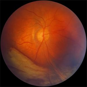
 Choroidal Hemangioma
Jun 18 2025 by Moazzam Parvez
Multicolor and infrared reflectance image of a 42 year old gentleman with a Choroidal hemangioma lesion temporal to the fovea complaining of distortion in his right eye . Fundus imaging revealed a well-circumscribed ,elevated, reddish orange lesion within the choroid involving the posterior pole temporally .
Photographer: Moazzam Parvez, Netralayam , Kolkata
Imaging device: Heidelberg Spectralis
Condition/keywords: Choroidal Hemangioma, tumor
-
 Complex Retinal Detachment with PVR and Starfold
Jun 6 2025 by Jenn Geelan
57 year old male with a Complex Retinoschisis related retinal detachment with PVR and a Posterior Star Fold
Photographer: Jenn Geelan, Retina-Vitreous Surgeons of CNY
Imaging device: Optos California
Condition/keywords: proliferative vitreoretinopathy (PVR), rare, Retinal Detachment, retinoschisis, Starfolds, subretinal fluid
-
 Diabetic Retinopathy
Jun 4 2025 by Paulina Araujo
The 55-degree central fundus photograph of the right eye demonstrates numerous hard exudates, dot intraretinal hemorrhages, and microaneurysms.
Photographer: Paulina D.Araujo Martínez, Asociación para Evitar la Ceguera en México I.A.P., Hospital Dr Luis Sánchez Bulnes.
Condition/keywords: diabetic retinopathy
-
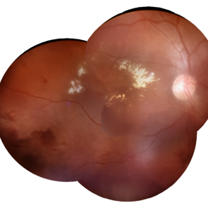 Macular Edema
Jun 4 2025 by Paulina Araujo
The composite fundus photograph of the right eye demonstrates circinate hard exudates in the thickened macular area, along with flame-shaped intraretinal hemorrhages along the inferior temporal arcade.
Photographer: Paulina D.Araujo Martínez, Asociación para Evitar la Ceguera en México I.A.P., Hospital Dr Luis Sánchez Bulnes.
Condition/keywords: macular edema
 Loading…