Search results (638 results)
-
 Angioid streak-associated choroidal neovasclar membranes
Dec 27 2016 by Young Hee Yoon, MD, PhD
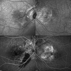
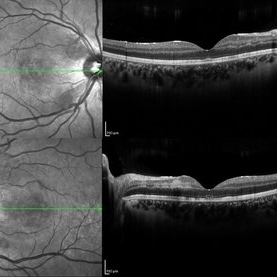
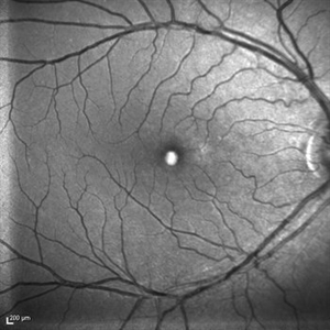
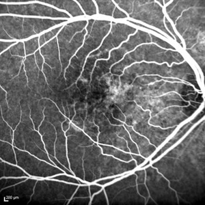
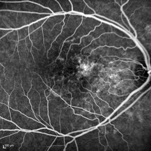
Optical coherence tomogaphs of an 67-year-old woman with CNVM associated with angioid streak in both eyes. (upper row : IR image) Irregular crak-like streaks (lower row : FAG image) Block fluorescence due to subretinal hemorrhage in her right eye and classic CNV in her left eye.
Photographer: Young Hee Yoon, University of Ulsan, Asan Medical Center, Seoul, Korea
Imaging device: Spectralis
Condition/keywords: angioid streaks, choroidal neovascularization (CNV)
-
 Angioid Streak-Associated Choroidal Neovasclar Membranes
Dec 27 2016 by Young Hee Yoon, MD, PhD
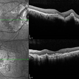
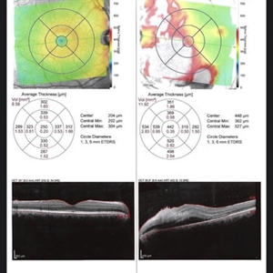
Optical coherence tomogaphs of an 74-year-old woman who received several anti-VEGF injections due to CNV associated with angioid streak in both eyes. There are diffuse CNVM in her right eye and subretinal scar in her left eye. Note the irregular crack in IR image of right eye and the focal Bruch's membrane dehiscence in corresponding B-scan image.
Photographer: Young Hee Yoon, University of Ulsan, Asan Medical Center, Seoul, Korea
Imaging device: Spectralis
Condition/keywords: angioid streaks, choroidal neovascularization (CNV)
-
 Autofluorescence of Ocular Hypotony
May 29 2013 by Zofia Anna Nawrocka (vel Michalewska), MD, PhD
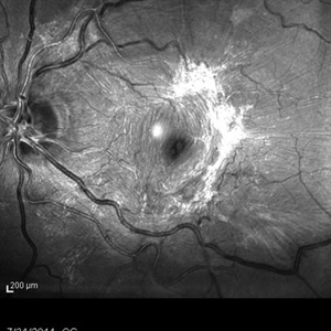
Autofluorescence image of a 75-year-old patient with hypotony, 2 weeks after trauma, 2 years after extracapsular cataract surgery.
Photographer: Zofia Michalewska, Ophthalmic Clinic "Jasne Blonia
Imaging device: Spectralis
Condition/keywords: hypotony
-
 Choroideremia
Oct 25 2024 by Poornachandra B, MS, FVRS
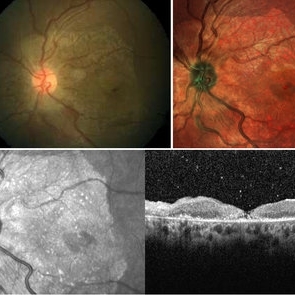
This is a multi color image of an 82 year old male with Choroideremia. Preserved island of macula with well defined borders.
Photographer: Mr Dhikshith
Imaging device: Spectralis
Condition/keywords: choroideremia, inherited retinal disease
-
 Coats Disease
Feb 7 2013 by Raj K. Maturi, MD
8-year-old male, flourescein angiogram, late phrase 4:22 minutes, OD.
Photographer: Stephan Morrow, Midwest Eye Institute Indianapolis Indiana
Imaging device: Heidelberg Spectralis
Condition/keywords: Spectralis
-
 Coats Disease
Feb 7 2013 by Raj K. Maturi, MD
8-year-old male, Heidelberg Spectralis OCT, OD.
Photographer: Stephan Morrow, Midwest Eye Institute Indianapolis Indiana
Imaging device: Heidelberg Spectralis
Condition/keywords: optical coherence tomography (OCT), Spectralis
-
 Coats Disease
Feb 7 2013 by Raj K. Maturi, MD
8-year-old male, flourescein angiogram, venous phrase, OD.
Photographer: Stephan Morrow, Midwest Eye Institute Indianapolis Indiana
Imaging device: Heidelberg Spectralis
Condition/keywords: Spectralis
-
 CRVO-associated Macular Edema
Jul 8 2012 by Jeffrey S. Heier, MD
Young physician with CRVO and macular edema
Imaging device: Spectralis
Condition/keywords: central retinal vein occlusion (CRVO), macular edema
-
 ERM
ERM
Aug 30 2018 by Dhaivat Shah
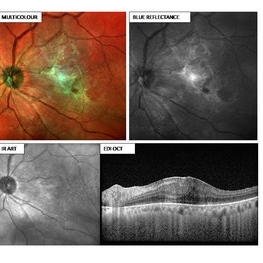
55-year-old female presented with left eye BCVA 6/24 N18, early cataract changes on slit lamp, fundus showing ERM with retinal thickening. Multi-color image (MCI) depicting an ERM (green hue) with retinal thickening. Note how beautifully the extent of ERM is captured, which can help the surgeon to decide the area of surgical peeling. The BR particularly provides details of the inner retina and the vitreoretinal interface, hence showing the ERM. This platform utilizes confocal technology and thus has unique advantages over CFP. MCI provides good image quality in hazy media and in small pupil. It does not use bright white light and thus is not discomforting to the patient. Images obtained with MCI have better contrast and sharper borders as compared to CFP. Definitely the new tech in for the next generation!
Photographer: Miss Moupiya Das
Imaging device: SPECTRALIS
Condition/keywords: blue reflectance, epiretinal membrane (ERM)
-
 FA-ICG
Jan 6 2016 by Jared Watson
FA-ICG
Photographer: Jared Watson COT/CRA
Imaging device: Spectralis
Condition/keywords: necrotizing retina, vasculitis
-
 Fluorescein Angiography of Ocular Hypotony
May 29 2013 by Zofia Anna Nawrocka (vel Michalewska), MD, PhD
Fluorescein angiography of a 7- year-old patient with hypotony, 2 weeks after trauma, 2 years after extracapsular cataract surgery.
Photographer: Zofia Michalewska, Ophthalmic Clinic "Jasne Blonia
Imaging device: Spectralis
Condition/keywords: hypotony
-
 Foveal Thinning Post Blunt Trauma
Aug 25 2018 by Dhaivat Shah
28-year-old male. Post blunt trauma with tennis ball. Fundus color photo shows large area of retinal thinning. Multi color image shows dull red color over fovea, depicting thinning. SD-OCT shows inner retinal ischemia and foveal thinning with early macular hole formation.
Imaging device: Spectralis
Condition/keywords: blunt trauma
-
 Hypotony
May 29 2013 by Zofia Anna Nawrocka (vel Michalewska), MD, PhD
Infrared image of a 75-year-old patient with hypotony, 2 weeks after trauma, 2 years after extracapsular cataract surgery.
Photographer: Zofia Michalewska, Ophthalmic Clinic "Jasne Blonia
Imaging device: Spectralis
Condition/keywords: hypotony
-
 Purtscher's Retinopathy
Mar 22 2021 by Marco Antonio Sauza
First OCT made after 3 weeks after chest and legs injury, with subretinal liquid/fluid, and a disorder of the nasal retina layers.
Photographer: Marco Sauza, hospital Ángeles, México
Imaging device: Spectralis
Condition/keywords: Purtscher's retinopathy
-
 Purtscher's Retinopathy
Mar 22 2021 by Marco Antonio Sauza
OCT after 3 weeks of car accident with Purtscher's retinopathy.
Photographer: Marco Sauza, hospital Ángeles, México
Imaging device: Spectralis
Condition/keywords: Purtscher's retinopathy
-
 Purtscher's Retinopathy
Mar 30 2018 by Olivia Rainey
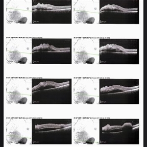
Bilateral OCTS of a 21-year-old female with Purtscher's Retinopathy affecting both eyes. Patient developed acute pancreatitis triggered by hypercalcemia and likely increased alcohol consumption.
Photographer: Olivia Rainey
Imaging device: Spectralis
Condition/keywords: acute pancreatitis, bilateral, optical coherence tomography (OCT), Purtscher's retinopathy
-
 Red Free Macular Pucker
Dec 5 2014 by Stuart Alfred, CRA, OCT-C
Red free image using the Spectralis.
Photographer: Stuart Alfred, CRA, OCT-C
Condition/keywords: macular pucker, red-free, Spectralis
-
 RPE Micro Rip in Central Serous Chorioretinopathy
Jun 26 2016 by Rameez N Hussain, MD
SD OCT image of a case of central serous retinopathy showing RPE micro rip (RPE leak).
Photographer: DR RAMEEZ N HUSSAIN
Imaging device: Dense scan mode - Heidelberg Spectralis
Condition/keywords: central serous chorioretinopathy (CSCR), focal laser, leakage, retinal pigment epithelium (RPE) tear, Spectralis
-
 Rubella Retinopathy 1
Jul 11 2013 by Raj K. Maturi, MD
48-year-old, deaf female, complains of blurry vision, mother had Rubella when she was pregnant
Photographer: Tom Steele, CRA
Imaging device: spectralis
Condition/keywords: optical coherence tomography (OCT), rubella retinopathy, Spectralis
-
 Rubella Retinopathy 10
Jul 11 2013 by Raj K. Maturi, MD
48-year-old, deaf female, complains of blurry vision, mother had Rubella when she was pregnant.
Photographer: Tom Steele, CRA
Imaging device: spectralis
Condition/keywords: IR, rubella retinopathy, Spectralis
-
 Rubella Retinopathy 11
Jul 11 2013 by Raj K. Maturi, MD
48-year-old, deaf female, complains of blurry vision, mother had Rubella when she was pregnant.
Photographer: Tom Steele, CRA
Imaging device: spectralis
Condition/keywords: red-free, rubella retinopathy, Spectralis
-
 Rubella Retinopathy 12
Jul 11 2013 by Raj K. Maturi, MD
48-year-old, deaf female, complains of blurry vision, mother had Rubella when she was pregnant.
Photographer: Tom Steele, CRA
Imaging device: Spectralis
Condition/keywords: autofluorescence imaging, rubella retinopathy, Spectralis
-
 Rubella Retinopathy 13
Jul 11 2013 by Raj K. Maturi, MD
48-year-old, deaf female, complains of blurry vision, mother had Rubella when she was pregnant.
Photographer: Tom Steele, CRA Midwest Eye Institute
Imaging device: Spectralis
Condition/keywords: rubella retinopathy, Spectralis
-
 Rubella Retinopathy 14
Jul 11 2013 by Raj K. Maturi, MD
48-year-old, deaf female, complains of blurry vision, mother had Rubella when she was pregnant.
Photographer: Tom Steele, CRA Midwest Eye Institute
Imaging device: Spectralis
Condition/keywords: rubella retinopathy, Spectralis
-
 Rubella Retinopathy 15
Jul 11 2013 by Raj K. Maturi, MD
48-year-old, deaf female, complains of blurry vision, mother had Rubella when she was pregnant.
Photographer: Tom Steele, CRA Midwest Eye Institute
Condition/keywords: rubella retinopathy, Spectralis
 Loading…