Search results (638 results)
-
 Wide-Field-OCT-montage
Jan 8 2018 by Netan Choudhry, MD, FRCS(C) FASRS
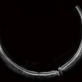
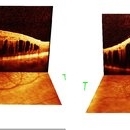
This is an SD-OCT montage image of a 55 year old male with optic neuropathy representing a wide-field OCT spanning 130 degrees.
Photographer: John Golding, Vitreous Retina Macula Specialists of Toronto
Imaging device: Heidelberg Spectralis OCT system
Condition/keywords: wide angle imaging
-
 Branch Retinal Artery Occlusion With Calcium Embolus at the Disc - Fundus Autofluorescence Imaging (FAF)
Apr 7 2018 by Rameez N Hussain, MD
Acute branch retinal artery occlusion with a calcium embolus at the disc which is hyper autofluorescent in fundus autofluorescence imaging (FAF) -resembles an LED light source ('LED sign').
Photographer: DR RAMEEZ N HUSSAIN
Imaging device: Heidelberg Spectralis
Condition/keywords: branch retinal artery occlusion (BRAO), embolus, fundus autofluorescence (FAF), retinal edema
-
 Oil Bubbles
Apr 27 2018 by Mark Lazcano
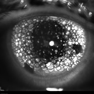
Infrared photograph of 56-year-old male with retinal detachment oil on lens.
Photographer: Mark Lazcano, University of Miami, Bascom Palmer Eye Institute
Imaging device: Heidelberg Spectralis
Condition/keywords: silicone oil
-
 Relentless Placoid Chorioretinitis
Jan 9 2019 by Janet Brazil
ICG photo of a 24-year-old male with Relentless placoid chorioretinitis
Photographer: Janet Atkinson, Eye Associates of New Mexico
Imaging device: Heidelberg HRA Spectralis
Condition/keywords: placoid retinal lesions
-
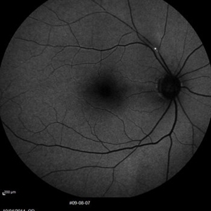 BRAO Rianto AF
Apr 12 2014 by Sjakon G Tahija, MD
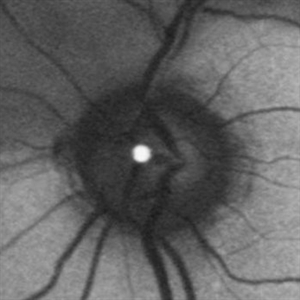
Auto fluorescence fundus image of a 70-year-old man with a superior temporal branch retinal artery occlusion. The emboli can be very clearly seen as the white dot of AF blockage.
Photographer: Avris Siahaan
Imaging device: Heidelberg Spectralis
Condition/keywords: branch retinal artery occlusion (BRAO)
-
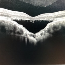 Choroidal Excavation
Jun 2 2019 by Nelson Chamma Capelanes, MD
SD-OCT of a 32-year-old woman showing a subfoveal choroidal excavation associated with chronic central serous chorioretinopathy.
Photographer: Nelson Chamma Capelanes, Promacula, Brazil
Imaging device: Heidelberg Spectralis SD-OCT
Condition/keywords: choroidal excavation, chronic central serous chorioretinopathy (CSCR), pachychoroid
-
 Silicon Oil
Apr 27 2018 by Giselle DeOliveira
Infrared photo of 75-year-old male with retinal detachment.
Photographer: Giselle DeOliveira, University of Miami, Bascom Palmer Eye Institute
Imaging device: Heidelberg Spectralis
Condition/keywords: infrared image, silicone oil
-
 Acute Macular Neuroretinopathy
Dec 11 2019 by Lauren Whaley
34-year-old female patient presented with changes in vision after recent upper respiratory infection. Referring doctor originally thought it was a blood pressure issue. She noticed a "C" shape in her vision. Infrared image was captured showing exactly what patient was describing! Doctor confirmed with this image that it was AMN.
Photographer: Lauren R. Whaley, COA
Imaging device: Heidelberg Spectralis
Condition/keywords: 30 degrees, acute macular neuroretinopathy, Heidelburg Spectralis, left eye, macula, near infrared autofluorescence (NIRAF)
-
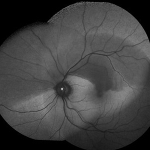
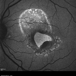
 AZOOR
AZOOR
Mar 19 2015 by Niloofar Piri, MD
#1: Fundus autofluorescence OD in a patient with AZOOR demonstrates characteristic peripapillary hypoAF as well as concentric rings of hypo and hyper AF in posterior pole .
Imaging device: Heidelberg Spectralis
Condition/keywords: acute zonal occult outer retinopathy (AZOOR)
-
 Branch Retinal Artery Occlusion With Calcium Embolus at the Disc - Fundus Autofluorescence Imaging (FAF)
Apr 7 2018 by Rameez N Hussain, MD
Acute branch retinal artery occlusion with a calcium embolus at the disc which is hyper autofluorescent in fundus autofluorescence Imaging (FAF) -resembles an LED light source ('LED sign').
Photographer: DR RAMEEZ N HUSSAIN
Imaging device: Heidelberg Spectralis
Condition/keywords: branch retinal artery occlusion (BRAO), embolus, fundus autofluorescence (FAF), retinal edema
-
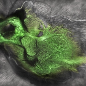 Green Goblin Detachment
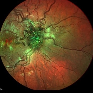
Jan 13 2022 by Netan Choudhry, MD, FRCS(C) FASRS
Tractional retinal detachment with macular hole in a 76-year-old female.
Photographer: John Golding BA, Vitreous Retina Macula Specialists of Toronto, OCTane Imaging Lab
Imaging device: Multicolor fundus photo taken on the Spectralis OCT2 (Heidelberg Engineering GmbH).
Condition/keywords: macular hole, Multispectral imaging, tractional retinal detachment
-
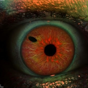 Iris
Iris
Apr 29 2019 by Stephanie Moolman
Multi-color images after Yag PI of iris.
Photographer: Stephanie Moolman, Dr Marissa Willemse, Pretoria, South Africa
Imaging device: Heidelberg Spectralis
Condition/keywords: glaucoma, iris, multicolor, NdYAG laser, peripheral iridotomy
-
 Lady in a dress
Feb 9 2023 by Shelby Helton
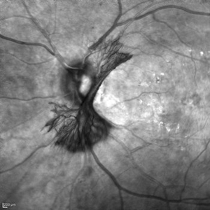
Fluorescein Angiography on a 67-year-old male with significant RPE changes secondary to a severe subretinal hemorrhage that required a vitrectomy with subretinal TPA in 2013.
Photographer: Shelby Helton
Imaging device: Heidelberg Spectralis
Condition/keywords: wet age-related macular degeneration (wet AMD)
-
 Choroideremia
Oct 25 2024 by Poornachandra B, MS, FVRS
This is a multi color image of an 82 year old male with Choroideremia. Preserved island of macula with well defined borders.
Photographer: Mr Dhikshith
Imaging device: Spectralis
Condition/keywords: choroideremia, inherited retinal disease
-
 Cuticular Drusen
Jan 17 2024 by John Lee
Heidelberg SD-OCT of a 65-year-old woman with age-related macular degeneration demonstrating classic sawtooth appearance of cuticular drusen.
Photographer: Natasha Vinson
Imaging device: Heidelberg Spectralis
Condition/keywords: age-related macular degeneration (AMD), cuticular drusen
-
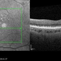
 Foveoschisis secondary to high myopia
Mar 13 2015 by Niloofar Piri, MD
Infrared and HD-OCT of the right eye in a 55-year-old African American female with high myopia (more than -6.00 D), BCVA: 20/25 OU Cartwheel appearance of the fovea in the infrared imaging is visible. HD- OCT demonstartes schisis in different layers of the retina (both NFL and OPL; notice stretching of the Muller cells); VMT is also present . Outer retinal layers are preserved which explains the good vision . She had the same findings in OS.
Photographer: Niloofar Piri, MD
Imaging device: Heidelberg Spectralis
Condition/keywords: high myopia, retinoschisis
-
 Macular Tear
May 14 2014 by Avris Romario Diparaja Siahaan
Blue autofluorescence (BAF) a 40-year-old man with macular tear (had a photocoagulation laser).
Photographer: Avris Romario Diparaja Siahaan
Imaging device: Heidelberg HRA + OCT Spectralis
Condition/keywords: autofluorescence imaging, macular hole
-
 NVI
NVI
Oct 24 2024 by Korey Starkey
Iris FA of a 74 year old male with neovascularization of the iris. Noted mild activity of NVI at the superior pupillary margin, recommending observation at time of visit.
Photographer: Korey Starkey
Imaging device: Heidelberg Spectralis
Condition/keywords: FA, Heidelburg Spectralis, Iris, iris fluorescein angiogram, neovascularization of iris (NVI), smokestack
-
 OCT Image of Epiretinal Membrane
Aug 29 2017 by Carolyn Daley
OCT photograph of a 64-year-old women with an epiretinal membrane in the right eye. Patient has not noticed any decline in vision so surgery was not recommended at this time.
Photographer: Carolyn Daley
Imaging device: Heidelberg Spectralis
Condition/keywords: epiretinal membrane (ERM), optical coherence tomography (OCT)
-
 Wyburn-Mason
Dec 9 2021 by Filip Kecer
Multi-color picture of an 18-year-old boy with Wyburn-Mason syndrome.
Photographer: Filip Kecer, National Institute of Childrens Diseases
Imaging device: Spectralis, Heidelberg Engineering
Condition/keywords: arteriovenous malformation, Wyburn-Mason
-
 X-Linked Retinoschisis
Nov 15 2019 by Nelson Chamma Capelanes, MD
SD-OCT of an 28-year-old man with X - linked retinoschisis.
Photographer: Nelson Capelanes, Promedica/Promacula Indaiatuba & UPO Oftalmologia São Paulo
Imaging device: Spectralis
Condition/keywords: x-linked retinoschisis (XLRS)
-
 "NVD Flower"
Oct 20 2023 by Daniel Davis, OCT-C
Infrared image of NVD (52F)
Imaging device: Heidelberg Spectralis
Condition/keywords: neovascularization of the disc (NVD)
-
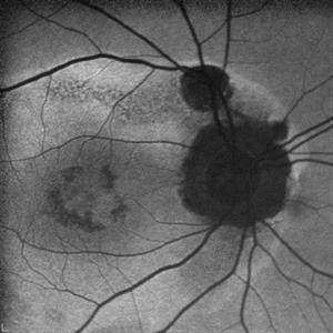
 Active Multi Focal Choroiditis
Jun 21 2025 by Moazzam Parvez
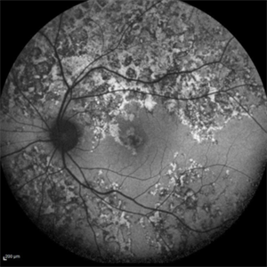
Auto fluorescence image of a 28 year old gentleman with active multifocal choroiditis in his left eye and healed choroiditic patches in the right eye.
Photographer: Moazzam Parvez , Netralayam , Kolkata
Imaging device: Heidelberg Spectralis
Condition/keywords: active, multifocal choroiditis
-
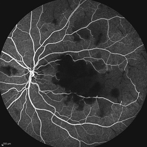 Acute Idiopathic Occlusive Retinal Vasculitis
May 31 2014 by Hamid Ahmadieh, MD
Mid- phase fluorescein angiogram of the left eye of a 28-year-old woman with acute drop of vision due to occlusive retinal vasculitis leading to extensive capillary nonperfusion and macular infarction.
Photographer: Naghmeh Nozhat, Negah Eye Center, Tehran
Imaging device: Heidelberg Spectralis
Condition/keywords: capillary nonperfusion, retinal vasculitis
-
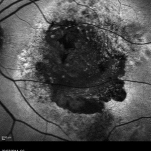 AMD
AMD
Jul 26 2014 by Avris Romario Diparaja Siahaan
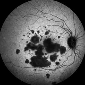
An autofluorescence image of a 78-year-old-man with an age-related macular degeneration on his both eyes.
Photographer: Avris Romario Diparaja Siahaan, Klinik Mata Nusantara
Imaging device: Heidelberg Spectralis
Condition/keywords: age-related macular degeneration (AMD), autofluorescence imaging
 Loading…