Search results (638 results)
-
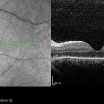
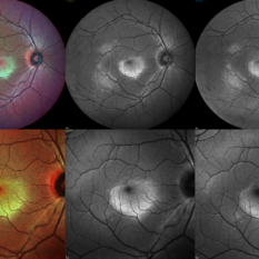
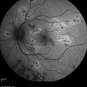
 Proliferative Diabetic Retinopathy
Jan 26 2026 by Daniel Davis, OCT-C
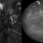
Multimodal-pseudocolor image of an 55-year-old male with OCT and OCTA overlay demonstrating neovascularization, the lower OCT shows subyaloid hemorrhage.
Photographer: Daniel Davis, The Retina Institute, St. Louis
Imaging device: Optos California - Fundus, Heidelberg Spectralis - OCT/A
Condition/keywords: multimodal imaging, NVE, OCT, OCT Angiography, PDR, proliferative diabetic retinopathy (PDR), SUBHYALOID HEMORRHAGE
-
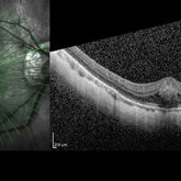
 Subretinal PFCL
Jan 25 2026 by Greeshma M G
Subretinal PFCL in the papillomacular bundle with outerretinal damage.
Imaging device: Centervue Eidon,Heidelberg Spectralis OCT
Condition/keywords: Outer retinal damage, Papillomacular bundle, Subretinal PFCL
-
 Branch Retinal Artery Occlusion
Dec 19 2025 by Gayathri M S
Multicolor Reflectance and Blue Reflectance of a 55 yr male patient with blurring since 1 month shows classical sectoral retinal whitening.
Photographer: Gayathri MS
Imaging device: Heidelberg Spectralis
Condition/keywords: blue reflectance, branch retinal artery occlusion (BRAO), multicolor
-
 Retinal Plumage
Nov 18 2025 by DR APOORVA JADHAV, MBBS , DNB
A 35 year-old male came in with diminished vision in right eye. On multicolor we can appreciate large pigmented choroidal mass lesion with surrounding srf which is reaching fovea. Diagnosed to be Choroidal Melanoma.
Photographer: Dr Apoorva Jadhav
Imaging device: Heidelberg Spectralis
Condition/keywords: choroidal melanoma, srf
-
 MEWDS
MEWDS
Oct 17 2025 by Jason Gayoski
32 year old female presenting to clinic with four day history of sudden onset unilateral left eye vision decrease with central scotoma upon awakening. VA OS 20/200 upon initial evaluation with wreath-like pattern of white dots surrounding macula OS. OD unaffected and asymptomatic.
Photographer: Jason Gayoski COA, Geisinger Ophthalmology
Imaging device: Heidelberg Spectralis
Condition/keywords: multiple evanescent white dot syndrome (MEWDS)
-
 Idiopathic CNVM
Sep 30 2025 by T. P . VIGNESH, MBBS,MS
SD-OCT of the left eye of 45 year old man with idiopathic CNVM.
Photographer: Sivanath
Imaging device: Heidelberg Spectralis
Condition/keywords: Idiopathic CNVM
-
 Large Sub-retinal Band
Aug 15 2025 by Caleb Westhouse
OCT and Optos imaging of a 58-year-old male with a macula-off retinal detachment, demonstrating a large subretinal band extending across the posterior pole. The OCT single-line scan provides a cross-sectional view of the subretinal band.
Photographer: Caleb Westhouse
Imaging device: Heidelberg Spectralis
Condition/keywords: Heidelburg Spectralis, OCT, Optos, Retinal detatatchment, Sub-retinal band
-
 Stargardt's Disease (Extensive)
Jul 16 2025 by Shivankar Sen, MS, FVRS
15-year-old female with complaints of defective vision for 6 years with best corrected visual acuity of 6/60; N36 in both eyes was found to have dystrophic macula with extensive spread out pigmentary bony spicules; On Confocal blue autofluorescence shows central hypo-autofluorescence and a heterogenous pisciform background with OCT showing extensive outer retinal layer disruption with genetic report confirming ABCA4 mutation and giving us definite diagnosis of Stargardt's Disease.
Photographer: Dr. N. Haindavi
Imaging device: Heidelberg Spectralis HRA+OCT
Condition/keywords: Blue autofluroscence, Stargardts Disease
-
 RPE Mottling
Jul 7 2025 by Moazzam Parvez
Fundus fluorescein image of a 62 year old gentleman in the early phase showing diffuse RPE mottling in the temporal aspect of the arcade.
Photographer: Moazzam Parvez
Imaging device: Heidelberg Spectralis
Condition/keywords: FA early phase, FFA, RPE mottling
-
 Combined Occlusion with TRD
Jun 30 2025 by Shivankar Sen, MS, FVRS
Posterior Pole and Ultra-wide field Fluorescein angiogram of a 79 yr. old one eyed male revealing arterial occlusion, grossly non-perfused peripheral retina with neovascularisation elsewhere and significant tractions at the posterior pole.
Photographer: Gayathri M S, Dr. Shivankar Sen MD
Imaging device: Heidelberg Spectralis HRA+OCT
Condition/keywords: arterial occlusion, Traction retinal detachment, Vein Occlusion
-
 BRVO-MCR-FFA
Jun 27 2025 by Gayathri M S
Case of impending Macular BRVO. 52 year old female on medication for Hypertension and Diabetes Mellitus since 2 years. BCVA 6/18,N6. IOP 16 mmHg. Multicolor Reflectance and Fundus Fluorescein Angiography picture shows mild dilated tortuous inferior vessels, small areas of capillary non perfusion and few microanurysms.
Photographer: Gayathri MS
Imaging device: Heidelberg spectralis
Condition/keywords: macular branch retinal vein occlusion (BRVO), multicolor
-
 CRAO With Cilio-retinal Sparing-MMI
Jun 25 2025 by Shivankar Sen, MS, FVRS
A 41 year old male came with complaints of Right eye blurring of vision since a day associated with watering and redness. He had no systemic illness, though gave a history of fall from bike 1 month back at the time of which he had blunt force trauma to the right side of the face. BCVA was 3/60, less than N36 in the right eye and 6/6, N6 in the left eye. Right eye had Marcus Gunn Pupil with clear lens, Left eye was within normal limits. IOP was normal; 16 in OD and 18 in OS. Retina evaluation revealed CRAO in the right eye with cilio-retinal artery sparing. Left eye was unremarkable Image Details Left to Right (Top 2 rows) Multicolor Reflectance Image (Blue-green enhanced 55 degree) revealing cilioretinal spared retinal stroma and a characteristic Cherry Red Spot; Green Reflectance showing corresopnding dark gray area with spared perfusion and black spot consistent with Cherry Red Spot on multicolor 2nd Row - 35 degree image (Multicolor Standard Reflectance and Green Reflectance) 3rd Row - SD-OCT revealing acute moderate CRAO findings with Middle retinal layer opacification and prominent middle limiting membrane (p-MLM) sign; Inner retinal layer opacification and prominent retinal pigment epithelium at the fovea with Diminished inner retinal layer stratification
Photographer: Gayathri M S
Imaging device: Heidelberg Spectralis HRA+OCT
Condition/keywords: CRAO with cilioretinal sparing, multicolor, multimodal imaging, OCT biomarkers, reflectance
-
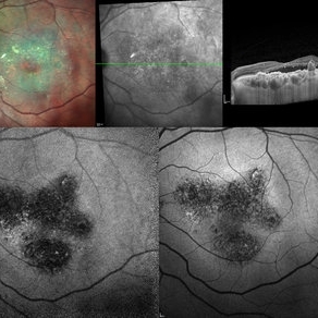
 Berlins Edema - Multimodal Imaging
Jun 25 2025 by Shivankar Sen, MS, FVRS
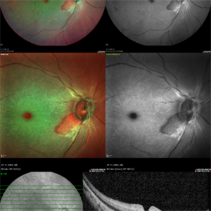
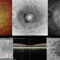
A 22 year old female came with history of injury to her left eye with a badminton racquet butt cap an hour before presentation On examination, she was found to have right eye 6/6;N6 vision and within normal limits, left eye 6/9;N6 vision, cells1+ in the anterior chamber, brisk pupillary response, no vitreous reaction and sub-clinical berlin's edema at the posterior pole. Multimodal imaging revealed frank boundaries of Berlin's edema more pronounced in the nasal parafoveal region. Figure details Top (Left to Right) Multicolor Reflectance showing bright yellow ring surrounding the perifovea; Blue Reflectance (Black on white contrast) showing corresponding black ring; Green Reflectance showing a characteristic white ring (all pronounced nasally); Bottom (Left-Right) Transverse structural OCT enface image showing white ring consistent with edema OCTA inner layer segmentation from ILM to GCL Transverse corresponding OCTA revealing faint hypo ring within perifoveal capillary bed
Photographer: Gayathri M S
Imaging device: Heidelberg Spectralis HRA+OCT
Condition/keywords: blue reflectance, En Face OCTA, enface imaging, multicolor, oct, reflectance
-
 Berlins
Jun 25 2025 by Shivankar Sen, MS, FVRS
A 22 year old female came with history of injury to her left eye with a badminton racquet butt cap an hour before presentation On examination, she was found to have right eye 6/6;N6 vision and within normal limits, left eye 6/9;N6 vision, cells1+ in the anterior chamber, brisk pupillary response, no vitreous reaction and sub-clinical berlin's edema at the posterior pole. Multimodal imaging revealed frank boundaries of Berlin's edema more pronounced in the nasal parafoveal region. Figure details Top (Left to Right) Multicolor Reflectance showing bright yellow ring surrounding the perifovea; Blue Reflectance (Black on white contrast) showing corresponding black ring; Green Reflectance showing a characteristic white ring (all pronounced nasally); Bottom (Left-Right) Transverse structural OCT enface image showing white ring consistent with edema OCTA inner layer segmentation from ILM to GCL
Photographer: Gayathri M S
Imaging device: Heidelberg Spectralis HRA+OCT
Condition/keywords: blue reflectance, En Face OCTA, multicolor
-
 Active Multi Focal Choroiditis
Jun 21 2025 by Moazzam Parvez
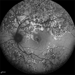
Auto fluorescence image of a 28 year old gentleman with active multifocal choroiditis in his left eye and healed choroiditic patches in the right eye.
Photographer: Moazzam Parvez , Netralayam , Kolkata
Imaging device: Heidelberg Spectralis
Condition/keywords: active, multifocal choroiditis
-
 Subretinal PFO
Jun 18 2025 by Korey Starkey
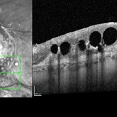
86-year-old patient had history for retinal detachment surgery x2 and intraocular injections for AMD performed elsewhere. Left eye has PVR developing and subretinal PFO. Due to guarded vision, opting to defer any further treatment at this time.
Photographer: Korey Starkey
Imaging device: Heidelberg
Condition/keywords: AMD, Heidelburg Spectralis, OCT, PFO, PVR, retinal detachment, silicone oil
-
 Choroidal Hemangioma
Jun 18 2025 by Moazzam Parvez
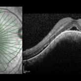
An OCT image of a 42 year old man presenting with a vision of 20/80 and complaining of distortion. OCT reveals serous retinal detachment with RPE alteration and disruption of outer retinal layers.
Photographer: Moazzam Parvez , Netralayam , Kolkata
Imaging device: Heidelberg Spectralis
Condition/keywords: Choroidal Hemangioma, Sub retinal fluid, tumor
-
 Choroidal Hemangioma
Jun 18 2025 by Moazzam Parvez
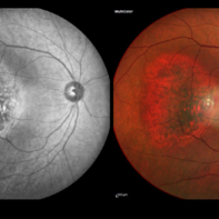
Multicolor and infrared reflectance image of a 42 year old gentleman with a Choroidal hemangioma lesion temporal to the fovea complaining of distortion in his right eye . Fundus imaging revealed a well-circumscribed ,elevated, reddish orange lesion within the choroid involving the posterior pole temporally .
Photographer: Moazzam Parvez, Netralayam , Kolkata
Imaging device: Heidelberg Spectralis
Condition/keywords: Choroidal Hemangioma, tumor
-
 Berlin's Edema
Jun 12 2025 by Shivankar Sen, MS, FVRS
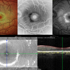
A 22 year old male came with history of sports injury to the right eye with the nose of shuttlecock while playing badminton. On examination, right eye anterior segment shows conjunctival congestion with brisk pupillary reaction and quiet anterior chamber. His best corrected visual acuity was 6/12; N6 in the right eye and 6/6; N6 in the left eye. Retinal examination revealed OD Berlin's Edema, OS within normal limits. Image Description (From Left to Right) Multicolor Reflectance (Blue-Green Enhanced) shows well defined yellowish discoloration Green reflectance and blue reflectance show corresponding whitish discoloration at the area of edema
Photographer: Dr. Shivankar Sen
Imaging device: Heidelberg Spectralis HRA+OCT
Condition/keywords: Shuttlecock Injury
-
 Neovascularization of the Disc
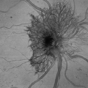
Jun 3 2025 by Scott D Walter, MD, MSc, FASRS
Near-infrared (NIR) en face OCT image showing neovascularization of the disc (NVD) in a patient with type II diabetes mellitus, complicated by proliferative diabetic retinopathy (PDR).
Imaging device: Heidelberg Spectralis
Condition/keywords: Diabetes, Heidelburg Spectralis, microaneurysms, Neovascularisation at the Disc (NVD), NEOVASCULARISATION OF DISC, OCT EN FACE, proliferative diabetic retinopathy (PDR)
-
 Central Bouquet Hemorrhage
May 31 2025 by Moazzam Parvez
OCT image of a 26 year old gentleman of right eye macula with a central foveolar cotton ball like lesion . Inward traction by Müller cells over CB causes upward displacement of foveal cones without major disturbance of ellipsoid zone (EZ) and ELM . Cotton ball sign is characterized by small, fuzzy subfoveal hyperreflective area between the inner segment ellipsoid zone (EZ) and the interdigitation zone (IZ) .
Photographer: Dr Moazzam Parvez , Netralayam , Kolkata
Imaging device: Heidelberg Spectralis
Condition/keywords: Central bouquet haemorrhage, macula, myopia
-
 Multi-modal Imaging of Type - 1 CNVM
May 30 2025 by Shivankar Sen, MS, FVRS
Multimodal Imaging of a case of Polypoidal Choroidal Vasculopathy Multicolor Reflectance showing multiple green-hyper-fringent lesions in the macular region (Up Left) Infra-red Autofluorescence and Blue Autofluorescence showing hypo-autofluorescent areas correspondingly revealing the exact extent of the sub-RPE Lesion (Down left and right respectively) Optical Coherence Tomography - Enhanced Depth Imaging showing Thumb-shaped Pigment Epithelial Detachment with presence of Sub-retinal fluid and Hyper-reflective foci (Top Right)
Photographer: Dr. Shivankar Sen
Imaging device: Heidelberg Spectralis HRA+OCT
Condition/keywords: Blue autofluroscence, CNVM, multicolor, near infrared autofluorescence (NIRAF), PCV, reflectance
-
 Active multifocal choroiditis
May 26 2025 by Moazzam Parvez
Auto fluorescence photograph of an 43 year old man with active choroiditic lesion present in the left eye with recurrence
Photographer: Dr Moazzam Parvez , Netralayam , Kolkata
Imaging device: Heidelberg Spectralis
Condition/keywords: active choroididtis, choroiditi
 Loading…