Search results (638 results)
-
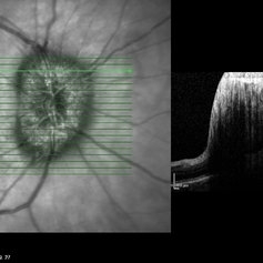
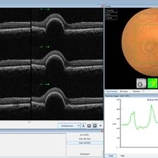 PED due to CSCR
Sep 2 2012 by Hamid Ahmadieh, MD
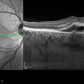
OCT image of a 37-year-old man with a serous PED secondary to CSCR.
Photographer: Hamid Ahmadieh, Ophthalmic Research Center, Labbafinejad Medical Center
Imaging device: Heidelberg Spectralis
Condition/keywords: central serous chorioretinopathy (CSCR), optical coherence tomography (OCT), pigment epithelial detachment (PED)
-
---thumb.jpg/image-square;max$300,300.ImageHandler) Geographic atrophy
Geographic atrophy
Aug 29 2012 by Young Hee Yoon, MD, PhD
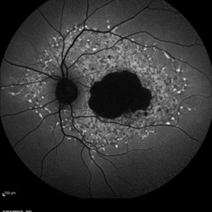
OCT image of an 78-year-old woman. Her best-corrected visual acuity was counting fingers at 30cm.
Photographer: Ji Hee Kim, Asan Medical Center
Imaging device: Heidelberg spectralis
Condition/keywords: dry age-related macular degeneration (dry AMD), geographic atrophy
-
 Optociliary Shunt Vessels in Old CRVO
Sep 8 2012 by Hamid Ahmadieh, MD
FA image of a 60-year-old woman with the history of central retinal vein occlusion.
Photographer: Hamid Ahmadieh, MD, Ophthalmic Research Center, Labbafinejad Medical Center, Shahid Beheshti University of Medical Sciences
Imaging device: Heidelberg Spectralis
Condition/keywords: central retinal vein occlusion (CRVO), shunts vessels
-
 Stargardts Disease in FAF
Sep 14 2012 by Michael P. Kelly, FOPS
This is a scanning laser ophthalmoscopic FAF image of a patient with Stargardts Disease captured with a Heidelberg Spectralis imaging unit. Note, besides the obvious hyper-autofluorescent areas centrally, the much smaller, and in greater number, pinpoints of hyper-autofluorescence extending from the vascular arcades into the mid-periphery.
Photographer: Michael P. Kelly, FOPS, Director, Duke Eye Center Labs, Duke Universtiy Hospital
Imaging device: Heidelberg Spectralis
Condition/keywords: fundus autofluorescence (FAF), Stargardt disease
-
 Angioid Streaks & CNV (Fig 3)
Aug 25 2012 by Hamid Ahmadieh, MD
Early phase ICG angiography imaging of a 53-year-old woman with a juxtafoveal CNV secondary to angioid streaks.
Photographer: Hamid Ahmadieh, Ophthalmic Research Center, Labbafinejad Medical Center
Imaging device: Heidelberg Spectralis
Condition/keywords: angioid streaks, choroidal neovascularization (CNV), indocyanine green (ICG) angiography
-
 Stage 1 Macular Hole
Jul 4 2012 by John T. Thompson, MD
Stage 1 macular hole with vitreomacular adhesion
Imaging device: Heidelberg Spectralis
Condition/keywords: macular hole, vitreomacular adhesion, vitreomacular traction (VMT)
-
 Macular Hole, Autofluorescence
Sep 14 2012 by Michael P. Kelly, FOPS
Fundus autofluorescence (FAF) of a macular hole captured using a Heidelberg Spectralis.
Photographer: Michael P. Kelly, FOPS, Director, Duke Eye Cneter Labs, Duke Universty Hospital
Imaging device: Heidelberg Spectralis
Condition/keywords: fundus autofluorescence (FAF), macular hole
-
 Papilledema
Sep 8 2012 by Hamid Ahmadieh, MD
OCT of the optic nerve head of the right eye of a 55-year-old woman with a malignant intracranial tumor.
Photographer: Hamid Ahmadieh, MD, Ophthalmic Research Center, Labbafinejad Medical Center, Shahid Beheshti University of Medical Sciences
Imaging device: Heidelberg Spectralis
Condition/keywords: malignant intracranial tumor, optical coherence tomography (OCT), papilledema
-
 Polypoidal Choroidal Vasculopathy
Aug 25 2012 by Hamid Ahmadieh, MD
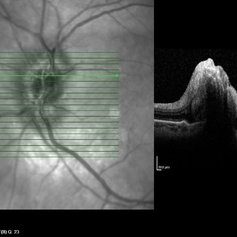
FA & ICG angiography imagings of a 73-year-old man with a peripapillary PCV.
Photographer: Hamid Ahmadieh, Ophthalmic Research Center, Labbafinejad Medical Center
Imaging device: Heidelberg Spectralis
Condition/keywords: indocyanine green (ICG) angiography, polypoidal choroidal vasculopathy (PCV)
-
 Polypoidal Choroidal Vasculopathy-OCT
Aug 27 2012 by Young Hee Yoon, MD, PhD
SD-OCT image of a 56-year-old woman. Her best-corrected visual acuity was 20/30.
Photographer: Kyoung Ree Kim, Asan Medical Center
Imaging device: Heidelberg Spectralis
Condition/keywords: polypoidal choroidal vasculopathy (PCV)
-
 Recurrent Central Serous Choroidopathy
Aug 21 2012 by Edwin H. Ryan, MD
EDI-OCT showing thickened choroid and subretinal fluid
Photographer: Edwin Ryan Jr. MD, VitreoRetinal Surgery, PA
Imaging device: Heidelberg Spectralis
Condition/keywords: central serous chorioretinopathy (CSCR), choroidal thickening, enhanced depth imaging
-
 Papilledema
Sep 8 2012 by Hamid Ahmadieh, MD
OCT of the optic nerve head of the left eye of a 55-year-old woman with a malignant intracranial tumor.
Photographer: Hamid Ahmadieh, MD, Ophthalmic Research Center, Labbafinejad Medical Center, Shahid Beheshti University of Medical Sciences
Imaging device: Heidelberg Spectralis
Condition/keywords: malignant intracranial tumor, optical coherence tomography (OCT), papilledema
-
 Late Stage Stargardt's Disease
Mar 13 2013 by Hamid Ahmadieh, MD
Autofluorescence imaging of the left eye of a 46-year-old man with decreased VA due to advanced Stargardt's disease.
Photographer: Nayereh Hadipoor, Negah Eye Center, Tehran
Imaging device: Heidelberg Spectralis
Condition/keywords: autofluorescence imaging, Stargardt disease
-
 Papilledema
Sep 8 2012 by Hamid Ahmadieh, MD
OCT of the optic nerve head of the right eye of a 55-year-old woman with a malignant intracranial tumor.
Photographer: Hamid Ahmadieh, MD, Ophthalmic Research Center, Labbafinejad Medical Center, Shahid Beheshti University of Medical Sciences
Imaging device: Heidelberg Spectralis
Condition/keywords: optical coherence tomography (OCT), papilledema
-
---thumb.jpg/image-square;max$300,300.ImageHandler) Tamoxifen Retinopathy- OCT
Tamoxifen Retinopathy- OCT
Aug 30 2012 by Young Hee Yoon, MD, PhD
OCT image of an 58-year-old woman with a bilateral tamoxifen maculopathy. She had taken tamoxifen for 24 months due to breast cancer. In spite of discontinuation 2 years ago, her macula remained unchanged. Her best-corrected visual acuity was 20/50 in the right and 20/100 in the left.
Photographer: Soon Tae Kim, Asan Medical Center
Imaging device: Heidelberg Spectralis
Condition/keywords: drug toxicity
-
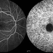
 Multifocal CSCR 2
Sep 2 2012 by Hamid Ahmadieh, MD
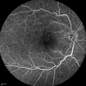
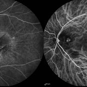
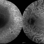
Early-phase FA and ICG angiograms of a 36-year-old man with an active multifocal CSCR.
Photographer: Hamid Ahmadieh, Ophthalmic Research Center, Labbafinejad Medical Center
Imaging device: Heidelberg Spectralis
Condition/keywords: central serous chorioretinopathy (CSCR), indocyanine green (ICG) angiography
-
Angioid Streaks & CNV (Fig 1)
Aug 25 2012 by Hamid Ahmadieh, MD
Fundus autofluorescence (FAF) of a 53-year-old woman with a juxtafoveal CNV secondary to angioid streaks.
Photographer: Hamid Ahmadieh, Ophthalmic Research Center, Labbafinejad Medical Center
Imaging device: Heidelberg Spectralis
Condition/keywords: angioid streaks, choroidal neovascularization (CNV), fundus autofluorescence (FAF)
-
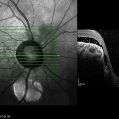 Coloboma of Disc & Choroid
Oct 6 2012 by Hamid Ahmadieh, MD
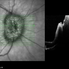
OCT image of a 25-year-old woman with serous retinal detachment secondary to coloboma of disc associated with coloboma of choroid.
Photographer: Hamid Ahmadieh, MD, Ophthalmic Research Center, Labbafinejad Medical Center, Shahid Beheshti University of Medical Sciences
Imaging device: Heidelberg Spectralis
Condition/keywords: coloboma of choroid, coloboma of optic disc, optical coherence tomography (OCT), serous retinal detachment
-
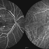
 PED due to CSCR 4
Sep 2 2012 by Hamid Ahmadieh, MD
Early phase FA & ICG images of a 37-year-old man with a serous PED secondary to CSCR
Photographer: Hamid Ahmadieh, Ophthalmic Research Center, Labbafinejad Medical Center
Imaging device: Heidelberg Spectralis
Condition/keywords: central serous chorioretinopathy (CSCR), indocyanine green (ICG) angiography, pigment epithelial detachment (PED)
-
 PED due to CSCR 2
Sep 2 2012 by Hamid Ahmadieh, MD
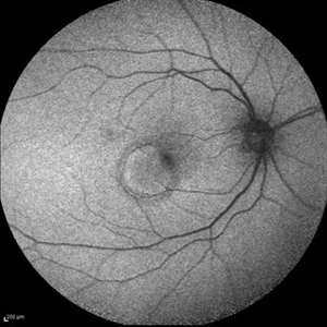
Autofluorescence imaging of a 37-year-old man with a serous PED secondary to CSCR.
Photographer: Hamid Ahmadieh, Ophthalmic Research Center, Labbafinejad Medical Center
Imaging device: Heidelberg Spectralis
Condition/keywords: autofluorescence imaging, central serous chorioretinopathy (CSCR), pigment epithelial detachment (PED)
-
 Myopic Choroidal Neovascularization
Aug 23 2012 by Gabriela Lopezcarasa Hernandez, MD
19-year-old male who complains of scotoma and metamorphopsias.
Photographer: Gabriela Lopezcarasa Hernandez, Macular Retina Consultores
Imaging device: Heidelberg Spectralis
Condition/keywords: choroidal neovascularization (CNV), myopia
-
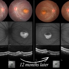 Vitelliform Macular Dystrophy or Best Disease
Dec 16 2016 by Young Hee Yoon, MD, PhD
Bilateral fundus photographs and autofluorescence images of 15-year-old girl who was diagnosed as vitelliform macular dystrophy or Best disease. Vitelliform macular lesion showed morphologic change during one year.
Photographer: Hyejin Jo, Sunghyun Kim, Heoni Hong, Minjung Chae, Mihwa Shin, Asan medical center, Seoul
Imaging device: Topcon TRC-500X fundus camera, Heidelberg HRA 2 autofluorescence, Heldelberg Spectralis OCT
Condition/keywords: Best disease, pseudohypopyon, scrambled-egg, vitelliform macular dystrophy
-
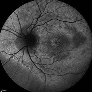 Cystoid Macular Edema (CME)
Sep 11 2012 by Hamid Ahmadieh, MD
Autofluorescence imaging of the left eye of a 17-year-old boy with chronic intermediate uveitis showing CME.
Photographer: Hamid Ahmadieh, MD, Ophthalmic Research Center, Labbafinejad Medical Center, Shahid Beheshti University of Medical Sciences
Imaging device: Heidelberg Spectralis
Condition/keywords: autofluorescence imaging, cystoid macular edema (CME), intermediate uveitis
-
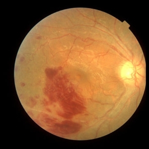 Behcet's Disease
Mar 13 2013 by Hamid Ahmadieh, MD
Color fundus photograph of the right eye of a 23-year-old man with retinal vasculitis and branch retinal vein occlusion (BRVO) due to Behcet's disease .
Photographer: Solmaz Shahmohammad, Negah Eye Center, Tehran
Imaging device: Heidelberg Spectralis
Condition/keywords: branch retinal vein occlusion (BRVO), retinal vasculitis
-
 Retinitis Pigmentosa
Sep 11 2012 by Hamid Ahmadieh, MD
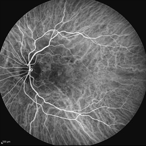
FA & ICG angiography images of a 40-year-old man with RP.
Photographer: Hamid Ahmadieh, MD, Ophthalmic Research Center, Labbafinejad Medical Center, Shahid Beheshti University of Medical Sciences
Imaging device: Heidelberg Spectralis
Condition/keywords: indocyanine green (ICG) angiography, retinitis pigmentosa
 Loading…