Search results (20 results)
-
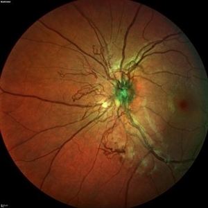
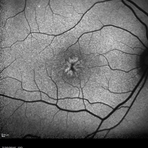
 Retinal Astrocytic Hamartoma
Feb 5 2025 by Rinat Sutiushev
Fundus photograph of a 42-year-old man with retinal astrocytic hamartoma type 3.
Photographer: Rinat Sutiushev, Ophthalmological center “Vision”, Saint Petersburg
Imaging device: Heidelberg Spectralis
Condition/keywords: retina
-
 Lady in a dress
Feb 9 2023 by Shelby Helton
Fluorescein Angiography on a 67-year-old male with significant RPE changes secondary to a severe subretinal hemorrhage that required a vitrectomy with subretinal TPA in 2013.
Photographer: Shelby Helton
Imaging device: Heidelberg Spectralis
Condition/keywords: wet age-related macular degeneration (wet AMD)
-
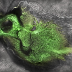
 Neovascular vessels
Sep 22 2022 by Filip Kecer
Multicolor widefield scan of a 16-year-old girl with a neovascularization from disc to vitreous space
Photographer: Filip Kecer, National Institute of Childrens Diseases
Imaging device: Spectralis, Heidelberg Engineering
Condition/keywords: neovascularization (NV), neovascularization at the disc, uveitis, vitreous
-
 Green Goblin Detachment
Jan 13 2022 by Netan Choudhry, MD, FRCS(C) FASRS
Tractional retinal detachment with macular hole in a 76-year-old female.
Photographer: John Golding BA, Vitreous Retina Macula Specialists of Toronto, OCTane Imaging Lab
Imaging device: Multicolor fundus photo taken on the Spectralis OCT2 (Heidelberg Engineering GmbH).
Condition/keywords: macular hole, Multispectral imaging, tractional retinal detachment
-
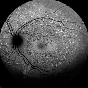 Fundus Flavimaculatus
Dec 9 2021 by Filip Kecer
Fundus autofluorescence of a 13-year-old girl with suspected Fundus flavumaculatus.
Photographer: Filip Kecer
Imaging device: Spectralis, Heidelberg Engineering
Condition/keywords: fundus flavimaculatus, Stargardt disease
-
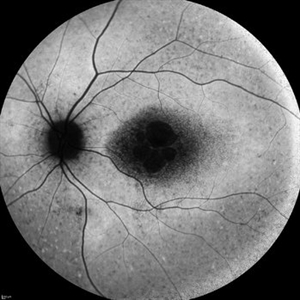 Stargardt Disease
Dec 9 2021 by Filip Kecer
Fundus autofluorescence of a 14-year-old girl with genetically confirmed Stargardt disease.
Photographer: Filip Kecer
Imaging device: Spectralis, Heidelberg Engineering
Condition/keywords: autofluorescence imaging, Stargardt disease
-
 Serous Retinal Detachment in Vogt Koyanagi Harada Patient
Apr 26 2021 by Pablo Baquero Ospina, MD
24-year-old woman with bilateral panuveitis and serous retinal detachment, headache and tinnitus.
Photographer: Pablo Baquero-Ospina, Asociación Para Evitar la Ceguera en México
Imaging device: Heidelberg Spectralis
Condition/keywords: serous retinal detachment, Vogt-Koyanagi-Harada
-
 Retinal Arteriovenous Malformation
Jun 6 2020 by Albert Li, MD, FASRS
Montaged infrared retinal imaging of a 37-year-old asymptomatic man with a grade II arteriovenous malformation (AVM) in the nasal mid-periphery. The presentation of the AVM can be classified with three categories. Grade 1 AVMs are characterized by an abnormal capillary plexus between the major communicating vessels. Grade 2 AVMs are defined by the direct arteriovenous communication without the interposition of arterioles or capillaries. Grade 3 AVMs are characterized by widespread, large caliber anastomosing vessels that are associated with decreased visual acuity and intracranial AVMs. Because retinal AVMs are mostly asymptomatic and non-progressive, further testing may not be indicated unless there are concomitant neurological signs and symptoms or if there is a strong clinical suspicion of a grade 3 retinal AVM. Observation was recommended for the patient in this image. On his most recent follow-up at four months, the patient remained asymptomatic with a stable appearance of the lesion.
Imaging device: Heidelberg Spectralis
Condition/keywords: arteriovenous anastomosis, arteriovenous malformation
-
 Acute Macular Neuroretinopathy
Dec 11 2019 by Lauren Whaley
34-year-old female patient presented with changes in vision after recent upper respiratory infection. Referring doctor originally thought it was a blood pressure issue. She noticed a "C" shape in her vision. Infrared image was captured showing exactly what patient was describing! Doctor confirmed with this image that it was AMN.
Photographer: Lauren R. Whaley, COA
Imaging device: Heidelberg Spectralis
Condition/keywords: 30 degrees, acute macular neuroretinopathy, Heidelburg Spectralis, left eye, macula, near infrared autofluorescence (NIRAF)
-
 Macular Pucker With Myelinated Nerve Fiber Layer
Nov 1 2018 by Kevin J. Blinder, MD, FASRS
Multi-color photo of macular pucker with myelinated nerve fiber layer.
Photographer: Jarrod Wehmeier
Imaging device: Heidelberg Spectralis
Condition/keywords: macular pucker
-
 Best Disease
Sep 28 2016 by Maciej Czepita
Color fundus image and fundus autofluorescence image of a 41-year-old male patient with Best disease (pseudohypopyon stage).
Photographer: Maciej Czepita, Pomeranian Medical University, Szczecin, Poland
Imaging device: Heidelberg Spectralis HRA+OCT
Condition/keywords: Best disease
-
 Retinal Dystrophy of 24-Year-Old Male/ AF OD
Nov 25 2015 by Zach Dupureur
Fluorescein angiography of a 24-year-old male. Juvenile retinoschisis on OCT. FA shows outer retinal staining. Could be associated with Goldman Farve Syndrome.
Photographer: Zach Dupureur OCT-C
Imaging device: Heidelberg Spectralis
Condition/keywords: Goldmann-Favre Syndrome, juvenile retinoschisis, retinal dystrophy
-
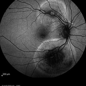
 Congenital Simple Hamartoma of RPE
Aug 3 2015 by Bindu Rajesh
OCT line scan through the hamartoma in a 26-year-old male, showing increased hyperreflectivity in the area of lesion with backshadowing and minimal protrusion into vitreous.
Imaging device: Heidelberg Spectralis
Condition/keywords: congenital, hamartoma, retinal pigment epithelium
-
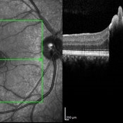
 Foveoschisis secondary to high myopia
Mar 13 2015 by Niloofar Piri, MD
Infrared and HD-OCT of the right eye in a 55-year-old African American female with high myopia (more than -6.00 D), BCVA: 20/25 OU Cartwheel appearance of the fovea in the infrared imaging is visible. HD- OCT demonstartes schisis in different layers of the retina (both NFL and OPL; notice stretching of the Muller cells); VMT is also present . Outer retinal layers are preserved which explains the good vision . She had the same findings in OS.
Photographer: Niloofar Piri, MD
Imaging device: Heidelberg Spectralis
Condition/keywords: high myopia, retinoschisis
-
 Optic Nerve Head Drusen
Feb 12 2015 by Timothy S Fuller, MD
Fundus autofluorescence image of a 34-year-old woman with striking, asymptomatic optic nerve head drusen.
Photographer: Nice Hesse, Texas Retina Associates
Imaging device: Heidelberg Spectralis
Condition/keywords: drusen of optic disc
-
 Suspected Multiple Evanescent White Dot Syndrome
Mar 3 2015 by Stuart Alfred, CRA, OCT-C
30 degree, late phase angiogram image of left fundus of a 28-year-old Caucasian female.
Photographer: Stuart Alfred, CRA, OCT-C, Midwest Eye Institute, Greenwood, Indiana
Imaging device: cSLO by Heidelberg engineering (Spectralis)
Condition/keywords: dry age-related macular degeneration (dry AMD), multiple evanescent white dot syndrome (MEWDS), punctate inner choroidopathy (PIC), uveitis
-
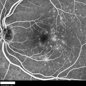
 Retinal Arterial Macroaneurysms
Jan 22 2015 by Darrell E. Baskin, MD
Fundus autofluorescence image of a 60-year-old woman with two retinal arterial macroaneurysms--one recent and one not.
Photographer: Darrell Baskin, Wilford Hall, Lackland Air Force Base, Texas
Imaging device: Heidelberg Spectralis
Condition/keywords: retinal arterial macroaneurysm
-
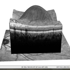 APMPPE With Serous Macular Detachment 3D SD-OCT
Jun 2 2014 by Rameez N Hussain, MD
3D SD-OCT of acute posterior multifocal placoid pigment epitheliopathy (APMPPE) with serous macular detachment.
Photographer: Rameez N Hussain MD, Vitreo Retinal Services, Giridhar Eye Institute, Cochin, India
Imaging device: Heidelberg Spectralis
Condition/keywords: acute posterior multifocal placoid pigment epitheliopathy (APMPPE), serous retinal detachment
-
 OCT Myopic Staphyloma With Schisis and ERM
Apr 24 2014 by Scott E. Pautler, MD
OCT of high myope with asymptomatic macular schisis.
Imaging device: Heidelberg Spectralis
Condition/keywords: foveal schisis, maculopathy, maculoschisis, optical coherence tomography (OCT), pathologic myopia, staphyloma
-
---thumb.jpg/image-square;max$300,300.ImageHandler) Serous Detachment of Retinal Epithelium
Serous Detachment of Retinal Epithelium
Nov 25 2013 by Alyssa Bristol
36-year-old man with serous detach of retinal epithelium.
Photographer: Alyssa Bristol, Chester County Eye Care
Imaging device: Heidelberg Spectralis
Condition/keywords: central serous retinopathy (CSR), retinal pigment epithelium
 Loading…