Search results (189 results)
-
 Eyes Too Celebrate Valentine’s Day
Jul 28 2024 by KANWALJEET HARJOT MADAN, M.S. (Ophthalmology); FAICO (Vitreous - Retina)
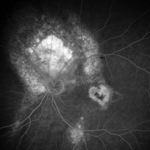
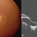
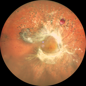
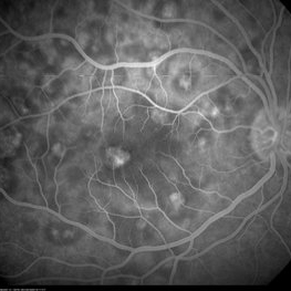
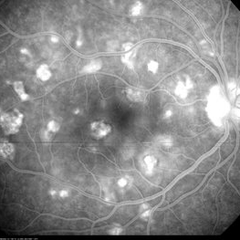
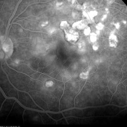
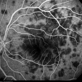
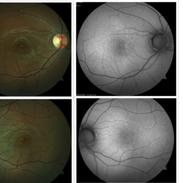
A 53 years male patient presented with decrease in vision in left eye for 6 months. His vison in left eye was counting fingers 1 meter. His vison in right eye was 20/20. Fundus examination in left eye depicted presence of large orange shaped elevated subretinal mass superior to optic disc with scar in macula. We made clinical diagnosis of Choroidal Hemangioma with macular scar. Fundus Fluorescein Angiography (FFA) in left eye revealed early fluorescence in area corresponding to Choroidal Hemangioma which persisted in late phases. Macular scar was “HEART” shaped on FFA which was very unique incident finding.
Photographer: Dr. Kanwaljeet Harjot Madan
Imaging device: Ziess Clarus
Condition/keywords: Choroidal Hemangioma, Fundus examination, Fundus Fluorescein Angiography
-
 Serpiginous Choroidopathy
Sep 24 2024 by Gustavo Uriel Fonseca Aguirre
Right fundus of a 32-year-old female patient diagnosed with serpiginous choroiditis.
Photographer: Gustavo U. Fonseca Aguirre, Fundación Hospital Nuestra Señora de la Luz, Ciudad de México
Condition/keywords: Fundus examination, serpiginous choroiditis
-
 Blau syndrome slide 2
Oct 22 2012 by Ronald C. Gentile, MD
Fundus examination revealed vascular sheathing with optic nerve edema and multiple focal areas of choroidal involvement.
Photographer: The New York Eye & Ear Infirmary Department of Medical Imaging
Condition/keywords: Blau syndrome
-
 Cone Rod Dystrophy slide 2
Oct 22 2012 by Ronald C. Gentile, MD
Fundus examination revealed spotty pigmentary changes in the macular area with some peripheral depigmentation of the retinal pigment epithelium.
Photographer: The New York Eye & Ear Infirmary Department of Medical Imaging
Condition/keywords: cone dystrophy, retinal pigment epithelium
-
 Deferoxamine Retinopathy Slide 2
Oct 22 2012 by Ronald C. Gentile, MD
Fundus examination revealed speckled pigmentation of the macula with disturbance of the retinal pigment epithelium and photoreceptors greater in the left eye compared to the right eye.
Photographer: The New York Eye & Ear Infirmary Department of Medical Imaging
Condition/keywords: angioid streaks, Deferoxamine retinopathy, Thalassemia intermedia
-
 Hemangioma
Oct 16 2012 by Anat Loewenstein, MD
Fundus examination of a 68 year old lady with decreased vision in her left eye for several months. VA 20/30 in her RE and counting fingers in the left eye. In the left eye there was a large red mass protruding and covering almost the entire optic nerve. Diagnosed as retinal hemangioma. The patient underwent low fluence PDT.
Photographer: Galit Yair-Pur
Condition/keywords: hemangioma
-
 Morning glory optic disc anomaly with retinal detachment
Sep 13 2022 by Min Kim, MD, PhD, MBA, FASRS
Fundus examination of this 5 year-old male shows large funneled optic nerve with conical excavation of the dysplastic optic disc. 360° macula-involving retinal detachment was observed. The best corrected visual acuity of the right eye was counting fingers 10cm.
Photographer: Min Kim, M.D.-Ph.D.-M.B.A. Gangnam Severance Hospital Yonsei University College of Medicine, Department of Ophthalmology
Imaging device: Optos Silverstone P200TxE
Condition/keywords: Morning Glory Anomaly, Morning Glory Syndrome
-
 Stargardt macular dystrophy slide 2
Oct 22 2012 by Ronald C. Gentile, MD
Fundus examination of the left eye had similar findings with centrally atrophic macula area with surrounding flecks.
Photographer: The New York Eye & Ear Infirmary Department of Medical Imaging
Condition/keywords: Stargardt disease
-
 Unilateral Macular Coloboma
Jul 29 2021 by Mihir Trivedi
Fundus examination of a 35-year-old man with focal areas of altered retinal pigment epithelium and subretinal yellowish lesion in the foveal area in the right eye. Left eye showed a punched out circumscribed lesion in the center of the macula with thin foveal roof suggestive probably of the internal limiting membrane. Macular coloboma is characterized by a sharply defined, oval or rounded, usually unilateral, atrophic lesions of varying size presenting rudimentary or absent retina, choroid and sclera located at the macula leading to decreased vision in the central area of the fundus. It can be associated with retinal dystrophy in the fellow eye, as was the case in our patient.
Photographer: Priyanshi Kambodi, RNC Eye Hospital, Valsad
Condition/keywords: macular coloboma
-
 "Untouched" Intra-ocular Foreign Body - Course Over 7 Years!
Jul 7 2020 by Deependra Vikram Singh, MD FASRS
25-yr-old male presented to our retina clinic in 2007 with history of Hammer and chisel injury to left eye 2 months back. On examination BCVA in left eye was 20/20. Slit-lamp examination revealed iris hole and fundus examination showed an encapsulated metallic intraocular foreign body (IOFB) close to inferior arcade in left eye. Patient was advised Vitreous surgery with IOFB removal. Patient, however did not turn up for Surgery and revisited our clinic after seven years in 2014. On examination his BCVA was 20/20 in left eye and IOFB has reduced in size with brown siderotic deposits seen over IOFB capsule. Examination revealed posterior sub-capsular cataract but no siderotic changes with intraocular pressure (IOP) also being recorded as normal. In view of good visual acuity and no siderotic changes, he was advised regular follow up and ERG. Since most IOFBs would get timely removed by vitreous surgery, this Image capturing the natural course of a metallic IOFB is rare.
Photographer: Deependra Vikram Singh, Eye-Q Hospitals, Gurugram, INDIA
Imaging device: Kowa and Zeiss
Condition/keywords: encapsulated intraocular foreign body
-
 A Classic Case of Retinal Ora Serrata Imaging
Jan 16 2025 by yuan duo
A 5-year-old girl, born full-term with no history of systemic disease, presented with poor vision since early childhood and underwent fundus examination. Anterior segments of both eyes showed no significant abnormalities. Fundus examination revealed retinal folds extending from the optic disc to the temporal peripheral retina, with blood vessels coursing through the folds (A, B). Avascular zones were observed in the peripheral retina, and the ora serrata’s boundaries were clearly visible, displaying dentate processes and bays (C, D). Retinal pigmentation was evident. Genetic testing confirmed the final diagnosis of bilateral Familial Exudative Vitreoretinopathy (FEVR).
Condition/keywords: Retinal Ora Serrata
-
 A rare case of a 45-year-old male with choroidal neovascular membrane in Familial Dominant Drusen (Doyne Honeycomb Drusen) in both eyes treated with intravitreal injections.
Nov 30 2022 by SHRADDHA ASHOK CHANDORKAR, DNB DO FVRS
A 45-year-old man presented with diminution of vision in both eyes with metamorphopsia, which was painless and gradually progressive in nature. BCVA at presentation were 6/40 and 6/36 for the right and left eye respectively. Anterior segment examination of both eyes was unremarkable. IOP were within normal limits. Fundus examination showed bilateral numerous yellowish white round closely spaced lesions extending radially from the vascular arcades till the periphery associated with an elevated grayish macular choroidal neovascular membrane (CNV) with multiple drusen in the macular area and posterior pole. Impression was Familial Dominant Drusen (Doyne Honeycomb Drusen) associated with CNVM, both eyes. Color fundus photograph and autofluorescence showed Familial Dominant Drusen with CNVM. Subsequently , the patient underwent periodic intravitreal injections of Ranibizumab in both eyes under guarded visual prognosis, for which he tolerated well.
Photographer: NATIONAL INSTITUTE OF OPHTHALMOLOGY, PUNE
Imaging device: ZEISS CLARUS
Condition/keywords: choroidal neovascular membrane (CNVM), Doyne's Honeycomb, FAMILIAL DOMINANT DRUSEN, IMIM (Online Mendelian Inheritance in Man), intravitreal injection, Malattia Leventinese
-
 Acute Syphilitic Posterior Placoid Chorioretinitis
May 4 2021 by RAFAEL REIS PEREIRA, MD
A 31-year-old patient with a complaint of photophobia and low visual acuity OD in the previous three weeks. BCVA was 20/60 and 20/20 The fundus examination revealed a placoid white lesion in the posterior pole and vitreous cells in the right eye. The left eye was unremarkable. Fluorescein angiography reveals hyperfluorescent plaque with distinctive “leopard spots” hypofluorescence.
Imaging device: Opto California
Condition/keywords: acute syphilitic posterior placoid chorioretinitis
-
 Angioid Streaks
Sep 29 2024 by Tejaswita Verma
Fundus photograph of a 35 year-old female with 6/6 vision in RE , unremarkable anterior segment and family history of angioid streaks and pseudoxanthoma elasticum in sister. Fundus examination revealed angioid streaks radiating from disc , sparing the fovea .Her Sister had received multiple anti VEGF injections for angioid streaks with CNVM.
Photographer: DR. TEJASWITA VERMA
Imaging device: MIRANTE
Condition/keywords: angioid streaks
-
 Annular Tractional Retinal Detachment
Jul 4 2024 by Hector Gabriel Moreno Solano, MD, MHA
52-year-old Hispanic female patient with a diagnosis of type II diabetes mellitus of 15 years of evolution, comes to the retina service for progressive visual loss in the right eye (single functional eye) with visual acuity of 20/100, Fundus examination reveals laser-modified proliferative diabetic retinopathy with activity + annular tractional retinal detachment with macular involvement.
Photographer: Hector Gabriel Moreno Solano, MD, MHA, HGZ #20 IMSS Puebla.
Imaging device: Mirante
Condition/keywords: macular detachment, proliferative diabetic retinopathy (PDR), tractional retinal detachment
-
 Anterior ischemic optic neuropathy slide 1
Oct 22 2012 by Ronald C. Gentile, MD
70-year-old women with acute loss of vision in the left eye. Review of symptoms was significant for temporal arteritis and ESR was very high. Fundus examination of the left eye had a swollen white optic nerve head with a few peri-papillary cotton wool spots.
Photographer: The New York Eye & Ear Infirmary Department of Medical Imaging
Condition/keywords: anterior ischemic optic neuropathy, choroidal ischemia, temporal arteritis
-
 APMPPE in a 21 Year-Old Female Patient
Oct 23 2015 by Roy Schwartz, MD
FA photograph of a 21-year-old, usually healthy, female, presenting with visual deterioration and photophobia in BE. Upon examination deep lesions were seen on fundus examination. FA showed hypofluorescent lesions (seen here at 2:28 minutes) that later became hyperfluorescent
Photographer: Galit Yair Pur
Condition/keywords: acute posterior multifocal placoid pigment epitheliopathy (APMPPE)
-
 APMPPE in a 21 Year-Old Female Patient
Oct 23 2015 by Roy Schwartz, MD
FA photograph of a 21-year-old, usually healthy, female, presenting with visual deterioration and photophobia in BE. Upon examination deep lesions were seen on fundus examination.
Photographer: Galit Yair Pur
Condition/keywords: acute posterior multifocal placoid pigment epitheliopathy (APMPPE)
-
 APMPPE in a 21-Year-Old Female Patient
Oct 23 2015 by Roy Schwartz, MD
FA photograph of a 21-year-old, usually healthy, female, presenting with visual deterioration and photophobia in BE. Upon examination deep lesions were seen on fundus examination. FA showed hypofluorescent lesions that later became hyperfluorescent (seen here at 7:40 minutes).
Photographer: Galit Yair-Pur
Condition/keywords: acute posterior multifocal placoid pigment epitheliopathy (APMPPE)
-
 APMPPE in a 21-Year-Old Female Patient
Oct 23 2015 by Roy Schwartz, MD
FA photograph of a 21-year-old, usually healthy, female, presenting with visual deterioration and photophobia in BE. Upon examination deep lesions were seen on fundus examination. FA showed hypofluorescent lesions that later became hyperfluorescent (seen here at 10:41 minutes).
Photographer: Galit Yair-Pur
Condition/keywords: acute posterior multifocal placoid pigment epitheliopathy (APMPPE)
-
 APMPPE in a 21-Year-Old Female Patient
Oct 23 2015 by Roy Schwartz, MD
FA photograph of a 21-year-old, usually healthy, female, presenting with visual deterioration and photophobia in BE. Upon examination deep lesions were seen on fundus examination. FA showed hypofluorescent lesions (seen here at 36 seconds) that later became hyperfluorescent
Photographer: Galit Yair Pur
Condition/keywords: acute posterior multifocal placoid pigment epitheliopathy (APMPPE)
-
 Asteroid Hyalosis
May 2 2023 by RAKESH SHAH, MS DNB FACS FRF FICO MBA
Routine fundus examination for 45 year-old male
Photographer: DR RAKESH SHAH
Condition/keywords: asteroid hyalosis
-
 Astrocytic Hamartoma
Apr 30 2015 by Mariam A Al-Feky, MD
A 15-year-old boy with history of seizures controlled on treatment. C/O: OD painless DV 10/7 ago (accidental discovery) O/E: BCVA OD: 6/60 ,, OS 6/6. AS: NAD OU. Pupil: RRR no RAPD OU. Fundus examination OD showed a retinitis like lesion with an overlying corkscrew vessel well evident on FFA with late leakage and CSR and OCT through the retinitis like lesion shows diffuse hypereflective thickeninig in the superficial NFL. Thorough history taking revealed that patient has seizures and MRI lesions suggestive of tuberous sclerosis. So this is exudative hamartoma secondary to tuberous sclerosis with marked resolution after single IVI of Lucentis. Retinitis like lesion with corkscrew vessels in FFA is typical together with the homogenous hypereflective thickening in the NFL.
Photographer: Mariam AL-Feky
Imaging device: Optical coherence tomography
Condition/keywords: astrocytic hamartoma
-
 Bilateral Benign Yellow Dot Maculopathy
May 6 2025 by Amol yuvraj ganvir
A 37-year-old female patient presented for a routine eye examination. Her best-corrected visual acuity was 6/6 in both eyes. Fundus examination revealed multiple small yellow dots over the macula in both eyes. FAF imaging demonstrated characteristic hyperautofluorescence corresponding to these dots.
Photographer: Dr. Amol Ganvir, Vitreo-Retina Fellow, Ishwar Eye Centre, Rohtak, Haryana
Imaging device: Visucam-Zeiss
Condition/keywords: Autoflourescence, yellow dots
 Loading…