 Initializing download.
-
 By KANWALJEET HARJOT MADAN, M.S. (Ophthalmology); FAICO (Vitreous - Retina)
Thind Eye Hospital, Jalandhar City (Punjab). India. - Uploaded on Jul 28, 2024.
- Last modified by Joshua Friedman on Jul 29, 2024.
- Rating
- Appears in
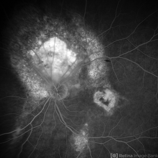
- HEART shaped Macular Scar (28th July 2024)
- Condition/keywords
- Fundus examination, Fundus Fluorescein Angiography, Choroidal Hemangioma
- Photographer
- Dr. Kanwaljeet Harjot Madan
- Imaging device
-
Fundus camera
Ziess Clarus - Description
- A 53 years male patient presented with decrease in vision in left eye for 6 months. His vison in left eye was counting fingers 1 meter. His vison in right eye was 20/20. Fundus examination in left eye depicted presence of large orange shaped elevated subretinal mass superior to optic disc with scar in macula. We made clinical diagnosis of Choroidal Hemangioma with macular scar. Fundus Fluorescein Angiography (FFA) in left eye revealed early fluorescence in area corresponding to Choroidal Hemangioma which persisted in late phases. Macular scar was “HEART” shaped on FFA which was very unique incident finding.


