 Initializing download.
-
 By Mariam A Al-Feky, MD
AIN SHAMS UNIVERSITY
Co-author(s): Mohamed Moghazy - Uploaded on Apr 30, 2015.
- Last modified by Caroline Bozell on Jun 2, 2015.
- Rating
- Appears in
- Miscellaneous
- Condition/keywords
- astrocytic hamartoma
- Photographer
- Mariam AL-Feky
- Imaging device
-
Fundus camera
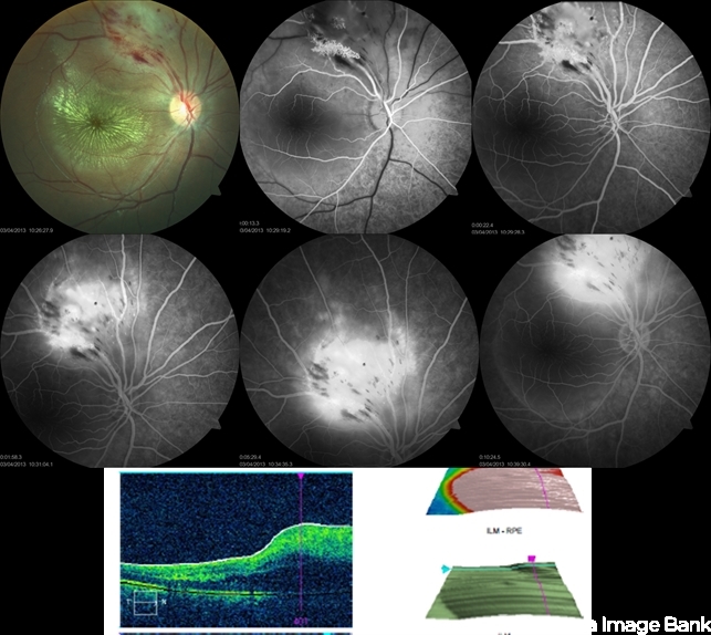
Optical coherence tomography - Description
- A 15-year-old boy with history of seizures controlled on treatment. C/O: OD painless DV 10/7 ago (accidental discovery) O/E: BCVA OD: 6/60 ,, OS 6/6. AS: NAD OU. Pupil: RRR no RAPD OU. Fundus examination OD showed a retinitis like lesion with an overlying corkscrew vessel well evident on FFA with late leakage and CSR and OCT through the retinitis like lesion shows diffuse hypereflective thickeninig in the superficial NFL. Thorough history taking revealed that patient has seizures and MRI lesions suggestive of tuberous sclerosis. So this is exudative hamartoma secondary to tuberous sclerosis with marked resolution after single IVI of Lucentis. Retinitis like lesion with corkscrew vessels in FFA is typical together with the homogenous hypereflective thickening in the NFL.


 FA - Astrocytic Hemartoma")