 Initializing download.
-
 By Ronald C. Gentile, MD
The New York Eye and Ear Infirmary of Mount Sinai - Uploaded on Oct 22, 2012.
- Last modified by Chayal Patel on Nov 20, 2012.
- Reviewed by Chayal Patel
- Rating
- Appears in
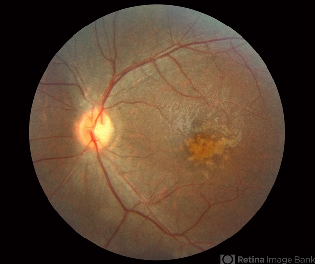
- Cone rod dystrophy
- Condition/keywords
- cone dystrophy, retinal pigment epithelium
- Photographer
- The New York Eye & Ear Infirmary Department of Medical Imaging
- Imaging device
- Fundus camera
- Description
- Fundus examination revealed spotty pigmentary changes in the macular area with some peripheral depigmentation of the retinal pigment epithelium.


