 Initializing download.
-
 By Mihir Trivedi
RNC Eye Hospital
Co-author(s): Yesha Bhavsar, RNC Eye Hospital, Valsad. Dr Shaheen Virani, RNC Eye Hospital, Valsad - Uploaded on Jul 29, 2021.
- Last modified by Caroline Bozell on Jul 30, 2021.
- Rating
- Appears in
- Miscellaneous
- Condition/keywords
- macular coloboma
- Photographer
- Priyanshi Kambodi, RNC Eye Hospital, Valsad
- Imaging device
- Optical coherence tomography system
- Description
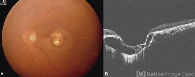
- Fundus examination of a 35-year-old man with focal areas of altered retinal pigment epithelium and subretinal yellowish lesion in the foveal area in the right eye. Left eye showed a punched out circumscribed lesion in the center of the macula with thin foveal roof suggestive probably of the internal limiting membrane. Macular coloboma is characterized by a sharply defined, oval or rounded, usually unilateral, atrophic lesions of varying size presenting rudimentary or absent retina, choroid and sclera located at the macula leading to decreased vision in the central area of the fundus. It can be associated with retinal dystrophy in the fellow eye, as was the case in our patient.


---thumb.jpg/image-square;max$79,0.ImageHandler "Neovascularization")


