Search results (46 results)
-
 Acute Idiopathic Occlusive Retinal Vasculitis
May 31 2014 by Hamid Ahmadieh, MD
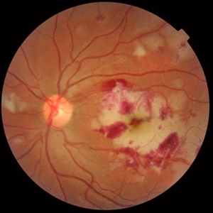
Color fundus photograph of the right eye of a 28-year-old woman with sudden drop of vision due to acute occlusive retinal vasculitis leading to extensive nerve fiber layer infarction and retinal hemorrhages.
Photographer: Naghmeh Nozhat, Negah Eye Center, Tehran
Condition/keywords: color fundus photograph, cotton wool spots, retinal hemorrhage, retinal ischemia
-
 Central Retinal Vein Occlusion
Sep 2 2012 by Hyung-Woo Kwak, MD
Multiple dense, dark, blotchy hemorrhages, cotton-wool spots, and pale optic disc are signs suggestive of retinal ischemia in CRVO.
Imaging device: Zeiss F450 plus
Condition/keywords: central retinal vein occlusion (CRVO)
-
 Acute Idiopathic Occlusive Retinal Vasculitis
May 31 2014 by Hamid Ahmadieh, MD
Color fundus photograph of the left eye of a 28-year-old woman with acute drop of vision due to occlusive retinal vasculitis leading to extensive nerve fiber layer infarction and retinal hemorrhages.
Photographer: Naghmeh Nozhat, Negah Eye Center, Tehran
Condition/keywords: color fundus photograph, cotton wool spots, retinal hemorrhage, retinal ischemia
-
 Hypertensive Retinopathy
Aug 24 2012 by Geoffrey G. Emerson, MD, PhD, FASRS
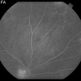
A 35-year-old man has headaches and decreased vision. The right eye measures 20/25 and the left eye measures 3/200. The blood pressure measures 180/110. This fluorescein angiogram shows dilated capillaries and capillary dropout in the central macula of the left eye.
Photographer: Geoffrey Emerson, MD, PhD, Retina Center, Minneapolis
Condition/keywords: hypertensive retinopathy, papilledema, retinal ischemia
-
 Wyburn Mason Racemose Angiomatosis
May 22 2016 by Olivia Rainey
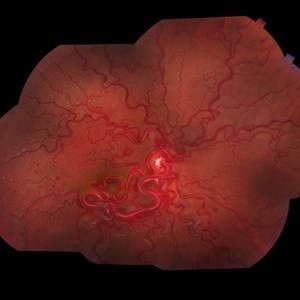
Color fundus montage of an 13-year-old female with arteriovenous malformation (Wyburn Mason Racemose Angiomatosis) affecting her right eye. The retinal arteriovenous malformation appears to be stable. She presented with NLP in the eye, strabismus, and peripheral retinal ischemia. She is at risk for neovascular complications; however, she is currently being treated with Sirolimus. Since she is on this systemically, there is no need to perform intraocular anti-VEGF injections or PRP laser. She also presented with optic atrophy affecting her left eye, secondary to chiasmal involvement of arteriovenous malformation. She has had a potential progressive visual field loss involving the temporal aspect of her visual field from the left eye. There is sector optic atrophy. Presumably, this is due to a compressive effect of her arteriovenous malformation on the nasal nerve fiber layer (corresponding to the temporal visual field) crossing to the right occipital cortex at the chiasm.
Photographer: Olivia Rainey
Imaging device: Topcon 50dx
Condition/keywords: arteriovenous malformation, color fundus photograph, color photo, montage, peripheral ischemia, Sirolimus
-
 Marked Retinal Ischemia in Patient with Mixed Connective Tissue Disease
Feb 26 2013 by Sharon Fekrat, MD FACS FASRS
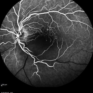
Fluorescein angiogram of the right eye of a 27-year-old female with mixed connective tissue disease and marked retinal ischemia. Panretinal laser photocoagulation (PRP) has been performed for neovascularization elsewhere (NVE).
Condition/keywords: mixed connective tissue disease, retinal ischemia
-
 PDR NVD NVE
Jul 21 2014 by Susanna S. Park, MD, PhD
Mid-transit view fluorecein angiogram of the right eye of a 59-year-old diabetic woman with minimal peripheral fundus changes suggestive of diabetic retinopathy showing diffuse leakage of the disc and focal leakage in the peripheral retina from neovascularization. Peripheral retinal ischemia and leaking retinal microaneurysms are also seen.
Photographer: Karishma Chandra, University of California Davis Eye Center
Condition/keywords: fluorescein leakage, neovascularization of the disc (NVD), proliferative diabetic retinopathy (PDR)
-
 Ischemic Proliferative Diabetic Retinopathy
Aug 22 2012 by Edwin H. Ryan, MD
Fundus photograph of 35-year-old poorly-controlled diabetic woman, 6/200 vision.
Photographer: Edwin Ryan Jr. MD, VitreoRetinal Surgery, PA
Condition/keywords: retinal ischemia
-
 Diabetic Retinopathy
Oct 2 2013 by Jerald A. Bovino, MD
This patient with long standing diabetes has peripheral non-perfusion.
Condition/keywords: retinal ischemia
-
 Ischemic Proliferative Diabetic retinopathy
Aug 22 2012 by Edwin H. Ryan, MD
Fluorescein angiogram of 35-year-old poorly-controlled diabetic woman, 6/200 vision.
Photographer: Edwin Ryan Jr. MD, VitreoRetinal Surgery, PA
Condition/keywords: retinal ischemia
-
---thumb.jpg/image-square;max$300,300.ImageHandler) Lupus Vasculitis Angiogram
Lupus Vasculitis Angiogram
Feb 13 2013 by From the Collections of Thomas M. Aaberg, MD and Thomas M. Aaberg Jr., MD
FA, lupus vasculitis angiogram.
Condition/keywords: lupus, obliterative peripheral vasculitis, retinal ischemia
-
 Retinal Ischemia, Edema, and Hemorrhages on the Infero-Temporal Macula
Aug 26 2019 by Narciso F. Atienza, MD, MBA, FASRS, FPCS, FPAO.
47-year-old female who came in with blurring of vision of the right eye of 2 weeks duration. She is hypertensive with poor control, taking Amlodipine irregularly. Denies any cardiac problem non-diabetic. Vision upon presentation was 20/400 (OD), 20/20 (OS) colored fundus photo of the right eye showing areas of retinal ischemia, edema and hemorrhages on the infero-temporal macula extending to the arcade.
Photographer: Narciso F Atienza, Jr. MD, MBA
Imaging device: Topcon TRC
Condition/keywords: edema, hemorrhage, inferotemporal arcade, retinal ischemia
-
 Multiple Myeloma with Cytomegalovirus Retinitis
Apr 5 2018 by Kim Barrett
Ultra-wide field fluorescein angiogram of a 77-year-old male with multiple myeloma. Patient's angiogram presented significant peripheral retinal ischemia and cystoid macular edema. Patient tested positive for polymerase chain reaction, confirming cytomegalovirus retinitis. Patient is being treated with intravitreal ganciclovir and his current vision is 20/200.
Photographer: Kim Barrett, COA
Imaging device: Optos
Condition/keywords: cystoid macular edema (CME), fluorescein angiogram (FA), fluorescein leakage, intravitreal ganciclovir, myeloma, peripheral ischemia, positive polymerase chain reaction (PCR), ultra-wide field imaging
-
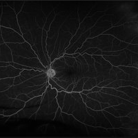
 Central Retinal Artery Occlusion
May 16 2017 by Olivia Rainey
Fluorescein angiogram of an 66-year-old female with a central retinal artery occlusion affecting her left eye.
Photographer: Olivia Rainey
Imaging device: Heidelberg Spectralis
Condition/keywords: 50 degrees, central retinal artery occlusion (CRAO), fluorescein angiogram (FA), left eye, mid phase, retinal ischemia
-
---thumb.jpg/image-square;max$300,300.ImageHandler) Primary Hyperoxaluria and Oxalosis
Primary Hyperoxaluria and Oxalosis
Jul 24 2013 by Hamid Ahmadieh, MD
Late phase FA image of the left eye of a 55-year-old man with primary hyperoxaluria and oxalosis. Profound leakage from disc due to NVD is visible. Vasoproliferative retinopathy has occurred secondary to retinal ischemia due to intravascular deposition of calcium oxalate crystals.
Photographer: Hanieh Payab, Ophthalmic Research Center, Tehran
Imaging device: Topcon Fundus Camera
Condition/keywords: oxalosis, primary hyperoxaluria, vasoproliferative retinopathy
-
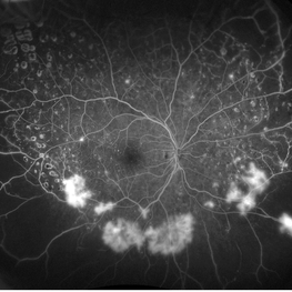
---thumb.jpg/image-square;max$300,300.ImageHandler) BRVO Late FA 102 Degree ART
BRVO Late FA 102 Degree ART
Apr 18 2014 by Susanna S. Park, MD, PhD
Wide angle fluorescein angiography late transit view taken of the left eye of a 45-year-old woman with branch retinal vein occlusion showing peripheral retinal ischemia with staining and leakage of the affected retinal veins.
Photographer: Karishma Chandra, UC Davis Eye Center
Condition/keywords: branch retinal vein occlusion (BRVO), fluorescein leakage, wide angle imaging
-
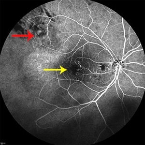 Radiation Retinopathy
Jun 5 2014 by Shlomit Schaal, MD, PhD, MHCM
Angiogram showing previously irradiated and regressed peripheral choroidal melanoma (red arrow), and the resulting central radiation retinopathy with retinal ischemia and remodeling of capillaries (yellow arrow).
Photographer: Shlomit Schaal MD, PhD, University of Louisville, Louisville, KY
Condition/keywords: radiation retinopathy
-
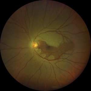
 Central Retinal Artery Occlusion With Cilioretinal Sparing
Apr 4 2018 by Soumya Venkatesh
Fundus photograph of a 23-year-old gentleman presenting with sudden loss of vision 2 days prior to presentation. He underwent all relevant investigations and found to have APLA positive. He also had dengue serology positive. On follow up, his retinal edema reduced unmasking the underlying hemorrhages( flame shaped).
Photographer: Soumya Harapanahalli Venkatesh, JSS university, Karnataka, India
Condition/keywords: central retinal artery occlusion (CRAO), cherry red spot, cilioretinal sparing, retinal ischemia
-
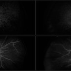
 Peripheral Retinal Ischemia
Apr 26 2018 by Olivia Rainey
Ultra-wide field fluorescein angiogram of a 55-year-old female with peripheral retinal ischemia affecting her left eye. CTA head and neck performed on 11/16/15 and showed calcified atherosclerotic plaque involving the intracranial internal carotid arteries with resulting luminal narrowing. Intracranial vertebral arteries have smooth luminal contours. CTA neck normal. Likely from internal carotid plaques. Sickle cell disease came back negative.
Photographer: Olivia Rainey
Imaging device: Optos California
Condition/keywords: fluorescein angiogram (FA), fluorescein leakage, left eye, Optos, retinal ischemia, ultra-wide field imaging
-
 Retinal Ischemia
Jul 11 2018 by Sarah Oelrich
Retinal Ischemia, PDR
Photographer: Sarah Oelrich CRA, Southeastern Retina Associates, Knoxville TN
Imaging device: Optos 200tx
Condition/keywords: ischemia
-
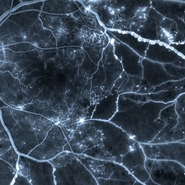 Venous Beading
Apr 30 2021 by Shivani Reddy, MD
This is a fluorescein angiogram image capturing a beautiful example of different stages of venous beading in diabetic retinopathy all in one frame. This patient also has various microangiopathic findings including microaneurysms, venous loops and capillary dropout. This patient is a 41 y/o male with a history of type 1 diabetes, presenting for his first eye exam in years.
Imaging device: Optos FA
Condition/keywords: capillary dropouts, nonproliferative diabetic retinopathy, proliferative diabetic retinopathy (PDR), retinal ischemia, venous beading
-
 Retinal Ischemia Secondary to Diabetic Retinopathy
Aug 29 2018 by Olivia Rainey
Fluorescein angiogram series of a 57-year-old male patient with proliferative diabetic retinopathy of the right eye. Patient has delayed AV transit with significant retinal ischemia and retinal capillary nonperfusion. The ischemia is extensive resulting in neovascularization of the iris and consequently neovascular glaucoma.
Photographer: Olivia Rainey
Imaging device: Optos
Condition/keywords: diabetes, disc hyperfluorescene, fluorescein angiogram (FA), non-perfusion, Optos, proliferative diabetic retinopathy (PDR), retinal ischemia, ultra-wide field imaging, vitreous hemorrhage
-
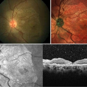 Foveal Thinning Post Blunt Trauma
Aug 25 2018 by Dhaivat Shah
28-year-old male. Post blunt trauma with tennis ball. Fundus color photo shows large area of retinal thinning. Multi color image shows dull red color over fovea, depicting thinning. SD-OCT shows inner retinal ischemia and foveal thinning with early macular hole formation.
Imaging device: Spectralis
Condition/keywords: blunt trauma
-
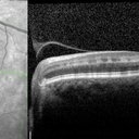 Paracentral Acute Middle Maculopathy (PAMM)
Oct 22 2019 by Jeffrey G. Gross, MD, FASRS
OCT of 75-year-old white male with 6 day history of acute vision loss. 20/40
Photographer: Tammy McLaughlin
Imaging device: Heidelberg Spectralis
Condition/keywords: paracentral acute middle maculopathy, retinal ischemia
-
 Central Retinal Artery Occlusion with Cilioretinal Sparing
Oct 28 2020 by Fang Helen Mi
Wide-field Clarus photography showing diffuse retinal ischemia and edema, with sparing of the cilioretinal artery region.
Condition/keywords: central retinal artery occlusion (CRAO), cilioretinal sparing
 Loading…