Search results (436 results)
-
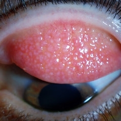 Giant Papillary Conjunctivitis, Left Upper Eyelid
Jul 22 2013 by Jason S. Calhoun
Contact lens wearer in for exam. Has rough feeling underneath both eyelids. Patient thought it was through SCL wear. Patient VA was 20/20. right eye, 20/30, left eye. Underneath the left upper eyelid, you can see papillary inflammation and redness.
Photographer: Jason S. Calhoun, Department of Ophthalmology, Mayo Clinic Jacksonville, Florida
Imaging device: TOPCON D-90 SL NIKON CAMERA
Condition/keywords: giant papillary conjunctivitis
-
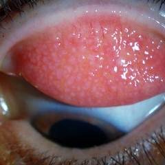
 Giant Papillary Conjunctivitis
Dec 13 2013 by Jason S. Calhoun
Patient wears soft contact lenses complained of irritation when the SCL would move. Inverted eyelid in both eyes and there was papillary +2 underneath the eyelid.
Photographer: Jason S. Calhoun, Ophthalmic Photographer, Department of Ophthalmology, Mayo Clinic Jacksonville
Imaging device: TOPCON D-90 SL NIKON CAMERA
Condition/keywords: giant papillary conjunctivitis
-
 Giant Papillary Conjunctivitis, Left Upper Eyelid
Jul 22 2013 by Jason S. Calhoun
Contact lens wearer, in for exam. Has rough feeling underneath both eyelids. Patient thought it was through SCL wear. Patient VA was 20/20. right eye, 20/30, left eye. Underneath the left upper eyelid, you can see papillary inflammation and redness.
Photographer: Jason S. Calhoun, Department of Ophthalmology, Mayo Clinic Jacksonville, Florida
Imaging device: TOPCON D-90 SL NIKON CAMERA
Condition/keywords: giant papillary conjunctivitis
-
 Anterior Chamber Intraocular Lens
Sep 20 2012 by Jeffrey G. Gross, MD, FASRS
AC-IOL, s/p PPV, lensectomy for dislocated crystalline lens, 20/20
Condition/keywords: anterior chamber, dislocated crystalline lens, intraocular lens (IOL), lensectomy
-
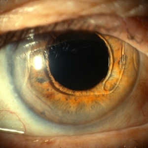
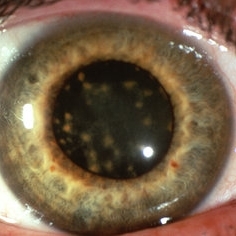 Siderosis
May 2 2013 by Henry J. Kaplan, MD
Iron deposition in the iris epithelium and sphincter and on lens epithelium in the same patient ; #2.
Condition/keywords: siderosis
-
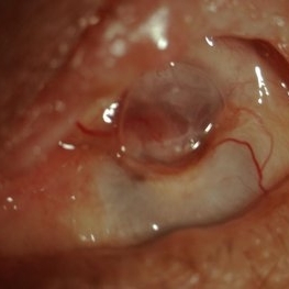
 Elschnig's Pearls
Sep 1 2015 by René Hernán Parada Vásquez
Fundus photograph of 58-year-old male with Elschnig's pearls, you can see the transparent clusters formed by proliferation of epithelial lens cells found in the remains of the capsule of the crystalline lens following cataract surgery.
Photographer: Parada René, ESO, Guatemala.
Condition/keywords: cataract surgery
-
 HLA-B27 Associated Uveitis
Jun 4 2014 by Henry J. Kaplan, MD
Severe anterior uveitis with fibrinous reaction and hypopyon formation related to HLA-B27. Notice the membrane on the lens surface.
Condition/keywords: acute anterior uveitis, HLA-B27, hypopyon
-
 Giant Papillary Conjunctivitis
Dec 13 2013 by Jason S. Calhoun
Patient wears soft contact lenses complained of irritation when the SCL would move. Inverted eyelid in both eyes and there was papillary +2 underneath the eyelid.
Photographer: Jason S. Calhoun, Ophthalmic Photographer, Department of Ophthalmology, Mayo Clinic Jacksonville
Imaging device: TOPCON D-90 SL NIKON CAMERA
Condition/keywords: giant papillary conjunctivitis
-
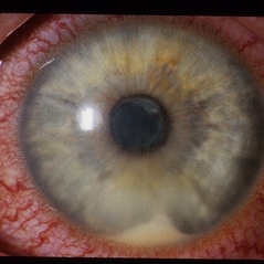
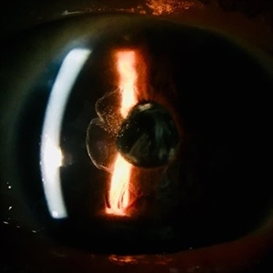
 Pseudoexfoliation Syndrome Ring
Sep 17 2015 by Jason S. Calhoun
Pseudoexfoliation syndrome ring on the lens capsule.
Photographer: Jason Calhoun, Mayo Clinic Jacksonville, Department of Opthalmolgy
Imaging device: Haag Striet Cannon D7
Condition/keywords: pseudoexfoliation glaucoma, pseudoexfoliation of lens capsule, pseudoexfoliation syndrome
-
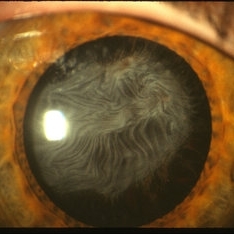
 Pseudoexfoliation of Lens Capsule
Oct 23 2012 by Larry Halperin, MD
Pseudoexfoliation of lens capsule
Condition/keywords: pseudoexfoliation of lens capsule
-
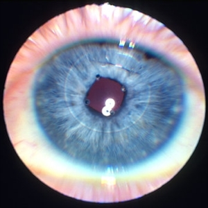 Iris clip IOL
Jan 11 2013 by Alex P. Hunyor, MD
Iris fixated IOL - note stainless steel suture.
Condition/keywords: intraocular lens (IOL), iris clip intraocular lens
-
 Anterior Capsule Opacification in Eye
Oct 11 2012 by Jeffrey G. Gross, MD, FASRS
Anterior capsule opacification in eye, s/p PPV lensectomy, without IOL.
Condition/keywords: anterior capsule opacification, lensectomy, without intraocular lens
-
 Optos Picture With Speculum: Dislocated Natural Lens
Oct 9 2018 by John S. King, MD
55-year-old white female with history of pathologic myopia+, lattice (laser), SB OU (1990s), and dislocated natural lenses OU that had been watched for years. In the fellow eye she developed phacolytic glaucoma and a PPV, PPL was performed. Plan for both eyes are monitoring. I wanted to get a good picture of her lens today with the optos machine, as the other pics had artifact from the lower lid. It worked out well to use a speculum in the left eye. Vision cc is 20/400 J1+ OD and 20/40 J2 OS; aphakic OU; vitreous clear OD; dislocated lens OS (see pic); retinas attached.
Photographer: Maisee Yang
Imaging device: Optos California
Condition/keywords: dislocated crystalline lens, pathologic myopia, scleral buckle, staphyloma
-
 Lens Feathering
Nov 9 2016 by Nimrod Dar
26-year-old man with lens feathering a day after a pars plana vitrectomy for rhegmatogenous retinal detachment.
Photographer: Nimrod Dar, M.D, Meir Ophthalmology Department
Condition/keywords: lens feathering, vitrectomy
-
 Traumatic Lens Dislocation Over Disc
Oct 19 2012 by Larry Halperin, MD
Traumatic lens dislocation over disc
Condition/keywords: lens dislocation
-
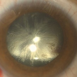
 Pseudoexfoliation Syndrome Ring
Sep 17 2015 by Jason S. Calhoun
Pseudoexfoliation syndrome ring on the lens.
Photographer: Jason Calhoun, Mayo Clinic Jacksonville, Department of Opthalmolgy
Imaging device: Haag Striet Cannon D7
Condition/keywords: pseudoexfoliation glaucoma
-
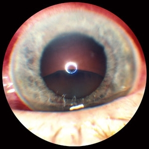
 Dislocated IOL With PI
Jul 14 2013 by Jason S. Calhoun
Subluxated lens with PI at 12-o'clock.
Photographer: Jason S. Calhoun, Department of Ophthalmology, Mayo Clinic Jacksonville, Florida
Imaging device: TOPCON D-90 SL NIKON CAMERA
Condition/keywords: dislocated posterior chamber intraocular lens (PCIOL), peripheral iridotomy
-
000---thumb.jpg/image-square;max$300,300.ImageHandler) Dropped IOL into the Vitreous Cavity
Dropped IOL into the Vitreous Cavity
Oct 7 2012 by Young Hee Yoon, MD, PhD
Fundus photograph of an 70-year-old man with a history of cataract operation 20 years ago. He visited our clinic with decreased visual acuity for 2 days.
Photographer: Yoon-hwa Kim, Asan Medical Center
Imaging device: Optomap, optos
Condition/keywords: intraocular lens dislocation
-
 Bergmeister's Papilla
Sep 29 2020 by Dhaivat Shah
Bergmeister's papilla is a small tuft of glial tissue which arises from the center of the optic disc, and represents a remnant of the fetal hyaloid artery. The hyaloid artery provides nutrition to the lens during development, and runs forward to the lens from the optic disc. At birth the hyaloid artery regresses, and is normally completely regressed by the time of birth. Bergmeister's papilla is frequently observed as an incidental clinical finding if this artery has an incomplete regression posteriorly. However, in the severe forms it can be associated with cataracts, persistence of the primitive vitreous, microphthalmia, vitreous hemorrhages and sometimes tractional retinal detachment, due to contraction of the residual fibro vascular tissue. Therefore, careful monitoring of vitreous thickening in the peripapillary areas, both by examining the ocular fundus, and especially by SD-OCT, is of considerable importance. Here we have one such of a 30 year old young male who came in for a routine checkup, in whom we noted a Bergmeister’s papilla. Due to its benign nature, patient was reassured and was asked to follow up yearly.
Condition/keywords: Bergmeister's Papillae
-
 Traumatic Dislocated Crystalline Lens
Oct 1 2012 by Jeffrey G. Gross, MD, FASRS
Traumatic dislocated crystalline lens.
Condition/keywords: dislocated crystalline lens
-
 Coats' Disease
Apr 27 2018 by Brenda Fallas
3-year-old boy with unilateral Coats' Disease fundus photo.
Photographer: Brenda Fallas, Bascom Palmer Eye Institute, Miami, FL
Imaging device: Retcam III 130 degree lens
Condition/keywords: Coats' disease, color fundus photograph, retinal telangiectasia
-
 Panophthalmitis
Jul 12 2014 by Philip J. Polkinghorne, MD
A 85-year-old lady who presented with an eroding intraocular lens. She had been initially treated with herbal medicines which failed to control the infection.
Photographer: Philip Polkinghorne
Condition/keywords: cataract surgery, endophthalmitis, panophthalmitis
-
 Dislocated PCIOL
Sep 14 2012 by Sharon Fekrat, MD FACS FASRS
Dislocated PCIOL
Photographer: Jim Crowell, Duke University Eye Center, Durham, NC
Condition/keywords: dislocated posterior chamber intraocular lens (PCIOL), posterior chamber intraocular lens (PCIOL)
-
---thumb.JPG/image-square;max$300,300.ImageHandler) Retinal Detachment With Dislocated IOL Lens
Retinal Detachment With Dislocated IOL Lens
Jun 30 2013 by Jason S. Calhoun
47-year-old male who had trauma to the right eye. Patient had retinal detachment surgery in the past (scleral buckle), to the right eye. Patient came in with another retinal detachment with dislocated PC IOL lens. Notice the haptics tearing the retina. Patient underwent vitrectomy with gas exchange. VA was hand motion 1 day post-op.
Photographer: Jason S. Calhoun, Mayo Clinic Jacksonville, Florida
Condition/keywords: dislocated posterior chamber intraocular lens (PCIOL), retinal tear
-
 Vitreous in AC
Jan 9 2018 by Andrea Arriola-Lopez, MD MSc
78-year-old male. Vision loss in OD. IOP 18 mmHg. Subluxated PCIOL and vitreous in anterior chamber was found.
Photographer: Andrea E. Arriola López MD MS
Condition/keywords: anterior chamber, dislocated intraocular lens (IOL), vitreous
 Loading…