Search results (436 results)
-
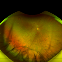
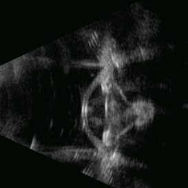
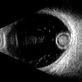 Morgagnian Ghost in the Deep
Jul 3 2025 by Gustavo Uriel Fonseca Aguirre
This B-mode para-axial ultrasound scan shows a posteriorly dislocated lens with cortical liquefaction, a dense nucleus, and an intact capsular bag. Vitreous bands are visible extending from the anterior to posterior segments. These findings were bilateral and not associated with trauma or prior surgery.
Photographer: Gustavo U. Fonseca Aguirre, Hospital Conde de Valenciana, Ciudad de México
Condition/keywords: ectopia lentis, morgagnian cataract
-
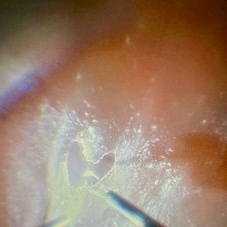 Love Through the Lens of Retinal Detachment
Jun 27 2025 by Claudio Brancato
The image depicts a case of rhegmatogenous retinal detachment where the vitreous was extremely adherent to the retina. The primary surgeon was performing membrane peeling using a surgical loop, while the assisting surgeon was captivated by the intricate procedure. In a moment of affectionate dedication, the primary surgeon carefully peeled the membrane to form a heart shape, symbolizing both his passion for surgery and perhaps a personal gesture towards the assisting surgeon. This delicate and precise maneuver highlights the complexity and artistry involved in vitreoretinal surgery, showcasing the blend of technical skill and emotional expression within the operating room.
Photographer: Claudio Brancato, ARNAS CIVICO Hospital, Palermo, Italy
Imaging device: Zeiss Artevo 800
Condition/keywords: finesse, peeling, proliferative vitreoretinopathy (PVR), Retina detachment
-
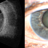
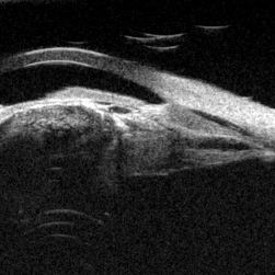
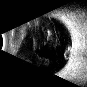
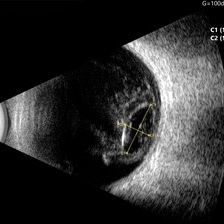 Ocular B-scan Ultrasound (Longitudinal Scan M6, gain 100 dB)
Jun 26 2025 by Hector Gabriel Moreno Solano, MD, MHA
B-scan ultrasound was performed in longitudinal section M6 with a gain of 100 dB. A hyperechoic structure with posterior acoustic shadowing is observed, consistent with lens luxation and condensed vitreous bands adjacent to the lens. The dislocated lens measures approximately 9.54 mm x 4.62 mm. The study was conducted following blunt ocular trauma caused by a golf ball. The remaining vitreous cavity appears anechoic, with no evidence of retinal detachment or other structural abnormalities in this section.
Photographer: Hector Gabriel Moreno Solano, Instituto Mexicano de Oftalmología “IMO I.A.P”
Imaging device: Quantel Medical
Condition/keywords: B scan ultrasound, lens luxation, ocular trauma
-
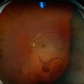
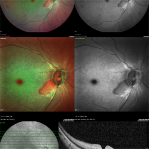 CRAO With Cilio-retinal Sparing-MMI
Jun 25 2025 by Shivankar Sen, MS, FVRS
A 41 year old male came with complaints of Right eye blurring of vision since a day associated with watering and redness. He had no systemic illness, though gave a history of fall from bike 1 month back at the time of which he had blunt force trauma to the right side of the face. BCVA was 3/60, less than N36 in the right eye and 6/6, N6 in the left eye. Right eye had Marcus Gunn Pupil with clear lens, Left eye was within normal limits. IOP was normal; 16 in OD and 18 in OS. Retina evaluation revealed CRAO in the right eye with cilio-retinal artery sparing. Left eye was unremarkable Image Details Left to Right (Top 2 rows) Multicolor Reflectance Image (Blue-green enhanced 55 degree) revealing cilioretinal spared retinal stroma and a characteristic Cherry Red Spot; Green Reflectance showing corresopnding dark gray area with spared perfusion and black spot consistent with Cherry Red Spot on multicolor 2nd Row - 35 degree image (Multicolor Standard Reflectance and Green Reflectance) 3rd Row - SD-OCT revealing acute moderate CRAO findings with Middle retinal layer opacification and prominent middle limiting membrane (p-MLM) sign; Inner retinal layer opacification and prominent retinal pigment epithelium at the fovea with Diminished inner retinal layer stratification
Photographer: Gayathri M S
Imaging device: Heidelberg Spectralis HRA+OCT
Condition/keywords: CRAO with cilioretinal sparing, multicolor, multimodal imaging, OCT biomarkers, reflectance
-
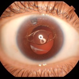
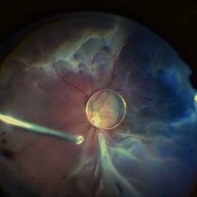
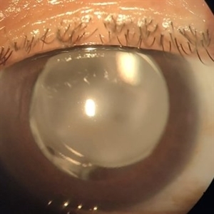
 Dislocated Cataractous Lens
Jun 19 2025 by Mrinali Gupta, MD, FASRS
Intraoperative image of a chronically dislocated cataractous lens. The patient underwent pars plana vitrectomy, lensectomy, and placement of an anterior chamber intraocular lens, with improvement in vision from Count Fingers to 20/20 without correction.
Photographer: Mrinali Gupta, MD
Imaging device: Intraoperative surgical video (Zeiss Lumera scope, Resight lens)
Condition/keywords: dislocated crystalline lens
-
 Massive Choroidal Melanoma
Jun 18 2025 by Corey R Lacher, MD
A 57-year-old patient presented with no light perception vision in her right eye. B-scan ultrasonography revealed evidence of a large choroidal melanoma. External photography demonstrated detached retina visible just posterior to the lens. The patient subsequently underwent enucleation, and histopathologic examination confirmed the diagnosis of choroidal melanoma. The tumor measured 24 mm anteroposteriorly, 24 mm horizontally, and 25 mm vertically.
Photographer: Beth Malpica
Condition/keywords: choroidal melanoma
-
 Commotio Retinae
Jun 10 2025 by CUI YUELING
The patient presented 2 hours after sustaining a left eye injury caused by a stick. Visual acuity in the left eye was 0.2 without improvement upon correction, and intraocular pressure measured 15 mmHg. Examination of the anterior segment revealed ciliary conjunctival injection accompanied by patchy subconjunctival hemorrhage. The corneal surface remained smooth, and the anterior chamber was deep with hyphema characterized by blood-tinged aqueous humor predominantly settled inferiorly. The pupil was slightly irregular, approximately 3 mm in diameter, with a superotemporal notch; pupillary light reflex was intact. The lens appeared clear. Fundus examination showed well-defined optic disc margins with normal coloration and a cup-to-disc ratio of 0.2. Retinal arteries and veins were normally distributed with an artery-to-vein ratio of 2:3. At the posterior pole, the foveal reflex exhibited concentric ripple-like changes centered on the fovea, accompanied by localized pigment attenuation and reduced reflex intensity. Irregular reflectivity was noted in the superotemporal and inferotemporal nerve fiber layers.
Photographer: Yueling Cui
Imaging device: Zeiss Clarus 500
Condition/keywords: commotio retinae
-
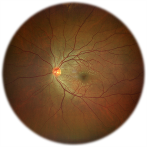
 Dislocated Intraocular Lens
Jun 4 2025 by Aditya S Kelkar, MS, FRCS, FASRS,FRCOphth
Fundus photograph of a 79-year-old man with a posteriorly dislocated intraocular lens in the inferior quadrant.
Photographer: Optom Chandrakanta Bhandare, National Institute of Ophthalmology, Pune
Imaging device: Optos Daytona
Condition/keywords: dislocated intraocular lens (IOL)
-
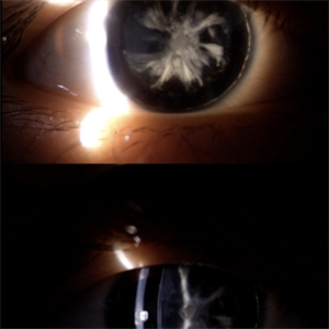
 Anterior Iris Claw Artisan Lens
May 14 2025 by Moazzam Parvez
Anterior segment image of a 40 year old gentleman with a anteriorly placed iris claw lens post retinal detachment surgery.
Photographer: Dr Moazzam Parvez, Netralayam , Kolkata
Imaging device: Topcon DC-4
Condition/keywords: Anteriorly placed iris claw lens
-
 Snowflake Lens
May 9 2025 by BENITO VERGARA, MD
Anterior polar cataract with subcapsular component and dense posterior plaque in a 20-year-old male with congenital aniridia. The image demonstrates a well-defined anterior polar opacity with associated subcapsular changes and a prominent posterior plaque, likely reflecting long-standing progression in the setting of congenital aniridia.
Photographer: Benito Vergara, Asociación Para Evitar la Ceguera en México.
Condition/keywords: aniridia, anterior subcapsular polar cataract
-
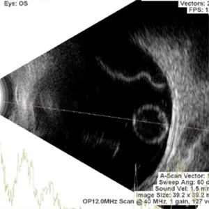 Pearl on a String
Apr 28 2025 by rohan jain
Ultrasound of LE of 22 years female showing dislocation of crystalline lens along with retinal detachment
Photographer: Dr. ROHAN JAIN
Condition/keywords: B scan ultrasound, dislocated crystalline lens, Retinal Detachment
-
 Cyclic Membrane
Apr 23 2025 by Gustavo Uriel Fonseca Aguirre
This UBM scan reveals pars planitis with characteristic findings: an inflammatory pupillary membrane, a cataractous lens, and cyclitic membrane causing ciliary body detachment and traction. The lens demonstrates spherical deformation due to zonular laxity from ciliary body traction.
Photographer: Gustavo U. Fonseca Aguirre, Hospital Conde de Valenciana, Ciudad de México
Condition/keywords: cyclic membrane, pars planitis
-
 Dislocated IOL
Apr 23 2025 by Anjana Mirajkar, MS Ophthalmology
A widefield imaging of the right eye of a 55 year old male showing dislocated IOL inferiorly.
Photographer: Dr. Anjana Mirajkar- HV desai eye hospital ,Pune
Imaging device: Optos
Condition/keywords: dislocated intraocular lens (IOL)
-
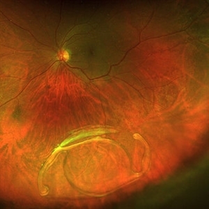
 Dislocation of the Crystalline Lens with a Retinal Detachment
Apr 21 2025 by Hrishikesh Naik, MS
An intraoperative screen grab shows a dislocation of the crystalline lens along with an associated rhegmatogenous retinal detachment in a case of Marfan’s syndrome. The case was managed by a combined PPV-SB procedure. A vitrectomy cutter is seen at the left.
Photographer: Hrishikesh Naik
Condition/keywords: intraoperative, lens dislocation, Marfan's syndrome, Retinal Detachment, vitrectomy
-
 PCR
PCR
Apr 17 2025 by Gustavo Uriel Fonseca Aguirre
B-mode transverse ultrasound scan of an eye with recent posterior capsule rupture during phacoemulsification shows hyperechoic punctate echoes in the vitreous (consistent with residual viscoelastic material) along with lens fragments in the subhyaloid space.
Photographer: Gustavo U. Fonseca Aguirre, Hospital Conde de Valenciana, Ciudad de México
Condition/keywords: Posterior capsule rupture (PCR)
-
 Posterior Polar Cataract
Apr 10 2025 by DR Rohit Gupta
52 year old male presented with the gradual. Painless, diminuition of vision. On slit lamp examination an onion peel appearance opacification of lens in central part was seen.
Photographer: Dr Rohit gupta
Condition/keywords: posterior capsule opacification, Posterior polar cataract
-
 Traumatic Posterior Capsular Rupture
Apr 9 2025 by Gustavo Uriel Fonseca Aguirre
Immersion B-mode ultrasound in a patient with blunt ocular trauma demonstrates an isolated posterior lens capsule rupture accompanied by phacodonesis.
Photographer: Gustavo U. Fonseca Aguirre, Hospital Conde de Valenciana, Ciudad de México
Condition/keywords: blunt trauma, Posterior Capsular Rupture
-
Eye of the Hurricane
Apr 9 2025 by Gustavo Uriel Fonseca Aguirre
Ultrasound biomicroscopy of a post-operative eye (status post trabeculectomy and phacoemulsification) reveals a patent ostium on the right side, along with an intraocular lens in position. A hyphema is observed displaying small convection currents, creating a circular motion pattern due to the temperature gradient between the iris and cornea. Notably, the blood flow can be seen circulating toward the trabeculectomy site.
Condition/keywords: hyphema, trabeculectomy
-
 Eye of the Hurricane
Apr 8 2025 by Gustavo Uriel Fonseca Aguirre
Ultrasound biomicroscopy of a post-operative eye (status post trabeculectomy and phacoemulsification) reveals a patent ostium on the right side, along with an intraocular lens in position. A hyphema is observed displaying small convection currents, creating a circular motion pattern due to the temperature gradient between the iris and cornea. Notably, the blood flow can be seen circulating toward the trabeculectomy site.
Photographer: Gustavo U. Fonseca Aguirre, Hospital Conde de Valenciana, Ciudad de México
Condition/keywords: Hyphema, trabeculectomy
-
 PCIOL Opacification
Mar 31 2025 by DR Rohit Gupta
A pseudophakic patient visiting after 6 months of cataract surgery. On slit lamp examination a complete hazy white PCIOL was seen, which is a rare complication after cataract surgery.
Photographer: Dr Rohit gupta
Imaging device: Samsung S21
Condition/keywords: posterior chamber intraocular lens (PCIOL)
-
 Ectopia Lentis
Mar 21 2025 by Gustavo Uriel Fonseca Aguirre
Cross-section of B-mode ultrasound showing the lens in the lower part of the vitreous cavity.
Photographer: Gustavo U. Fonseca Aguirre, Hospital Conde de Valenciana, Ciudad de México
Condition/keywords: ectopia lentis
-
 Lacteocrumenasia
Mar 11 2025 by Gustavo Uriel Fonseca Aguirre
A 75-year-old female with a history of cataract surgery with intraocular lens implantation 20 years ago presented with progressive visual loss. On slit lamp examination, opaque material was found in the capsular bag behind the intraocular lens. Ultrasound biomicroscopy revealed hyperechoic material contained in the temporal-posterior sector of the capsular bag corresponding to lacteocrumenasia.
Photographer: Gustavo U. Fonseca Aguirre, Hospital Conde de Valenciana, Ciudad de México
Condition/keywords: Lacteocrumenasia, ultrasound biomicroscopy
-
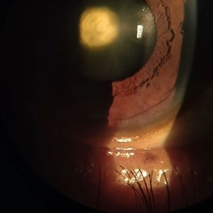
 Dislocated Lens
Jan 30 2025 by Kimberly Wakester
Fundus photograph of a 37-year-old man with an anteriorly dislocated lens in the left eye. The natural lens has displaced anteriorly in the AC secondary to trauma to the eye. There is also a Macular hole present with vitreous hemorrhage. Patient was recommended to proceed with lensectomy, iris repair and MH repair in the left eye.
Photographer: Kimberly Wakester, COA
Imaging device: Topcon TRC-50DX
Condition/keywords: dislocated lens, iridodialysis
-
 Macular Hole
Jan 30 2025 by Kimberly Wakester
Fundus photograph of a 37-year-old man with an anteriorly dislocated lens in the left eye. The natural lens has displaced anteriorly in the AC secondary to trauma to the eye. There is also a Macular hole present with vitreous hemorrhage. Patient was recommended to proceed with lensectomy, iris repair and MH repair in the left eye.
Photographer: Kimberly Wakester, COA
Imaging device: Optos California
Condition/keywords: dislocated lens, macular hole, vitreous hemorrhage
-
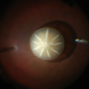
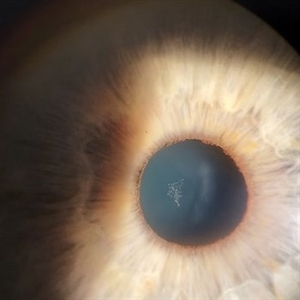 Epicapsular Stars
Jan 28 2025 by Korey Starkey
Epicapsular stars and cataract noted in natural lens of 68-year-old patient.
Photographer: Korey Starkey
Imaging device: Slit lamp camera
Condition/keywords: cataract, chicken tracks, epicapsular stars, slit lamp photography
 Loading…