Search results (18 results)
-
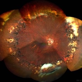
 Dislocated Crystalline Lens
Mar 19 2024 by Annaka Gooding
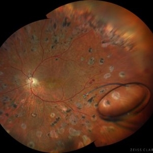
Ultra Wide field fundus photography of a 70 year old male who presented to clinic with a sudden increase of vision due to dropped crystalline lens secondary to severely dense cataract. Patient reported seeing a full black circle in his inferior visual field. Patient's visual acuity at time of visit was 20/100 with a +5.00 diopter lens. The physician recommended surgical intervention, and discussed surgery for PPV/PPL/IOL implantation with an ACIOL.
Photographer: Annaka Gooding, CPO
Imaging device: Optos California RGB
Condition/keywords: dislocated crystalline lens, fundus photography, inferior retina, OPTOS CALIFORNIA RGB, Right Eye, Ultra-wide field retinal imaging
-
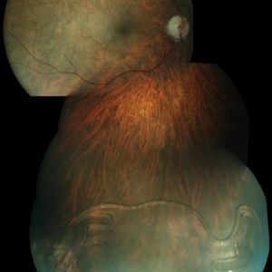
 Dislocated Lens, Posterior OD
Jan 26 2024 by Corey Grant
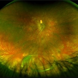
OPTOS California photo presents a 71 year old male patient with a dislocated lens, posterior in the right eye. Presented on 1/26/24 with posteriorly dislocated SN60WF with a Soemmerring ring. Associated retinal hemorrhage within retinoschisis as well. This will result in a PPV/IOL exchange/SFIOL/STK for the right eye.
Photographer: Corey Grant, Ophthalmic Imager, Retina Specialist of Michigan
Imaging device: OPTOS California
Condition/keywords: color photo, IOL, OD, Optos, OPTOS CALIFORNIA, pars plana vitrectomy (PPV), retina
-
 Intraocular lens luxated to the vitreous cavity
Jun 24 2023 by Mariam Cernichiaro-Espinosa, MD
Three-piece intraocular lens luxated to the vitreous cavity in a patient with photocoagulated diabetic retinopathy after blunt trauma.
Photographer: Mariam Cernichiaro-Espinosa, Asociación para Evitar la Ceguera en México, I.A.P. Mexico City, Mexico.
Imaging device: Zeiss Clarus
Condition/keywords: diabetic retinopathy, intraocular lense in vitreous, lens luxation
-
 Fraternal Twins
May 22 2023 by Gustavo M. Hüning, MD, MBA, FASRS
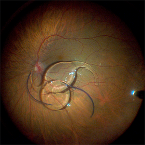
Intrasurgical photograph using a non-contact system and 3D visualization system of a 65-year-old woman who suffered an ocular trauma.
Photographer: Gustavo M. Hüning, Hüning Clínica do Olhar, Santa Maria - Brazil
Imaging device: Alcon Luxor combined with Alcon nGenuity
Condition/keywords: dislocated intraocular lens (IOL), implant, pars plana vitrectomy (PPV)
-
 Dislocated Lens
Apr 26 2023 by Chloe Hanifan
Ultra wide field fundus photograph of a 41-year-old male with a dislocated lens affecting his right eye. IOL noted inferior vitreous base and vitrectomy surgery for removal of IOL was recommended. Patient has history of retinitis pigmentosa as well. Patient's vision at the time of presentation was counting fingers at 2 feet.
Photographer: Chloe Hanifan
Imaging device: Optos California
Condition/keywords: dislocated lens, fundus photography, Optos, pseudocolor, retinitis pigmentosa, ULTRA WIDE FIELD
-
 Inflammatory pupillary membrane in patient with endophthalmitis
Jan 28 2023 by Kingston Rodolfo Ureña-Wong, MD, Opht, MSc
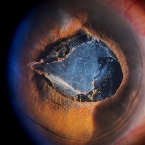
Anterior segment photography of a 54-year-old woman with post phacoemulsification endophthalmitis. She did not improve after first intravitreal antibiotics injection and develop an inflammatory pupillary membrane. After two vitrectomies, and a complete three intravitreal injections scheme, we decided to remove the intraocular lens and capsules.
Photographer: Marco Antonio Rubio-Atonal,UNAM, Asociación para evitar la ceguera en México
Imaging device: Zeiss Clarus 700
Condition/keywords: endophthalmitis, pupillary membranes
-
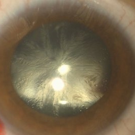
 Wrinkled Anterior Capsule 40X zoom
Feb 18 2023 by Ahmed Abbas Hashmi, OD
Imprint of Iris Pigmentation on Anterior Lens Surface with wrinkled anterior capsule
Photographer: Ahmed Abbas Hashmi
Condition/keywords: lens opacity
-
 Posteriorly dislocated IOL
Oct 22 2022 by Vishal Agrawal, MD, FRCS,FACS,FASRS
67 yr old male , post PPV for retinal detachment ( 5 years ) presented with sudden DOV . On examination posteriorly dislocated 4 loop haptic iol - bag complex was noted .
Photographer: Pankaj
Imaging device: CLARUS 700
Condition/keywords: dropped intraocular lens (IOL)
-
 Spontaneously Dropped Lens in a Congenital Rubella Syndrome
Apr 30 2022 by NEIFFER RABELO
Intraoperative photograph of a 68-year-old patient with congenital rubella syndrome and light perception visual acuity since childhood. The image shows a pigmentary retinopathy and the lens spontaneously displaced into the vitreous cavity. The patient sought care complaining of a total and sporadic loss of vision that was hindering her in daily tasks. Surgery was indicated to remove the lens.
Photographer: Rodrigo Dos Anjos Versiani - Retina Institute - Belo Horizonte - Brazil
Imaging device: ZEISS OPMI LUMERA 700
Condition/keywords: dropped nucleus, retina surgery, rubella retinopathy
-
 Traumatic Lens Drop in Vitreous
Dec 15 2020 by Manish Nagpal, MD, FRCS (UK), FASRS
Patient had come to us status post blunt trauma with the lens dislocated in inferior vitreous.
Photographer: Gayathri Mohan, Retina Fellow, Retina Foundation, Ahmedabad, India
Imaging device: Mirante CSLO
Condition/keywords: dropped nucleus, lens dislocation, traumatic cataract
-
 Dislocated-P/C IOL Bag Complex
Nov 27 2018 by Maria H. Berrocal, MD
85-year-old who underwent phaco IOL 15 years prior, who noticed loss of vision OS.
Photographer: Thaylan Calderon, Berrocal & Associates, San Juan, PR
Imaging device: Optos
Condition/keywords: dislocated intraocular lens (IOL)
-
 Optos Picture With Speculum: Dislocated Natural Lens
Oct 9 2018 by John S. King, MD
55-year-old white female with history of pathologic myopia+, lattice (laser), SB OU (1990s), and dislocated natural lenses OU that had been watched for years. In the fellow eye she developed phacolytic glaucoma and a PPV, PPL was performed. Plan for both eyes are monitoring. I wanted to get a good picture of her lens today with the optos machine, as the other pics had artifact from the lower lid. It worked out well to use a speculum in the left eye. Vision cc is 20/400 J1+ OD and 20/40 J2 OS; aphakic OU; vitreous clear OD; dislocated lens OS (see pic); retinas attached.
Photographer: Maisee Yang
Imaging device: Optos California
Condition/keywords: dislocated crystalline lens, pathologic myopia, scleral buckle, staphyloma
-
 Retinal Cavernous Hemangioma
Nov 30 2018 by Brenda Fallas
2-year-old female with retinal cavernous hemangioma.
Photographer: Brenda Fallas
Imaging device: Retcam 130 lens
Condition/keywords: cluster of grapes, retinal angioma
-
 Dislocated IOL in Vitreous Cavity
Apr 17 2018 by S. Natarajan, MD, FASRS, FRCS (GLASGOW) , FICO, D.Sc, FELA
Fundus photograph of an 61-year-old male with dislocated IOL in vitreous cavity.
Photographer: Ashwini Borde
Imaging device: Carl Zeiss 450 plus IR
Condition/keywords: dislocated intraocular lens (IOL)
-
 Coats' Disease
Apr 27 2018 by Brenda Fallas
3-year-old boy with unilateral Coats' Disease fundus photo.
Photographer: Brenda Fallas, Bascom Palmer Eye Institute, Miami, FL
Imaging device: Retcam III 130 degree lens
Condition/keywords: Coats' disease, color fundus photograph, retinal telangiectasia
-
 Bullous RD With Dislocated Lens
Apr 3 2018 by Navneet Mehrotra, DNB
Dislocated clear lens and associated retinal detachment in a young patient with Marfan's syndrome.
Photographer: Navneet Mehrotra
Imaging device: Sony 3 chip camera
Condition/keywords: dislocated crystalline lens, Marfan's syndrome
-
 IOL With BAG in Vitreous of Myopic Eye
Apr 14 2017 by Manish Nagpal, MD, FRCS (UK), FASRS
50-year-old male having myopia presented with a IOL in vitreous within its bag.
Photographer: Pooja Barot
Condition/keywords: intraocular lens (IOL), intraocular lense in vitreous, intraocular lense with bag, myopia
-
 Lens Feathering
Nov 9 2016 by Nimrod Dar
26-year-old man with lens feathering a day after a pars plana vitrectomy for rhegmatogenous retinal detachment.
Photographer: Nimrod Dar, M.D, Meir Ophthalmology Department
Condition/keywords: lens feathering, vitrectomy
 Loading…