Search results (436 results)
-
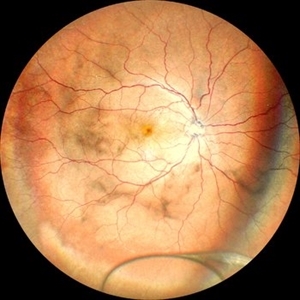
 Coats' Disease
Apr 27 2018 by Brenda Fallas
3-year-old boy with unilateral Coats' Disease fundus photo.
Photographer: Brenda Fallas, Bascom Palmer Eye Institute, Miami, FL
Imaging device: Retcam III 130 degree lens
Condition/keywords: Coats' disease, color fundus photograph, retinal telangiectasia
-
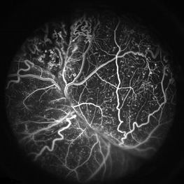 Coats' Disease FA
Apr 27 2018 by Brenda Fallas
3-year-old boy with unilateral Coats' Disease FA photo.
Photographer: Brenda Fallas, Bascom Palmer Eye Institute, Miami, FL
Imaging device: Retcam III 130 degree lens
Condition/keywords: Coats' disease, FA early phase, fluorescein angiogram (FA), retinal telangiectasia
-
 Retinoblastoma
Apr 27 2018 by Brenda Fallas
2-year-old boy with stage D+ retinoblastoma of the right eye.
Photographer: Brenda Fallas, Bascom Palmer Eye Institute, Miami, FL
Imaging device: RETCAM III 130 degree lens montage
Condition/keywords: tumor, tumor seeding
-
 Inflammatory pupillary membrane in patient with endophthalmitis
Jan 28 2023 by Kingston Rodolfo Ureña-Wong, MD, Opht, MSc
Anterior segment photography of a 54-year-old woman with post phacoemulsification endophthalmitis. She did not improve after first intravitreal antibiotics injection and develop an inflammatory pupillary membrane. After two vitrectomies, and a complete three intravitreal injections scheme, we decided to remove the intraocular lens and capsules.
Photographer: Marco Antonio Rubio-Atonal,UNAM, Asociación para evitar la ceguera en México
Imaging device: Zeiss Clarus 700
Condition/keywords: endophthalmitis, pupillary membranes
-
 Oil Bubbles
Apr 27 2018 by Mark Lazcano
Infrared photograph of 56-year-old male with retinal detachment oil on lens.
Photographer: Mark Lazcano, University of Miami, Bascom Palmer Eye Institute
Imaging device: Heidelberg Spectralis
Condition/keywords: silicone oil
-
 Dropped IOL
Apr 5 2018 by Mohamed Tawfik, MD
Intra operative photo of a case of dropped IOL after phaco+IOL.
Photographer: Mohamed A,Tawfik
Imaging device: Intra operative Photography Screen shoot
Condition/keywords: dropped intraocular lens (IOL)
-
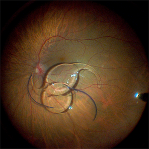
 Intraocular lens luxated to the vitreous cavity
Jun 24 2023 by Mariam Cernichiaro-Espinosa, MD
Three-piece intraocular lens luxated to the vitreous cavity in a patient with photocoagulated diabetic retinopathy after blunt trauma.
Photographer: Mariam Cernichiaro-Espinosa, Asociación para Evitar la Ceguera en México, I.A.P. Mexico City, Mexico.
Imaging device: Zeiss Clarus
Condition/keywords: diabetic retinopathy, intraocular lense in vitreous, lens luxation
-
 Spontaneously Dropped Lens in a Congenital Rubella Syndrome
Apr 30 2022 by NEIFFER RABELO
Intraoperative photograph of a 68-year-old patient with congenital rubella syndrome and light perception visual acuity since childhood. The image shows a pigmentary retinopathy and the lens spontaneously displaced into the vitreous cavity. The patient sought care complaining of a total and sporadic loss of vision that was hindering her in daily tasks. Surgery was indicated to remove the lens.
Photographer: Rodrigo Dos Anjos Versiani - Retina Institute - Belo Horizonte - Brazil
Imaging device: ZEISS OPMI LUMERA 700
Condition/keywords: dropped nucleus, retina surgery, rubella retinopathy
-
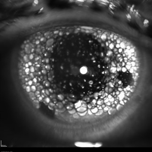
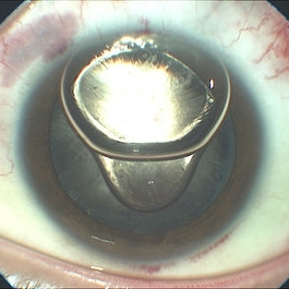
 Wrinkled Anterior Capsule 40X zoom
Feb 18 2023 by Ahmed Abbas Hashmi, OD
Imprint of Iris Pigmentation on Anterior Lens Surface with wrinkled anterior capsule
Photographer: Ahmed Abbas Hashmi
Condition/keywords: lens opacity
-
 Dropped Crystalline Lens
Mar 8 2019 by Abdulaziz A. Alshamrani, MD
A 15-year-old female with congenital glaucoma complaining of acute diminution of vision after a blunt trauma.
Condition/keywords: crystalline lens, dropped nucleus, ora serrata
-
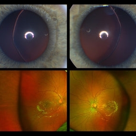 Ectopia Lentis
Jan 21 2021 by Jamin S. Brown, MD
This image serial demonstrates a patient with simple ectopia lentis. Anterior segment photographs in the upper panel demonstrate nasally subluxated crystalline lenses. Widefield fundus photography shows a "pseudo-buckle" which is the result of an optical effect due to the lens subluxation (artifactual image enlargement). Also note the juvenile macular reflex in this young patient. Ectopia lentis can present isolated ("simple") or in combination with various systemic defects (Marfan's syndrome, Weil-Marchesani syndrome or Ehlers-Danlos syndrome to name a few). Isolated ectopia lentis can be hereditary and causative genes have been identified as ADAMTSL4 located on chromosome 4 and FBN1 gene located on chromosome 15. Defects in the genes cause weakness in the zonular fibers which can lead to lens dislocation. Lastly, various ocular disorders such as Aniridia, Axenfeld-Rieger, Pseudoexfoliation or Trauma may also result in lens dislocation or subluxation.
Photographer: Stefanie Palmer CRA, Retina Vitreous Surgeons of CNY
Condition/keywords: dislocated lens, ectopia lentis
-
Eye of the Hurricane
Apr 9 2025 by Gustavo Uriel Fonseca Aguirre
Ultrasound biomicroscopy of a post-operative eye (status post trabeculectomy and phacoemulsification) reveals a patent ostium on the right side, along with an intraocular lens in position. A hyphema is observed displaying small convection currents, creating a circular motion pattern due to the temperature gradient between the iris and cornea. Notably, the blood flow can be seen circulating toward the trabeculectomy site.
Condition/keywords: hyphema, trabeculectomy
-
 Eye of the Hurricane
Apr 8 2025 by Gustavo Uriel Fonseca Aguirre
Ultrasound biomicroscopy of a post-operative eye (status post trabeculectomy and phacoemulsification) reveals a patent ostium on the right side, along with an intraocular lens in position. A hyphema is observed displaying small convection currents, creating a circular motion pattern due to the temperature gradient between the iris and cornea. Notably, the blood flow can be seen circulating toward the trabeculectomy site.
Photographer: Gustavo U. Fonseca Aguirre, Hospital Conde de Valenciana, Ciudad de México
Condition/keywords: Hyphema, trabeculectomy
-
 Fraternal Twins
May 22 2023 by Gustavo M. Hüning, MD, MBA, FASRS
Intrasurgical photograph using a non-contact system and 3D visualization system of a 65-year-old woman who suffered an ocular trauma.
Photographer: Gustavo M. Hüning, Hüning Clínica do Olhar, Santa Maria - Brazil
Imaging device: Alcon Luxor combined with Alcon nGenuity
Condition/keywords: dislocated intraocular lens (IOL), implant, pars plana vitrectomy (PPV)
-
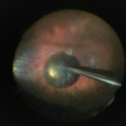 PPV retained cataract
Apr 19 2023 by Denica Rodriguez
A 46-year-old male with hypermature dense cataract. Patient got a piece of metal in his eye when he was 5 years old and was not able to see since. Patient was having cataract surgery and phacodonesis was present. The lens dropped to the back of the eye. Patient had to have another surgery to do vitrectomy. The lens removal was done with a fragmatome handpiece.
Photographer: Denica Rodriguez COA, ST
Imaging device: Zeiss Microscope with resight
Condition/keywords: cataract, dropped nucleus, fragmatome, lens capsule, ocular trauma, pars plana vitrectomy (PPV), retained lens fragments, Retina, retina surgery, traumatic cataract
-
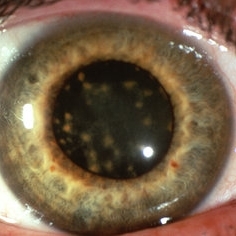
 Pseudoexfoliation Syndrome
Sep 17 2015 by Jason S. Calhoun
Pseudoexfoliation on lens.
Photographer: Jason Calhoun, Mayo Clinic Jacksonville, Department of Opthalmolgy
Imaging device: Haag Striet Cannon D7
Condition/keywords: pseudoexfoliation glaucoma, pseudoexfoliation of lens capsule
-
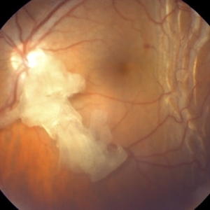 Retained Lens Fragment
Mar 2 2014 by Homayoun Tabandeh, MD, FASRS
Retained lens fragment, choroidal detachment, and serous retinal detachment post cataract surgery
Condition/keywords: retained lens fragments
-
 Siderosis
May 2 2013 by Henry J. Kaplan, MD
Iron deposition in the iris epithelium and sphincter and on lens epithelium in the same patient ; #2.
Condition/keywords: siderosis
-
 4 Point Scleral Fixation Akreos AO60 With Gore Tex Suture
May 21 2021 by Jesus Lozano, MD
Anterior segment photo of a 54-year-old man after 4 point scleral fixation Akreos AO60 with Gore Tex suture plus PPV who had a severe traumatic iris defect and was aphakic after ocular trauma.
Photographer: Luigi Zinn, Hadassah Medical Center, Jerusalem.
Condition/keywords: aphakia, cornea rupture, lens, penetrating trauma
-
 Anterior Segment Gas Bubble and PFC Interface
Jun 21 2018 by Maria Stephanie R. Jardeleza, MD
Anterior segment photographs of 30-year-old male who underwent superior rhegmatogenous retinal detachment repair with intraocular gas tamponade. Perfluorocarbon was used to flatten the macula to prevent a macular fold and was removed during PFC/air exchange. Post operative week two visit shows gas migration into the anterior chamber with retained PFC on the posterior aspect of the gas bubble/anterior surface of the lens. Patient had been maintaining face down positioning.
Photographer: Andy Zepeda, COA, Retina Clinic, San Antonio Eye Center, San Antonio, TX
Condition/keywords: retained perfluorocarbon, vitreous substitutes
-
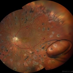
 Ciliary Body Melanoma
Nov 2 2024 by Virginia Gebhart
53 year old male with a large mass behind the lens as well as prominent scleral vessels. Clinical exam and ultrasound findings consistent with melanoma. Pt will be scheduled for enucleation pending CT scan results. Edit: Sadly patient has canceled all appointments and has requested no further contact
Photographer: Virginia Gebhart, Retina Consultants of Carolina
Imaging device: Optos California
Condition/keywords: ciliary body mass, ciliary body melanoma, ciliary body tumor
-
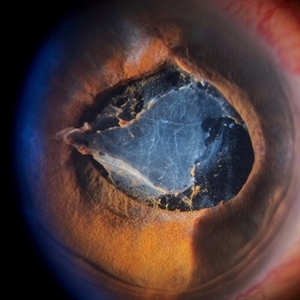
 Dislocated Brown Cataract with a Chorioretinal Coloboma
Sep 8 2021 by Ram Sudarshan
A 44 year-old male with dislocated brown cataract along with a chorioretinal coloboma.
Photographer: Dr.Sivadarshan
Condition/keywords: Brown cataract, chorioretinal coloboma, d, dislocated lens
-
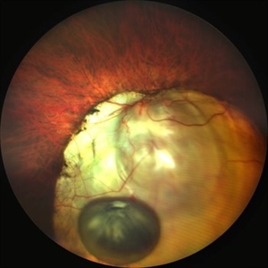
 Dislocated Cataractous Lens
Feb 11 2024 by Anjana Mirajkar, MS Ophthalmology
A wide field image of LE of a 40 year old male showing inferior dislocation of crystalline lens which is cataractous in vitreous cavity.
Photographer: Dr. Anjana Mirajkar -Retina Foundation, Ahmedabad
Imaging device: Mirante-Nidek
Condition/keywords: dislocated crystalline lens
-
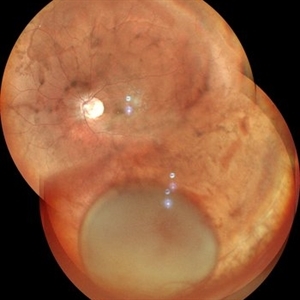
 Dislocated Crystalline Lens
Mar 19 2024 by Annaka Gooding
Ultra Wide field fundus photography of a 70 year old male who presented to clinic with a sudden increase of vision due to dropped crystalline lens secondary to severely dense cataract. Patient reported seeing a full black circle in his inferior visual field. Patient's visual acuity at time of visit was 20/100 with a +5.00 diopter lens. The physician recommended surgical intervention, and discussed surgery for PPV/PPL/IOL implantation with an ACIOL.
Photographer: Annaka Gooding, CPO
Imaging device: Optos California RGB
Condition/keywords: dislocated crystalline lens, fundus photography, inferior retina, OPTOS CALIFORNIA RGB, Right Eye, Ultra-wide field retinal imaging
-
 Dislocated Intraocular Lens
Nov 15 2024 by Tejaswita Verma
Fundus image of a spontaneously posteriorly dislocated IOL 10 years following surgery. Other eye had a subluxated opacified IOL.
Photographer: DR. TEJASWITA VERMA
Imaging device: MIRANTE
Condition/keywords: dislocated intraocular lens (IOL)
 Loading…