Search results (436 results)
-
 4 Point Scleral Fixation Akreos AO60 With Gore Tex Suture
May 20 2021 by Jesus Lozano, MD
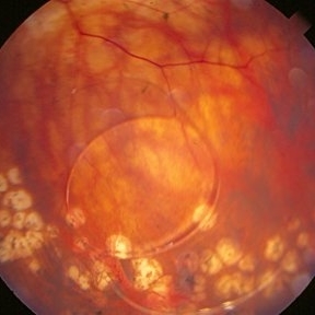
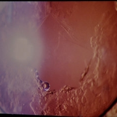
Optos Silverstone fundus image of a 54-year-old man after 4 point scleral fixation Akreos AO60 with Gore Tex suture plus PPV who had a severe traumatic iris defect and was aphakic after ocular trauma.
Photographer: Yair Bet Yosef, Hadassah Medical Center. Israel
Imaging device: Optos Silverstone
Condition/keywords: aphakia, globe perforation, lens, pars plana vitrectomy (PPV), penetrating trauma, vitreous hemorrhage
-
 4 Point Scleral Fixation Akreos AO60 With Gore Tex Suture
May 21 2021 by Jesus Lozano, MD
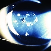
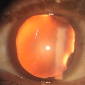
Anterior segment photo of a 54-year-old man after 4 point scleral fixation Akreos AO60 with Gore Tex suture plus PPV who had a severe traumatic iris defect and was aphakic after ocular trauma.
Photographer: Luigi Zinn, Hadassah Medical Center, Jerusalem.
Condition/keywords: aphakia, cornea rupture, lens, penetrating trauma
-
 Air Bubbles behind Posterior Capsule and Intra-Ocular Lens in the Anterior Hyaloid Phase during Vitrectomy Retina Surgery
Apr 28 2023 by Veer Singh, MS, FVRS, FMRF, FICO (Retina)
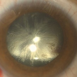
Air Bubbles behind Posterior Capsule and Intra-Ocular Lens in the Anterior Hyaloid Phase during Vitrectomy Retina Surgery | Intra-Operative Still
Photographer: Dr. Veer Singh
Condition/keywords: air bubbles, anterior hyaloid, hyaloid, Intra-Operative Still, lens, vitrectomy
-
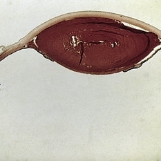
 Extruded Lens
-
 Eyeball Dissection
Jan 12 2021 by Niu Yongyi
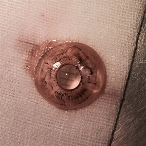
Dissecting an eyeball. Complete lens and vitreous body.
Photographer: Yongyi Niu, Guangdong Provincial People's Hospital
Condition/keywords: eyeball, lens, vitreous
-
 Eyeball Dissection
Jan 12 2021 by Niu Yongyi
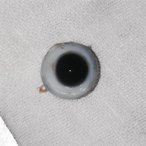
Dissecting an eyeball. Complete lens and vitreous body.
Photographer: Yongyi Niu, Guangdong Provincial People's Hospital
Condition/keywords: eyeball, lens, vitreous
-
 Eyeball Dissection
Jan 12 2021 by Niu Yongyi
Dissecting an eyeball. Complete lens and vitreous body.
Photographer: Yongyi Niu, Guangdong Provincial People's Hospital
Condition/keywords: eyeball, lens, vitreous
-
 Lens Bite
May 30 2016 by Andrea Arriola-Lopez, MD MSc
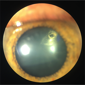
32-year-old man underwent a vitrectomy due hemovitreous. There was a dense retrolental membrane, while aspirating it, a little bite to lens was done. We decided not to performed lensectomy. VA is 20/30 two months later.
Photographer: Andrea E. Arriola-López MD MSc
Condition/keywords: lens, lens opacity
-
 Lens displacement
Jun 1 2015 by Andrea Arriola-Lopez, MD MSc
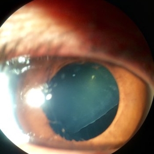
Anterior segment photograph of a 24-year-old female with crystalline lens displacement, showing zonula fibers opposite to the direction of the lens.
Photographer: Andrea Elizabeth Arriola, MSc. Asociacion para evitar la ceguera en México IAP
Condition/keywords: dislocated crystalline lens, lens
-
 LIO Dipped in the Vitreo
Aug 29 2016 by JEFFERSON R SOUSA, Tecg.º (Biomedical Systems Technology)
Patient Male, 51-years-old, with treatment with laser photocoagulation in myopic degeneration peripheral. Did FEC. suffered trauma (elbow) and had LIO dipped in the víteo.
Photographer: JEFFERSON R SOUSA - Institute Dr. Suel Abujamra / São Paulo - Brazil
Imaging device: Topcon TRC-50VT, Film, Kodak Ektachrome 160 - ASA 100 / 35mm, field of 35 degrees. Flash 100.
Condition/keywords: lens, myopic degeneration
-
 Slide 4-26
Feb 20 2019 by Lancaster Course in Ophthalmology
Congenital aniridia. Clinical appearance showing the equator of the lens and some of the zonules.
Condition/keywords: aniridia, lens, zonules
-
 Slide 4-29
Feb 20 2019 by Lancaster Course in Ophthalmology
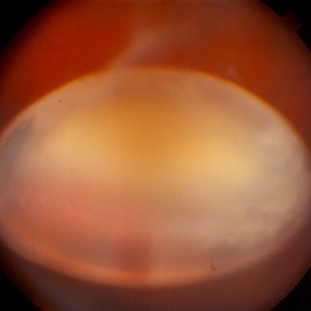
Persistent hyperplastic primary vitreous. Clinical appearance showing a dense posterior cataract. With the pupil dilated, the ciliary processes extend forward toward the apex of the lens.
Condition/keywords: cataract, ciliary, lens, vitreous
-
 Slide 4-31
Feb 20 2019 by Lancaster Course in Ophthalmology
Persistent hyperplastic primary vitreous showing the area around the posterior capsule of the lens. Note the rupture of the capsule by the contracting fibrous membrane, and the extensive cataract. (Courtesy of T. Makley, M.D.)
Condition/keywords: cataract, lens, vitreous
-
 Slide 7-74
Feb 25 2019 by Lancaster Course in Ophthalmology
Vossius ring showing a ring of pigment on the surface of the lens following blunt trauma.
Condition/keywords: lens, trauma, Vossius ring
-
 Slide 7-77
Feb 25 2019 by Lancaster Course in Ophthalmology
Lens dislocated into the anterior chamber.
Condition/keywords: dislocated lens, lens
-
 Traumatic Lens Drop
Nov 25 2020 by Gayathri Mohan
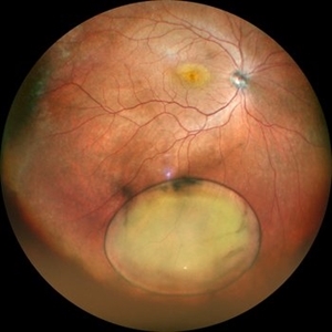
Widefield color fundus photograph of a 58-year-old man with traumatic lens drop post blunt trauma.
Photographer: Gayathri Mohan
Imaging device: Mirante, Nidek
Condition/keywords: blunt trauma, lens, trauma
-
 Ectopia Lentis et Pupillae
Sep 24 2024 by Christian A Leal, MD
Fundus photograph of a 4 year old child with ectopia lentis et pupillae showing the crystalline lens dislocated into the vitreous cavity.
Photographer: Baker Hubbard, MD; Emory Eye Center
Condition/keywords: lens dislocation
-
 Lens After YAG Capsulotomy
Feb 19 2015 by H. Michael Lambert, MD
Color photo of lens after YAG laser.
Condition/keywords: laser, posterior chamber intraocular lens (PCIOL)
-
 Lens Coloboma
Oct 17 2018 by Mehul A Shah
35-year-old male presented with diminished vision. On examination he was having nuclear sclerosis and lens coloboma.
Photographer: MEHUL SHAH
Condition/keywords: coloboma
-
 Lens Drop
Aug 21 2023 by Harsh Vardhan Singh, MS
Intra-operative image of Posterior lens dislocation as a complication of cataract surgery
Photographer: Harsh Vardhan Singh
Condition/keywords: lens dislocation, Lens Drop
-
 Lens Feathering
Nov 9 2016 by Nimrod Dar
26-year-old man with lens feathering a day after a pars plana vitrectomy for rhegmatogenous retinal detachment.
Photographer: Nimrod Dar, M.D, Meir Ophthalmology Department
Condition/keywords: lens feathering, vitrectomy
-
 Lens Luxation
Aug 29 2016 by JEFFERSON R SOUSA, Tecg.º (Biomedical Systems Technology)
Patient, 65-years-old, male, suffered trauma blunt (the jackpot) in the right eye. Ultrasound of the eye found dislocation the total of the crystalline.
Photographer: JEFFERSON R SOUSA - Institute Dr. Suel Abujamra / São Paulo - Brazil
Imaging device: Topcon TRC-50VT, Film Kodak Ektachrome 160 - ASA 100 / 35mm, field of 35 degrees. Flash 100.
Condition/keywords: dislocated lens, lens luxation
-
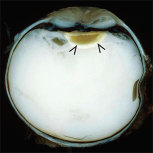 Lens-Induced Uveitis
May 18 2020 by McGill University Health Centre
In lens-induced uveitis, lens protein in the anterior chamber causes a zonal granulomatous response, which occurs usually 1 day to 3 weeks after capsule rupture. This may be associated with sympathetic ophthalmia. This enucleation specimen shows an indented cornea, accompanied by complete hypopyon occupying the anterior chamber with intense vitreitis. Note that the translucent vitreous has become whitish, and the lens surface is irregular with decoloration of the peripheral areas (arrows).
Condition/keywords: uveitis
-
 Lense Opacity
Aug 1 2013 by From the Collections of Thomas M. Aaberg, MD and Thomas M. Aaberg Jr., MD
Lense opacity.
Condition/keywords: lens opacity
-
 Lense Opacity
Aug 1 2013 by From the Collections of Thomas M. Aaberg, MD and Thomas M. Aaberg Jr., MD
Lense opacity.
Condition/keywords: lens opacity
 Loading…