File number: 26804
Comments
-
 Mitzy E Torres Soriano, MD (November 18 2016)
I referred the patient to a rheumatologist who after clinical evaluation and laboratory tests made a diagnosis of SLE.
-
 Wesam Safwat (November 16 2016)
Please sir , can you discuss how to reach diagnosis???
Sign in to comment.
 Initializing download.
-
 By Mitzy E Torres Soriano, MD
Co-author(s): Federico Furno Sola, Pilar Lucena - Uploaded on Nov 14, 2016.
- Last modified by Caroline Bozell on Jan 27, 2017.
- Image of the week
-
Jan 29, 2017
View all images of the week - Rating
- Appears in
- Miscellaneous
- Condition/keywords
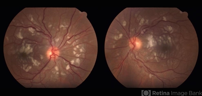
- systemic lupus erythematosus, cotton wool spots, vasculopathy, occlusive vasculitis, occlusive retinal vasculitis
- Photographer
- Grupo Laser Vision, Rosario, Argentina
- Imaging device
- Fundus camera
- Description
- 25-year-old female patient with systemic lupus erythematosus. Photographs show cotton wool spots, intraretinal hemorrhages and vascular tortuosity. FA demonstrated retinal vasculitis and OCT revealed cystoid macular edema. In this case diagnosis of SLE was made after ocular manifestation.


