Search results (109 results)
-
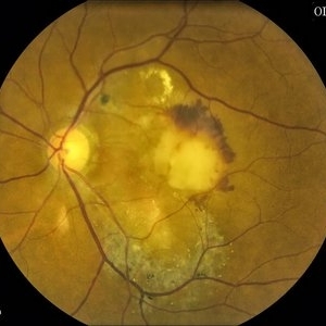
 Disciform Scar
Jul 13 2013 by Jason S. Calhoun
Chorioretinal scar inferior temporal in the right eye of a middle aged patient.
Photographer: Jason S. Calhoun, Department of Ophthalmology, Mayo Clinic Jacksonville, Florida
Condition/keywords: chorioretinal scar
-
 Central Serous Chorioretinopathy (CSC)
Oct 16 2012 by S. Natarajan, MD, FASRS, FRCS (GLASGOW) , FICO, D.Sc, FELA
Middle-aged male came with small PED 4 months back; now this has progressed to a larger PED with SRF underneath the fovea.
Photographer: Prof. Dr. S. Natarajan
Condition/keywords: central serous chorioretinopathy (CSCR), central serous retinopathy (CSR), pigment epithelial detachment (PED), subretinal fibrosis
-
 Elevated Lesion
Nov 9 2012 by Norman Byer
This photograph and the next are two views of a very interesting elevated lesion in a 45-year-old man. This photograph shows the immense value of closely scrutinizing the profile of the indented area. Note that in the middle of the slide there is a sudden break in the continuity of the dark convex shadow that lies just behind the crest of the scleral indentation. If the elevated tissue is "filmy" or "wispy" or filamentous as in this case, it raises a strong suspicion that a retinal break is present just behind it.
Condition/keywords: elevated retinal lesion, elevated tissue, retinal break, scleral indentation
-
 Pattern Dystrophy slide 1
Oct 22 2012 by Ronald C. Gentile, MD
Asymptomatic middle-age man with normal vision and a multifocal pattern dystrophy. The pattern dystrophy simulates Stargardt disease/fundus flavimaculatus with irregular yellow-white flecks scattered throughout the posterior pole. Some lesions extend beyond the retinal vascular arcades.
Photographer: The New York Eye & Ear Infirmary Department of Medical Imaging
Condition/keywords: pattern macular dystrophy
-
 Retinoschisis Detachment
Nov 9 2012 by Norman Byer
Combined retinoschisis detachment, so-called schisis detachment, in a 47-year-old woman. The large outer layer hole in the center has a posterior yellow border which represents the position of the outer layer. Please observe superior to the hole the dark convexity of the scleral indentation. Just below the hole at the middle of the slide and going to the left the yellow zone comes to lie right against the inner layer and a fluid filled cavity lies deep to the outer layer. At this point, therefore, there is a true neurosensory detachment of the retina. On the right side of the hole, the yellow line slants up and to the right and lies close to the pigment epithelium. On the right side of the photograph, the original schisis cavity can be seen separating the yellow line of the outer layer above from the inner retinal layer below. The mechanism of this detachment is that some of the fluid from the schisis cavity passes through the outer layer hole and detaches the outer layer. This lesion has not been treated and has remained exactly the same for 13 years. A similar symmetrical "schisis-detachment" is present in the fellow eye.
Condition/keywords: neurosensory detachment of retina, outer layer hole, pigment epithelium, retinoschisis, schisis detachment, scleral indentation
-
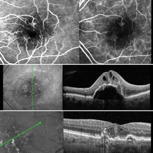
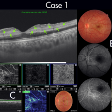 Figure-1 Paracentral Acute Middle Maculopathy (PAMM)
Dec 21 2018 by Fawwaz F Al Mamoori, MD, Medical Retina Consultant
25-year-old male patient medically free, had sudden deterioration in his left eye vision. Visual acuity on presentation was counting fingers at 3 meter distance. Marked Relative Pupillary Afferent Defect (RAPD) was detected and fundoscopic exam showed abnormal foveal reflex. SS OCT B scan: showed a hypereflectivity of the inner plexiform layer (IPL), inner nuclear layer (INL) and OPL layer (fig 1, A).FA images were normal (fig 1, B). Angiography shows remarkable perifoveal capillary drop out within middle retinal layer correlating with perfusion density map which reveals significant decrease in capillary density at the same level (Fig 1, C). Enface ads more proof to PAMM by delineating ischemic distribution in a fern like pattern of hyper reflective areas within DCP (fig1, D).
Photographer: Dr.Fawwaz Al Mamoori (Al Mamoori Eye Clinic)
Imaging device: Triton Swept Source OCT (TOPCON)
Condition/keywords: optical coherence tomography (OCT), paracentral acute middle maculopathy
-
 Pseudo Retinal Break
Nov 9 2012 by Norman Byer
This large reddish area in the nasal periphery of this left eye is actually an enclosed ora bay. For other examples of this, see slide pair 5 and 6. This developmental abnormality could easily be confused with a retinal break. A very unusual feature of this photograph is the presence of a tiny true retinal break at the far right end of the enclosed ora bay and lying just to the left of the yellow zone in the middle of the photograph
Condition/keywords: enclosed ora bay, pseudo retinal break, reddish areas
-
 Paracentral Acute Middle Maculopathy (PAMM)
Mar 21 2019 by Jonathan C. Tsui, MD
26-year-old female with hypertension presenting with chief complaint of "darkening" in her nasal visual field in the right eye. No flashes, floaters, or vision loss. Va 20/60 and nasal VF defect OD. SD-OCT demonstrated hyperreflectivity in the INL consistent with paracentral acute middle maculopathy. She was referred to her PCP for blood pressure optimization and a cardiovascular work-up. She returned for follow-up two months later with 20/80 OD, 20/20 OS. Repeat SD-OCT demonstrated inner retinal atrophy.
Photographer: Zellers, Diane
Condition/keywords: paracentral acute middle maculopathy
-
---thumb.jpg/image-square;max$300,300.ImageHandler) Pattern Dystrophy
Pattern Dystrophy
Aug 7 2013 by From the Collections of Thomas M. Aaberg, MD and Thomas M. Aaberg Jr., MD
Middle aged patient with yellow macular pigment pattern.
Condition/keywords: butterfly dystrophy, pattern macular dystrophy
-
---thumb.jpg/image-square;max$300,300.ImageHandler) Adult Vitelliform Dystrophy
Adult Vitelliform Dystrophy
Apr 1 2013 by Henry J. Kaplan, MD
Fundus photograph of a middle aged patient with mild decreased vision and bilateral macular vitelliform lesion #1.
Condition/keywords: adult vitelliform dystrophy, vitelliform lesion, vitelliform macular dystrophy
-
 Lattice Lesion
Nov 9 2012 by Norman Byer
This lattice lesion in a 27-year-old woman shows an interesting change in the middle of the lesion. The predominant feature on the left side of the lesion is the snailtrack appearance while the right side of the lesion shows mainly a reddish crater. Note the many yellow dots above the surface of the retina which are actually located in the vitreous condensation which surrounds the pocket of liquified vitreous over the lesion.
Condition/keywords: lattice lesion, reddish crater, snail track, vitreous condensation, vitreous liquefaction
-
 Acute Necrotizing Retinal Vasculitis as Onset of Systemic Lupus Erythematosus.
Sep 3 2016 by ADRIANO FERREIRA
A 28-year-old white man was referred to the rheumatology clinic with gradually and rapid deterioration of the vision (both eyes). In this picture, we can observe cotton wool spots in the papillomacular area and extensive hemorrhages in posterior polo and in the middle periphery. Hard exudates are present in macular area (macular edema)
Photographer: Claudio Zett Lobo
Imaging device: TRC50DXi TOPCON
Condition/keywords: systemic lupus erythematosus (SLE) vasculitis, vasculitis
-
 Polymorphous Vitelliform Maculopathy
Jul 11 2013 by Eric M. Shrier, DO
38-year-old Middle Eastern female referred for IFN screening (Hep. c +).
Condition/keywords: idiopathic, polymorphous exudative vitelliform maculopathy
-
 Giant Retinal Tear With RD
Jun 29 2013 by Jason S. Calhoun
Middle aged patient comes in with sudden loss of vision inferior, nasally. Patient presents plus 2 APD in the right eye. Fundus exam reveals a giant retinal tear at 10-11 o'clock with RD. Vitrectomy with laser and gas exchange of C3F8 scheduled.
Photographer: Jason S. Calhoun, Mayo Clinic Jacksonville, Florida
Imaging device: TOPCON TRC 50-EX
Condition/keywords: retinal tear
-
 EDI OCT Macular Hole
Jun 29 2013 by Jason S. Calhoun
Enhanced depth imaging OCT shows a macular hole with no traction on middle aged female.
Photographer: Jason S. Calhoun, Mayo Clinic Jacksonville, Florida
Imaging device: TOPCON TRC 50-EX/CIRRUS HD OCT
Condition/keywords: macular hole
-
 Branch Retinal Vein Occlusion
Oct 17 2012 by Sharon Fekrat, MD FACS FASRS
Fluorescein angiography of an inferior perfused branch retinal vein occlusion in the left eye of a middle aged male with hypertension. The foveal avascular zone is irregular. Subretinal hemorrhage is present.
Photographer: John Reaves, Ophthalmic Photographer, Durham VA Medical Center Eye Clinic Imaging Suite, Durham, NC
Imaging device: Fluorescein Angiography
Condition/keywords: branch retinal vein occlusion (BRVO), subretinal hemorrhage
-
 Retinoschisis
Nov 9 2012 by Norman Byer
This is a different view of the previous case taken with scleral indentation. There is a very large outer layer hole on the left side of the photograph with prominent yellow rolled posterior borders and a small yellow nubbin in the middle, which is probably the remnant of a former bridge between two smaller holes. This case has not been treated and has not progressed during four year’s observation. In fact, it has gotten measurably smaller in extent.
Condition/keywords: intact inner layer, outer layer hole, retinoschisis, rolled edges of retina, scleral indentation
-
 Retinal Angiomatous Proliferation RAP
Mar 11 2020 by RAFAEL REIS PEREIRA, MD
Retinal angiomatous proliferation (RAP) is a unique variant of neovascular age-related macular degeneration. Published studies have estimated that up to 15% of patients with neovascular age-related macular degeneration have RAP. Clinical features frequently associated with RAP include bilateral disease, presence of pigment epithelial detachments, and reticular pseudodrusen. RAP is more frequently associated with the development of retinal pigment epithelial tears and geographic atrophy that can lead to severe vision loss. We present a stereo fluorescein angiography and ICG (upper right and left image respectively) and OCT of left and right eye (middle and inferior image) of a RAP choroidal neovascularization in an 89-year-old patient.
Photographer: Rafael Reis Pereira
Imaging device: HRA Heildelberg Spectralis
Condition/keywords: retinal angiomatous proliferation (RAP)
-
 Full Thickness Macular Hole With ERM
Feb 26 2014 by Sharon Fekrat, MD FACS FASRS
Middle aged woman with a full thickness macular hole in the left eye associated with an epiretinal membrane.
Photographer: Michael P Kelly, Ophthalmic Photographer, Duke Eye Imaging, Duke Eye Center
Condition/keywords: epiretinal membrane (ERM), macular hole
-
 Macular Drusen
Jul 14 2013 by Jason S. Calhoun
Fundus shows honeycomb shape drusen in middle aged female.
Photographer: Jason S. Calhoun, Department of Ophthalmology, Mayo Clinic Jacksonville, Florida
Imaging device: TOPCON TRC 50-EX
-
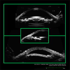 Oversized ACIOL
Apr 10 2019 by Scott D Walter, MD, MSc, FASRS
A patient with a corneal diameter of 11.5mm was implanted with an MTA4UO anterior chamber intraocular lens (ACIOL). The patient subsequently developed cystoid macular edema (CME) with petalloid angiographic leakage and secondary glaucoma. Ultrasound biomicroscopy (UBM) demonstrates appropriate centration of the ACIOL optic (top), with both haptics embedded in the anterior chamber angle (middle), and anterior bowing of the midperipheral iris (bottom). Placement of an MTA3UO lens would likely have avoided this complication.
Imaging device: Ellex 50MHz anterior B-scan probe
Condition/keywords: cystoid macular edema (CME), dislocated anterior chamber intraocular lens (ACIOL), glaucoma disorders of the lens, lrvine-Gass syndrome, uveitis glaucoma hyphaema (UGH) syndrome
-
 Choroidal Neovascularization (CNV)
Jul 13 2013 by Jason S. Calhoun
Active CNV in middle aged black female. Proceeded with Avastin injection.
Photographer: Jason S. Calhoun, Department of Ophthalmology, Mayo Clinic Jacksonville, Florida
Imaging device: TOPCON TRC 50-EX
Condition/keywords: choroidal neovascularization (CNV)
-
 Retinal Detachment Repair in Patient With a Coloboma
Jun 29 2018 by Gareth Lema, MD, PhD
15-year-old boy with RD from a temporal giant retinal tear after blunt trauma. An encircling band was placed and shave vitrectomy was done. This photo was taken after silicone oil had been removed. There is a haze in the middle of the image due to a cataract.
Photographer: Sandra Boglione, Ross Eye Institute, University at Buffalo Jacobs School of Medicine, Buffalo, NY
Imaging device: Optos
Condition/keywords: chorioretinal coloboma, encircling scleral buckle
-
 Progressive Bifocal Chorioretinal Atrophy
Feb 1 2015 by Andree Henaine-Berra, MD
Fluorescein angiography of the left eye of an 13-year-old female patient with poor vision, high myopia and nystagmus. The image shows a hypofluorescent area corresponding to the area of chorioretinal atrophy. Some middle sized choroidal vessels can still be observed.
Photographer: Andree Henaine-Berra, MD
Condition/keywords: chorioretinal atrophy
-
 Figure-2 Paracentral Acute Middle Maculopathy (PAMM)
Dec 21 2018 by Fawwaz F Al Mamoori, MD, Medical Retina Consultant
70-year-old female patient known to have hypertension, presented with acute deterioration of left eye vision, best corrected visual acuity was 6/60. Fundoscopic exam showed abnormal foveal reflex whiting .SS-OCT B scan showed also a hypereflectivity of the inner plexiform layer (IPL), inner nuclear layer (INL) and OPL layer(figure-2, A). FA images were also normal(figure-2 B). Segmented angiographic images elucidate ischemia and capillary drop out predominantly at the level of DCP but less severe than Case 1 (fig 2, C). Correspondingly, Enface highlights hyper reflective areas in a fern like distribution in the middle retina at similar depth of ischemic lesions demonstrated on B scans and OCTA (fig 2, D)
Photographer: Dr.Fawwaz Al Mamoori (Al Mamoori Eye Clinic)
Imaging device: Triton Swept Source OCT (TOPCON)
Condition/keywords: optical coherence tomography (OCT), paracentral acute middle maculopathy
 Loading…