Search results (109 results)
-
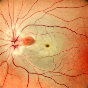
 Shooting Stars
Jul 9 2025 by Majda Hadziahmetovic, MD
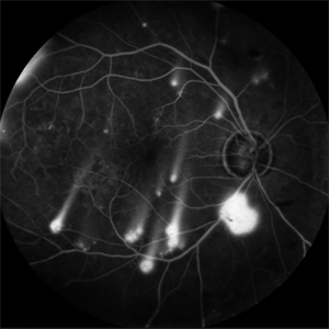
Fluorescein angiography image demonstrating multiple areas of neovascularization in a middle-aged male patient with long-standing diabetes.
Condition/keywords: proliferative diabetic retinopathy (PDR)
-
 Acute Necrotizing Retinal Vasculitis as Onset of Systemic Lupus Erythematosus.
Sep 3 2016 by ADRIANO FERREIRA
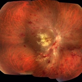
A 28-year-old white man was referred to the rheumatology clinic with gradually and rapid deterioration of the vision (both eyes). In this picture, we can observe cotton wool spots in the papillomacular area and extensive hemorrhages in posterior polo and in the middle periphery. Hard exudates are present in macular area (macular edema)
Photographer: Claudio Zett Lobo
Imaging device: TRC50DXi TOPCON
Condition/keywords: systemic lupus erythematosus (SLE) vasculitis, vasculitis
-
 Branch Retinal Vein Occlusion
Oct 17 2012 by Sharon Fekrat, MD FACS FASRS
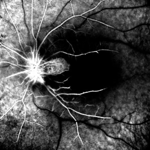
Fluorescein angiography of an inferior perfused branch retinal vein occlusion in the left eye of a middle aged male with hypertension. The foveal avascular zone is irregular. Subretinal hemorrhage is present.
Photographer: John Reaves, Ophthalmic Photographer, Durham VA Medical Center Eye Clinic Imaging Suite, Durham, NC
Imaging device: Fluorescein Angiography
Condition/keywords: branch retinal vein occlusion (BRVO), subretinal hemorrhage
-
 Cilioretinal Artery Sparing CRAO
May 1 2025 by Tejaswita Verma
Fundus photo of a middle aged male with CRAO partially sparing cilioretinal artery and papillomacular bundle. Vision 6/60.
Photographer: Dr. Tejaswita Verma
Imaging device: MIRANTE
Condition/keywords: CRAO with cilioretinal sparing
-
 Combined Traction and Rhegmatogenous Retinal Detachment From Proliferative Diabetic Retinopathy
Mar 27 2025 by Nikhil K Bommakanti, MD
A middle-aged patient presented with a combined traction and rhegmatogenous retinal detachment.
Condition/keywords: Active PDR Tractional retinal Detachment, PDR, Retinal Detachment, rrd, TRD
-
 CRAO Sparing Cilioretinal Artery
May 1 2025 by Tejaswita Verma
Fundus photo of a middle aged male with 6/60 vision in left eye showing CRAO partially sparing cilioretinal artery.
Photographer: Dr. Tejaswita Verma
Imaging device: MIRANTE
Condition/keywords: cilioretinal sparing, CRAO
-
 Elevated Lesion
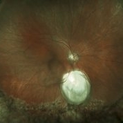
Nov 9 2012 by Norman Byer
This photograph and the next are two views of a very interesting elevated lesion in a 45-year-old man. This photograph shows the immense value of closely scrutinizing the profile of the indented area. Note that in the middle of the slide there is a sudden break in the continuity of the dark convex shadow that lies just behind the crest of the scleral indentation. If the elevated tissue is "filmy" or "wispy" or filamentous as in this case, it raises a strong suspicion that a retinal break is present just behind it.
Condition/keywords: elevated retinal lesion, elevated tissue, retinal break, scleral indentation
-
 Full Thickness Macular Hole With ERM
Feb 26 2014 by Sharon Fekrat, MD FACS FASRS
Middle aged woman with a full thickness macular hole in the left eye associated with an epiretinal membrane.
Photographer: Michael P Kelly, Ophthalmic Photographer, Duke Eye Imaging, Duke Eye Center
Condition/keywords: epiretinal membrane (ERM), macular hole
-
 Intravitreal Ozurdex Implant
Apr 3 2025 by Tejaswita Verma
Fundus photo of a middle-aged diabetic male showing Ozurdex implant in situ with laser marks.
Photographer: Tejaswita Verma
Imaging device: MIRANTE
Condition/keywords: dexamethasone implant, ozurdex
-
 Maternally-Inherited Diabetes and Deafness (MIDD) Syndrome
Jan 12 2025 by Niloofar Piri, MD
Fundus Autofluorescence image of right posterior pole in a 43 year old female who was referred for diabetic retinopathy evaluation, demonstrated multiple patches of hypoautrofluorescence surrounding the nerve and fovea. Please note that central fovea is spared. Granular hyper and hypoauto fluorescence is present in the macula and peripapillary region. She was noted to have hearing loss as well and after further evaluation was diagnosed with MIDD syndrome.
Condition/keywords: Maternally inherited diabetes and deafness (MIDD), Maternally-inherited-diabetes-and-deafness-(MIDD) syndrome, Mitochondrial Disorder
-
 MIDD (Maternally Inherited Diabetes and Deafness) - Left AF
Nov 30 2024 by John S. King, MD
Both right and left eyes have symmetrical ring of mottled hypo/hyper AF around the fovea and disc. The HyperAF areas correspond to RPE deposits on OCT as well as areas of blockage on FA, and drusenoid deposits seen on fundus photos 57 yo WF referred for AMD vs Pattern Dystrophy that was diagnosed 10 years ago. Reported some slow progressive vision loss in both eyes for distance and near. Denies nyctalopia or hemeralopia. Background medical history includes HTN, CVD, and DM. No family history of eye problems. Denied pentosan use. Anterior segment showed moderate cataracts (OD>OS). Posterior segment exam showed macular changes and mild NPDR. The macular appearance showed a symmetrical, paramacular ring of fleck-like drusenoid material with some faint focal areas of RPE hyperplasia. Fundus Photos, AF, OCT were performed as well as a gene test. Further questioning showed revealed that her mother and maternal grandmother had both diabetes mellitus and sensorineural hearing loss. The patient developed diabetes in her teens, and some high frequency hearing loss in her early twenties. She had not had a previous genetic test or diagnosis of MIDD. Gene testing is pending for the mitochondrial component. Invitae's retinal panel, which does not include mitochondrial disorders, only showed a variant of uncertain significance, HMCN1. I discussed this case with Dr. Freund, and it is similar to a the case report : Inoue M, Kiss S, Freund KB. MACULAR PIGMENT RINGS AS THE PRESENTING FINDING OF MITOCHONDRIAL MYOPATHY, ENCEPHALOPATHY, LACTIC ACIDOSIS, AND STROKELIKE EPISODES. Retin Cases Brief Rep. 2015 Fall;9(4):260-4. doi: 10.1097/ICB.0000000000000182. PMID: 26200388.
Photographer: Grace Melton and Carley Gunn
Imaging device: Clarus
Condition/keywords: Macular Dystrophy, Maternally Inherited Diabetes and Deafness, MIDD, Mitochondrial Disorder
-
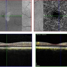 PAMM-OCTA
Nov 29 2023 by Daniel Davis, OCT-C
OCT-A of a 30 yo female with PAMM OD.
Photographer: Daniel Davis, OCT-C
Imaging device: Heidelberg Spectralis
Condition/keywords: OCTA, paracentral acute middle maculopathy
-
Paracentral Acute Middle Maculopathy
Nov 29 2023 by Daniel Davis, OCT-C
30 yo female OCT with Paracentral Acute Middle Maculopathy (PAMM) OD VA OD: sc20/60+1
Condition/keywords: OCT, PAMM
-
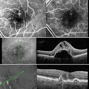 Retinal Angiomatous Proliferation RAP
Mar 11 2020 by RAFAEL REIS PEREIRA, MD
Retinal angiomatous proliferation (RAP) is a unique variant of neovascular age-related macular degeneration. Published studies have estimated that up to 15% of patients with neovascular age-related macular degeneration have RAP. Clinical features frequently associated with RAP include bilateral disease, presence of pigment epithelial detachments, and reticular pseudodrusen. RAP is more frequently associated with the development of retinal pigment epithelial tears and geographic atrophy that can lead to severe vision loss. We present a stereo fluorescein angiography and ICG (upper right and left image respectively) and OCT of left and right eye (middle and inferior image) of a RAP choroidal neovascularization in an 89-year-old patient.
Photographer: Rafael Reis Pereira
Imaging device: HRA Heildelberg Spectralis
Condition/keywords: retinal angiomatous proliferation (RAP)
-
 Polymorphous Vitelliform Maculopathy
Jul 11 2013 by Eric M. Shrier, DO
38-year-old Middle Eastern female referred for IFN screening (Hep. c +).
Condition/keywords: idiopathic, polymorphous exudative vitelliform maculopathy
-
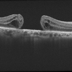 EDI OCT Macular Hole
Jun 29 2013 by Jason S. Calhoun
Enhanced depth imaging OCT shows a macular hole with no traction on middle aged female.
Photographer: Jason S. Calhoun, Mayo Clinic Jacksonville, Florida
Imaging device: TOPCON TRC 50-EX/CIRRUS HD OCT
Condition/keywords: macular hole
-
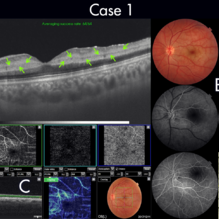 Figure-1 Paracentral Acute Middle Maculopathy (PAMM)
Dec 21 2018 by Fawwaz F Al Mamoori, MD, Medical Retina Consultant
25-year-old male patient medically free, had sudden deterioration in his left eye vision. Visual acuity on presentation was counting fingers at 3 meter distance. Marked Relative Pupillary Afferent Defect (RAPD) was detected and fundoscopic exam showed abnormal foveal reflex. SS OCT B scan: showed a hypereflectivity of the inner plexiform layer (IPL), inner nuclear layer (INL) and OPL layer (fig 1, A).FA images were normal (fig 1, B). Angiography shows remarkable perifoveal capillary drop out within middle retinal layer correlating with perfusion density map which reveals significant decrease in capillary density at the same level (Fig 1, C). Enface ads more proof to PAMM by delineating ischemic distribution in a fern like pattern of hyper reflective areas within DCP (fig1, D).
Photographer: Dr.Fawwaz Al Mamoori (Al Mamoori Eye Clinic)
Imaging device: Triton Swept Source OCT (TOPCON)
Condition/keywords: optical coherence tomography (OCT), paracentral acute middle maculopathy
-
 Paracentral Acute Middle Maculopathy
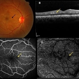
Oct 25 2019 by Gayathri Mohan
Multimodal images of a case of a 29-year-old female with paracentral acute middle maculopathy. A-color fundus photograph showing multiple confluent white retinal patches. B- On OCT the acute lesions of PAMM characteristically appear as placoid, hyperreflective bands at the level of the INL C-Fundus fluorescein angiography showing a capillary nonperfusion area D-flow void areas in deep capillary plexus
Photographer: Akshar Soni
Imaging device: Heidelberg, Nidek
Condition/keywords: fundus albipunctatus, optical coherence tomography (OCT), paracentral acute middle maculopathy
-
 Retinal Detachment Repair in Patient With a Coloboma
Jun 29 2018 by Gareth Lema, MD, PhD
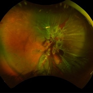
15-year-old boy with RD from a temporal giant retinal tear after blunt trauma. An encircling band was placed and shave vitrectomy was done. This photo was taken after silicone oil had been removed. There is a haze in the middle of the image due to a cataract.
Photographer: Sandra Boglione, Ross Eye Institute, University at Buffalo Jacobs School of Medicine, Buffalo, NY
Imaging device: Optos
Condition/keywords: chorioretinal coloboma, encircling scleral buckle
-
 Acromegaly
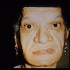
Jul 29 2013 by H. Michael Lambert, MD
Acromegaly, chronic disease of middle life characterized by overgrowth of bone, connective tissue and viscera in response to prolonged and excessive secretion of growth hormone. Visual loss due to chiasmal compression and less frequently diabetic retinopathy.
Condition/keywords: acromegaly
-
 Acute Retinal Necrosis
Mar 26 2019 by Gary R. Cook, MD, FACS
Middle-aged white female with ARN OS showing additional involvement within 2 weeks. Patient was seen prior to the availability of anti-viral therapies.
Imaging device: Topcon VT-50
Condition/keywords: acute retinal necrosis
-
 Acute Retinal Necrosis
Mar 26 2019 by Gary R. Cook, MD, FACS
Middle-aged white female with peripheral retinal lesions of acute retinal necrosis OS at presentation.
Imaging device: Topcon VT-50
Condition/keywords: acute retinal necrosis
-
---thumb.jpg/image-square;max$300,300.ImageHandler) Adult Vitelliform Dystrophy
Adult Vitelliform Dystrophy
Apr 1 2013 by Henry J. Kaplan, MD
Fundus photograph of a middle aged patient with mild decreased vision and bilateral macular vitelliform lesion #1.
Condition/keywords: adult vitelliform dystrophy, vitelliform lesion, vitelliform macular dystrophy
-
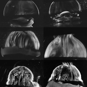 Age-Related Differences in the Structure of the Human Vitreous Body
Sep 1 2020 by J. Sebag, MD, FACS, FRCOphth, FARVO
Dark-field slit microscopy was performed on fresh, unfixed, post-mortem human eyes that had undergone dissection to peel off the sclera, choroid, and retina. The vitreous body remains attached to the anterior segment which is seen below, while the posterior pole is above in these images. The top panel demonstrates the absence of internal vitreous structures that scatter light in youth (left image from an 11 year-old girl, right image from a 14 year-old boy. The middle panel demonstrates light scattering from linear, fibrous structures that have an antero-posterior orientation with insertions into the vitreous base peripherally and the posterior vitreous cortex, typical in middle age (left image from a 56 year-old and right image from a 59 year-old). The bottom panel illustrates advance fibrous liquefaction in old age (88-year-old subject). [From Sebag J, Niemeyer M, Koss M: Anomalous PVD and vitreoschisis. In: Vitreous – in Health & Disease (J. Sebag, ed.) Springer, New York, 2014, pg. 245; image © Springer Nature, reprinted with permission]
Condition/keywords: vitreous
-
 Before and After Vitrectomy
Nov 17 2023 by Bradley T. Smith, MD, FASRS
Middle age male diabetic retinopathy and resolving exudate following repair of tractional detachment with membrane peeling.
Condition/keywords: coats-like response, Diabetes, fibrotic neovascularization, fibrovascular proliferation, pars plana vitrectomy (PPV), proliferative diabetic retinopathy (PDR), tractional retinal detachment
 Loading…