File number: 14338
Comments
-
 James B. Soque, CRA, OCT-C, COA, FOPS (March 24 2014)
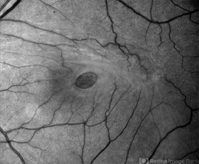
This Red Free left eye image of a Macular Hole is of excellent quality and shows wonderful detail and light balance, from this fundus camera. The posterior hyaloid can be seen superiorly providing the insuing tangental traction.
Sign in to comment.
 Initializing download.
-
 By Sharon Fekrat, MD FACS FASRS
Duke University School of Medicine - Uploaded on Feb 26, 2014.
- Last modified by Caroline Bozell on Feb 26, 2014.
- Rating
- Appears in
- Miscellaneous
- Condition/keywords
- macular hole, epiretinal membrane (ERM)
- Photographer
- Michael P Kelly, Ophthalmic Photographer, Duke Eye Imaging, Duke Eye Center
- Imaging device
- Fundus camera
- Description
- Middle aged woman with a full thickness macular hole in the left eye associated with an epiretinal membrane.


