Search results (109 results)
-
 MIDD
MIDD
May 26 2023 by Virginia Gebhart
51-year-old female with dry AMD, advanced atrophic without subfoveal involvement OU. Genetic testing confirmed MIDD (maternal inherited diabetes and deafness) which is a mitochondrial inherited dystrophy. Unaware of any family hx of macular degeneration.
Photographer: Virginia Gebhart, Retina Consultants of Carolina
Imaging device: Topcon TRC 50DX
Condition/keywords: advanced geographic atrophy, geographic atrophy
-
 MIDD (Maternally Inherited Diabetes and Deafness)
Feb 25 2025 by Virginia Gebhart
53 year old female with confirmed MIDD (genetic testing at Emory). Vision is stable with progressing GA but still central sparing OU. No evidence of choroidal neovascularization. Moderate myopia.
Photographer: Virginia Gebhart, Retina Consultants of Carolina
Imaging device: Topcon 50DX
Condition/keywords: geographic atrophy, Maternally inherited diabetes and deafness (MIDD), MIDD
-
 MIDD (Maternally Inherited Diabetes and Deafness) - Left AF
Nov 30 2024 by John S. King, MD
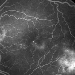
Both right and left eyes have symmetrical ring of mottled hypo/hyper AF around the fovea and disc. The HyperAF areas correspond to RPE deposits on OCT as well as areas of blockage on FA, and drusenoid deposits seen on fundus photos 57 yo WF referred for AMD vs Pattern Dystrophy that was diagnosed 10 years ago. Reported some slow progressive vision loss in both eyes for distance and near. Denies nyctalopia or hemeralopia. Background medical history includes HTN, CVD, and DM. No family history of eye problems. Denied pentosan use. Anterior segment showed moderate cataracts (OD>OS). Posterior segment exam showed macular changes and mild NPDR. The macular appearance showed a symmetrical, paramacular ring of fleck-like drusenoid material with some faint focal areas of RPE hyperplasia. Fundus Photos, AF, OCT were performed as well as a gene test. Further questioning showed revealed that her mother and maternal grandmother had both diabetes mellitus and sensorineural hearing loss. The patient developed diabetes in her teens, and some high frequency hearing loss in her early twenties. She had not had a previous genetic test or diagnosis of MIDD. Gene testing is pending for the mitochondrial component. Invitae's retinal panel, which does not include mitochondrial disorders, only showed a variant of uncertain significance, HMCN1. I discussed this case with Dr. Freund, and it is similar to a the case report : Inoue M, Kiss S, Freund KB. MACULAR PIGMENT RINGS AS THE PRESENTING FINDING OF MITOCHONDRIAL MYOPATHY, ENCEPHALOPATHY, LACTIC ACIDOSIS, AND STROKELIKE EPISODES. Retin Cases Brief Rep. 2015 Fall;9(4):260-4. doi: 10.1097/ICB.0000000000000182. PMID: 26200388.
Photographer: Grace Melton and Carley Gunn
Imaging device: Clarus
Condition/keywords: Macular Dystrophy, Maternally Inherited Diabetes and Deafness, MIDD, Mitochondrial Disorder
-
 MIDD (Maternally Inherited Diabetes and Deafness) - Left FA (7 min)
Nov 30 2024 by John S. King, MD
Both eyes had similar FA findings. There was no dark choroid or signs of leakage. Granular staining around the fovea and disc were present, and the HypoAF areas corresponded to the drusenoid deposits that showed HyperAF. Mild MAs present due to NPDR 57 yo WF referred for AMD vs Pattern Dystrophy that was diagnosed 10 years ago. Reported some slow progressive vision loss in both eyes for distance and near. Denies nyctalopia or hemeralopia. Background medical history includes HTN, CVD, and DM. No family history of eye problems. Denied pentosan use. Anterior segment showed moderate cataracts (OD>OS). Posterior segment exam showed macular changes and mild NPDR. The macular appearance showed a symmetrical, paramacular ring of fleck-like drusenoid material with some faint focal areas of RPE hyperplasia. Fundus Photos, AF, OCT were performed as well as a gene test. Further questioning showed revealed that her mother and maternal grandmother had boith diabetes mellitus and sensorineural hearing loss. The patient developed diabetes in her teens, and some high frequency hearing loss in her early twenties. She had not had a previous genetic test or diagnosis of MIDD. Gene testing is pending for the mitochondrial component. Invitae's retinal panel, which does not include mitochondrial disorders, only showed a variant of uncertain significance, HMCN1. I discussed this case with Dr. Freund, and it is similar to a the case report : Inoue M, Kiss S, Freund KB. MACULAR PIGMENT RINGS AS THE PRESENTING FINDING OF MITOCHONDRIAL MYOPATHY, ENCEPHALOPATHY, LACTIC ACIDOSIS, AND STROKELIKE EPISODES. Retin Cases Brief Rep. 2015 Fall;9(4):260-4. doi: 10.1097/ICB.0000000000000182. PMID: 26200388.
Photographer: Grace Melton and Carley Gunn
Imaging device: Clarus
Condition/keywords: Macular Dystrophy, Maternally Inherited Diabetes and Deafness, MIDD, Mitochondrial Disorder
-
 MIDD (Maternally Inherited Diabetes and Deafness) - Left FP
Nov 30 2024 by John S. King, MD
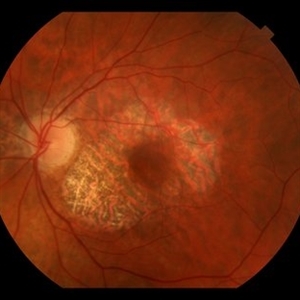
Both the right and left Eye have fairly symmetrical, extrafoveal drusenoid-like flecks and focal and faint areas of RPE hyperplasia (in addition to mild NPDR and PPA) 57 yo WF referred for AMD vs Pattern Dystrophy that was diagnosed 10 years ago. Reported some slow progressive vision loss in both eyes for distance and near. Denies nyctalopia or hemeralopia. Background medical history includes HTN, CVD, and DM. No family history of eye problems. Denied pentosan use. Anterior segment showed moderate cataracts (OD>OS). Posterior segment exam showed macular changes and mild NPDR. The macular appearance showed a symmetrical, paramacular ring of fleck-like drusenoid material with some faint focal areas of RPE hyperplasia. Fundus Photos, AF, OCT were performed as well as a gene test. Further questioning showed revealed that her mother and maternal grandmother had both diabetes mellitus and sensorineural hearing loss. The patient developed diabetes in her teens, and some high frequency hearing loss in her early twenties. She had not had a previous genetic test or diagnosis of MIDD. Gene testing is pending for the mitochondrial component. Invitae's retinal panel, which does not include mitochondrial disorders, only showed a variant of uncertain significance, HMCN1. I discussed this case with Dr. Freund, and it is similar to a the case report : Inoue M, Kiss S, Freund KB. MACULAR PIGMENT RINGS AS THE PRESENTING FINDING OF MITOCHONDRIAL MYOPATHY, ENCEPHALOPATHY, LACTIC ACIDOSIS, AND STROKELIKE EPISODES. Retin Cases Brief Rep. 2015 Fall;9(4):260-4. doi: 10.1097/ICB.0000000000000182. PMID: 26200388.
Photographer: Grace Melton and Carley Gunn
Imaging device: Clarus
Condition/keywords: Macular Dystrophy, Maternally Inherited Diabetes and Deafness, MIDD, Mitochondrial Disorder
-
 MIDD (Maternally Inherited Diabetes and Deafness) - OCT OD
Nov 30 2024 by John S. King, MD
OCT shows mild RPE deposit inferiorly (corresponds to area of FA blockage and HyperAF) and a focal area of iRORA with loss of EZ more superiorly (possibly due to regression of RPE deposit). No choroidal thickening (like in pachychoroid pigment epitheliopathy or cscr) 57 yo WF referred for AMD vs Pattern Dystrophy that was diagnosed 10 years ago. Reported some slow progressive vision loss in both eyes for distance and near. Denies nyctalopia or hemeralopia. Background medical history includes HTN, CVD, and DM. No family history of eye problems. Denied pentosan use. Anterior segment showed moderate cataracts (OD>OS). Posterior segment exam showed macular changes and mild NPDR. The macular appearance showed a symmetrical, paramacular ring of fleck-like drusenoid material with some faint focal areas of RPE hyperplasia. Fundus Photos, AF, OCT were performed as well as a gene test. Further questioning showed revealed that her mother and maternal grandmother had both diabetes mellitus and sensorineural hearing loss. The patient developed diabetes in her teens, and some high frequency hearing loss in her early twenties. She had not had a previous genetic test or diagnosis of MIDD. Gene testing is pending for the mitochondrial component. Invitae's retinal panel, which does not include mitochondrial disorders, only showed a variant of uncertain significance, HMCN1. I discussed this case with Dr. Freund, and it is similar to a the case report : Inoue M, Kiss S, Freund KB. MACULAR PIGMENT RINGS AS THE PRESENTING FINDING OF MITOCHONDRIAL MYOPATHY, ENCEPHALOPATHY, LACTIC ACIDOSIS, AND STROKELIKE EPISODES. Retin Cases Brief Rep. 2015 Fall;9(4):260-4. doi: 10.1097/ICB.0000000000000182. PMID: 26200388.
Photographer: Grace Melton and Carley Gunn
Imaging device: Zeiss Cirrus
Condition/keywords: Macular Dystrophy, Maternally Inherited Diabetes and Deafness, MIDD, Mitochondrial Disorder
-
 MIDD (Maternally Inherited Diabetes and Deafness) - OCT OS
Nov 30 2024 by John S. King, MD
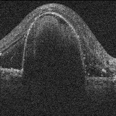
Magnified section of radial scan through the left eye showing a focal nodular RPE deposit that corresponds to a focal drusenoid deposit in temporal macula, that HypoFLs and HyperAFs. Choroid not significantly thickened or thinned, and the nodular thickening may be just above a large outer choroid vessel?) 57 yo WF referred for AMD vs Pattern Dystrophy that was diagnosed 10 years ago. Reported some slow progressive vision loss in both eyes for distance and near. Denies nyctalopia or hemeralopia. Background medical history includes HTN, CVD, and DM. No family history of eye problems. Denied pentosan use. Anterior segment showed moderate cataracts (OD>OS). Posterior segment exam showed macular changes and mild NPDR. The macular appearance showed a symmetrical, paramacular ring of fleck-like drusenoid material with some faint focal areas of RPE hyperplasia. Fundus Photos, AF, OCT were performed as well as a gene test. Further questioning showed revealed that her mother and maternal grandmother had both diabetes mellitus and sensorineural hearing loss. The patient developed diabetes in her teens, and some high frequency hearing loss in her early twenties. She had not had a previous genetic test or diagnosis of MIDD. Gene testing is pending for the mitochondrial component. Invitae's retinal panel, which does not include mitochondrial disorders, only showed a variant of uncertain significance, HMCN1. I discussed this case with Dr. Freund, and it is similar to a the case report : Inoue M, Kiss S, Freund KB. MACULAR PIGMENT RINGS AS THE PRESENTING FINDING OF MITOCHONDRIAL MYOPATHY, ENCEPHALOPATHY, LACTIC ACIDOSIS, AND STROKELIKE EPISODES. Retin Cases Brief Rep. 2015 Fall;9(4):260-4. doi: 10.1097/ICB.0000000000000182. PMID: 26200388.
Photographer: Grace Melton and Carley Gunn
Imaging device: Zeiss Cirrus
Condition/keywords: Macular Dystrophy, Maternally Inherited Diabetes and Deafness, MIDD, Mitochondrial Disorder
-
 MIDD (Maternally Inherited Diabetes and Deafness) - Right AF
Nov 30 2024 by John S. King, MD
Both right and left eyes have symmetrical ring of mottled hypo/hyper AF around the fovea and disc. The HyperAF areas correspond to RPE deposits on OCT as well as areas of blockage on FA, and drusenoid deposits seen on fundus photos. Disc drusen in right eye present as HyperAF spot 57 yo WF referred for AMD vs Pattern Dystrophy that was diagnosed 10 years ago. Reported some slow progressive vision loss in both eyes for distance and near. Denies nyctalopia or hemeralopia. Background medical history includes HTN, CVD, and DM. No family history of eye problems. Denied pentosan use. Anterior segment showed moderate cataracts (OD>OS). Posterior segment exam showed macular changes and mild NPDR. The macular appearance showed a symmetrical, paramacular ring of fleck-like drusenoid material with some faint focal areas of RPE hyperplasia. Fundus Photos, AF, OCT were performed as well as a gene test. Further questioning showed revealed that her mother and maternal grandmother had both diabetes mellitus and sensorineural hearing loss. The patient developed diabetes in her teens, and some high frequency hearing loss in her early twenties. She had not had a previous genetic test or diagnosis of MIDD. Gene testing is pending for the mitochondrial component. Invitae's retinal panel, which does not include mitochondrial disorders, only showed a variant of uncertain significance, HMCN1. I discussed this case with Dr. Freund, and it is similar to a the case report : Inoue M, Kiss S, Freund KB. MACULAR PIGMENT RINGS AS THE PRESENTING FINDING OF MITOCHONDRIAL MYOPATHY, ENCEPHALOPATHY, LACTIC ACIDOSIS, AND STROKELIKE EPISODES. Retin Cases Brief Rep. 2015 Fall;9(4):260-4. doi: 10.1097/ICB.0000000000000182. PMID: 26200388.
Photographer: Grace Melton and Carley Gunn
Imaging device: Clarus
Condition/keywords: Macular Dystrophy, Maternally Inherited Diabetes and Deafness, MIDD, Mitochondrial Disorder
-
 MIDD (Maternally Inherited Diabetes and Deafness) - Right FA (4 min)
Nov 30 2024 by John S. King, MD
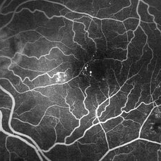
Both eyes had similar FA findings. There was no dark choroid or signs of leakage. Granular staining around the fovea and disc were present, and the HypoAF areas corresponded to the drusenoid deposits that showed HyperAF. Mild MAs present due to NPDR 57 yo WF referred for AMD vs Pattern Dystrophy that was diagnosed 10 years ago. Reported some slow progressive vision loss in both eyes for distance and near. Denies nyctalopia or hemeralopia. Background medical history includes HTN, CVD, and DM. No family history of eye problems. Denied pentosan use. Anterior segment showed moderate cataracts (OD>OS). Posterior segment exam showed macular changes and mild NPDR. The macular appearance showed a symmetrical, paramacular ring of fleck-like drusenoid material with some faint focal areas of RPE hyperplasia. Fundus Photos, AF, OCT were performed as well as a gene test. Further questioning showed revealed that her mother and maternal grandmother had boith diabetes mellitus and sensorineural hearing loss. The patient developed diabetes in her teens, and some high frequency hearing loss in her early twenties. She had not had a previous genetic test or diagnosis of MIDD. Gene testing is pending for the mitochondrial component. Invitae's retinal panel, which does not include mitochondrial disorders, only showed a variant of uncertain significance, HMCN1. I discussed this case with Dr. Freund, and it is similar to a the case report : Inoue M, Kiss S, Freund KB. MACULAR PIGMENT RINGS AS THE PRESENTING FINDING OF MITOCHONDRIAL MYOPATHY, ENCEPHALOPATHY, LACTIC ACIDOSIS, AND STROKELIKE EPISODES. Retin Cases Brief Rep. 2015 Fall;9(4):260-4. doi: 10.1097/ICB.0000000000000182. PMID: 26200388.
Photographer: Grace Melton and Carley Gunn
Imaging device: Clarus
Condition/keywords: Macular Dystrophy, Maternally Inherited Diabetes and Deafness, MIDD, Mitochondrial Disorder
-
 MIDD (Maternally Inherited Diabetes and Deafness) - Right FP
Nov 30 2024 by John S. King, MD
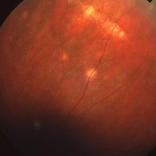
Both the right and left Eye have fairly symmetrical, extrafoveal drusenoid-like flecks and focal and faint areas of RPE hyperplasia (in addition to mild NPDR and PPA) 57 yo WF referred for AMD vs Pattern Dystrophy that was diagnosed 10 years ago. Reported some slow progressive vision loss in both eyes for distance and near. Denies nyctalopia or hemeralopia. Background medical history includes HTN, CVD, and DM. No family history of eye problems. Denied pentosan use. Anterior segment showed moderate cataracts (OD>OS). Posterior segment exam showed macular changes and mild NPDR. The macular appearance showed a symmetrical, paramacular ring of fleck-like drusenoid material with some faint focal areas of RPE hyperplasia. Fundus Photos, AF, OCT were performed as well as a gene test. Further questioning showed revealed that her mother and maternal grandmother had both diabetes mellitus and sensorineural hearing loss. The patient developed diabetes in her teens, and some high frequency hearing loss in her early twenties. She had not had a previous genetic test or diagnosis of MIDD. Gene testing is pending for the mitochondrial component. Invitae's retinal panel, which does not include mitochondrial disorders, only showed a variant of uncertain significance, HMCN1. I discussed this case with Dr. Freund, and it is similar to a the case report : Inoue M, Kiss S, Freund KB. MACULAR PIGMENT RINGS AS THE PRESENTING FINDING OF MITOCHONDRIAL MYOPATHY, ENCEPHALOPATHY, LACTIC ACIDOSIS, AND STROKELIKE EPISODES. Retin Cases Brief Rep. 2015 Fall;9(4):260-4. doi: 10.1097/ICB.0000000000000182. PMID: 26200388.
Photographer: Grace Melton and Carley Gunn
Imaging device: Clarus
Condition/keywords: Macular Dystrophy, Maternally Inherited Diabetes and Deafness, MIDD, Mitochondrial Disorder
-
 Acute Retinal Necrosis
Mar 26 2019 by Gary R. Cook, MD, FACS
Middle-aged white female with ARN OS showing additional involvement within 2 weeks. Patient was seen prior to the availability of anti-viral therapies.
Imaging device: Topcon VT-50
Condition/keywords: acute retinal necrosis
-
 Acute Retinal Necrosis
Mar 26 2019 by Gary R. Cook, MD, FACS
Middle-aged white female with peripheral retinal lesions of acute retinal necrosis OS at presentation.
Imaging device: Topcon VT-50
Condition/keywords: acute retinal necrosis
-
 Before and After Vitrectomy
Nov 17 2023 by Bradley T. Smith, MD, FASRS
Middle age male diabetic retinopathy and resolving exudate following repair of tractional detachment with membrane peeling.
Condition/keywords: coats-like response, Diabetes, fibrotic neovascularization, fibrovascular proliferation, pars plana vitrectomy (PPV), proliferative diabetic retinopathy (PDR), tractional retinal detachment
-
---thumb.JPG/image-square;max$300,300.ImageHandler) Best's Disease
Best's Disease
Jul 12 2013 by Jason S. Calhoun
Middle-aged male complained of blurry vision or spot in vision in the right eye. Fluorescence angiogram shows hypofluorescent staining in the mid and late phases. Notice the egg-yolk shape on the macula in the fundus photo.
Photographer: Jason S. Calhoun, Department of Ophthalmology, Mayo Clinic Jacksonville, Florida
Condition/keywords: Best disease
-
 Bilateral RD and Final Myopic Maculopahty Stage
Aug 11 2018 by Matias Iglicki, MD
Middle age male with myopic macular degeneration. This patient have bilateral vitrectomy plus sclera buckle due to a bilateral RD, here you can see the post op. Retinas have been re attached right eye with silicon oil tamponade, left eye c3f8 tamponade plus LASER and buckle indentation.
Photographer: matias iglicki MD certifed Teacher of Ophthalmology. University of Buenos Aires
Imaging device: DAYTONA
Condition/keywords: myopic degeneration, post-op
-
 Central Serous Chorioretinopathy (CSC)
Oct 16 2012 by S. Natarajan, MD, FASRS, FRCS (GLASGOW) , FICO, D.Sc, FELA
Middle-aged male came with small PED 4 months back; now this has progressed to a larger PED with SRF underneath the fovea.
Photographer: Prof. Dr. S. Natarajan
Condition/keywords: central serous chorioretinopathy (CSCR), central serous retinopathy (CSR), pigment epithelial detachment (PED), subretinal fibrosis
-
 DME With Outer Hole
Jul 1 2014 by John S. King, MD
Middle age African American female with C-DME. Mild-mod central leakage on FA.
Photographer: Wayne A Ladlee Jr
Imaging device: FA
Condition/keywords: diabetic macular edema
-
 DME With Outer Hole
Jul 1 2014 by John S. King, MD
Middle age African American female with C-DME. Mild-mod central leakage on FA.
Photographer: Wayne A Ladlee Jr
Imaging device: FA
Condition/keywords: diabetic macular edema
-
 Dominant Drusen
Mar 26 2019 by Gary R. Cook, MD, FACS
Middle-aged white male with a wedge-shaped pattern of drusen deposits in temporal macula; Dominant drusen; VA = 20/20.
Condition/keywords: hereditary drusen
-
 Dominant Drusen
Mar 26 2019 by Gary R. Cook, MD, FACS
Middle-aged white male with a similar wedge-shaped pattern of drusen deposits in temporal macula OS; dominant drusen; VA = 20/25.
Condition/keywords: hereditary drusen
-
 Full Thickness Macular Hole With ERM
Feb 26 2014 by Sharon Fekrat, MD FACS FASRS
Middle aged woman with a full thickness macular hole in the left eye associated with an epiretinal membrane.
Photographer: Michael P Kelly, Ophthalmic Photographer, Duke Eye Imaging, Duke Eye Center
Condition/keywords: epiretinal membrane (ERM), macular hole
-
 Giant Retinal Tear With RD
Jun 29 2013 by Jason S. Calhoun
Middle aged patient comes in with sudden loss of vision inferior, nasally. Patient presents plus 2 APD in the right eye. Fundus exam reveals a giant retinal tear at 10-11 o'clock with RD. Vitrectomy with laser and gas exchange of C3F8 scheduled.
Photographer: Jason S. Calhoun, Mayo Clinic Jacksonville, Florida
Imaging device: TOPCON TRC 50-EX
Condition/keywords: retinal tear
-
 Linear Histoplasmosis
Mar 27 2019 by Gary R. Cook, MD, FACS
Middle-aged white male with presumed ocular histoplasmosis (POHS) demonstration single astrophic histo spots and a linear histo streak in the superior periphery.
Imaging device: Topcon VT-50
Condition/keywords: atrophic spot, ocular histoplasmosis syndrome (OHS), presumed ocular histoplasmosis syndrome (POHS)
-
---thumb.jpg/image-square;max$300,300.ImageHandler) Pattern Dystrophy
Pattern Dystrophy
Aug 7 2013 by From the Collections of Thomas M. Aaberg, MD and Thomas M. Aaberg Jr., MD
Middle aged patient with yellow macular pigment pattern.
Condition/keywords: butterfly dystrophy, pattern macular dystrophy
-
 PCV
PCV
Jul 1 2014 by John S. King, MD
Middle age Mediterranean female with acute decrease in vision mainly characterized as paracentral scotoma. Suboptimal response to short course of anti-VEGF; responded well to PDT. This is the initial scan via the polypoidal PED inf-nasal.
Photographer: Wayne A Ladlee Jr
Imaging device: Cirrus
Condition/keywords: polypoidal choroidal vasculopathy (PCV)
 Loading…