Search results (46 results)
-
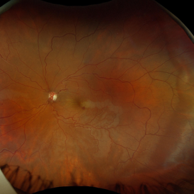
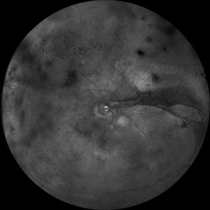
 Ocular Ischemic Syndrome/ Severe NPDR
Oct 6 2021 by Becca Harris
53 year old female with Severe NPDR and Ocular Ischemic Syndrome.
Photographer: Becca Harris
Imaging device: Optos California
Condition/keywords: Diabetic Retinopathy, left eye, nonproliferative diabetic retinopathy, ocular ischemic syndrome, optos, retinal ischemia
-
 Central Retinal Artery Occlusion With Cilioretinal Sparing
Apr 4 2018 by Soumya Venkatesh
Fundus photograph of a 23-year-old gentleman presenting with sudden loss of vision 2 days prior to presentation. He underwent all relevant investigations and found to have APLA positive. He also had dengue serology positive. On follow up, his retinal edema reduced unmasking the underlying hemorrhages( flame shaped).
Photographer: Soumya Harapanahalli Venkatesh, JSS university, Karnataka, India
Condition/keywords: central retinal artery occlusion (CRAO), cherry red spot, cilioretinal sparing, retinal ischemia
-
 Acute Idiopathic Occlusive Retinal Vasculitis
May 31 2014 by Hamid Ahmadieh, MD
Color fundus photograph of the right eye of a 28-year-old woman with sudden drop of vision due to acute occlusive retinal vasculitis leading to extensive nerve fiber layer infarction and retinal hemorrhages.
Photographer: Naghmeh Nozhat, Negah Eye Center, Tehran
Condition/keywords: color fundus photograph, cotton wool spots, retinal hemorrhage, retinal ischemia
-
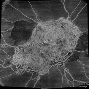
 Venous Beading
Apr 30 2021 by Shivani Reddy, MD
This is a fluorescein angiogram image capturing a beautiful example of different stages of venous beading in diabetic retinopathy all in one frame. This patient also has various microangiopathic findings including microaneurysms, venous loops and capillary dropout. This patient is a 41 y/o male with a history of type 1 diabetes, presenting for his first eye exam in years.
Imaging device: Optos FA
Condition/keywords: capillary dropouts, nonproliferative diabetic retinopathy, proliferative diabetic retinopathy (PDR), retinal ischemia, venous beading
-
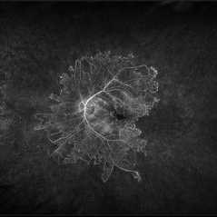
 Who Stole My Blood Supply?
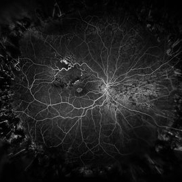
Jan 25 2025 by Muna Bhende, MD
Superficial capillary plexus slab montage image of a young female diabetic with florid proliferation . There is no flow in the capillaries anterior to the tangle of new vessels indicating severe retinal ischemia.
Photographer: Mohanapriya L , Medical Research Foundation, Sankara Nethralaya, Chennai, India
Imaging device: PLEX elite 9000
Condition/keywords: florid type PDR, OCTA
-
 Acute Idiopathic Occlusive Retinal Vasculitis
May 31 2014 by Hamid Ahmadieh, MD
Color fundus photograph of the left eye of a 28-year-old woman with acute drop of vision due to occlusive retinal vasculitis leading to extensive nerve fiber layer infarction and retinal hemorrhages.
Photographer: Naghmeh Nozhat, Negah Eye Center, Tehran
Condition/keywords: color fundus photograph, cotton wool spots, retinal hemorrhage, retinal ischemia
-
 Branch Retinal Vein Occlusion
Dec 9 2020 by Olivia Rainey
Ultra-widefield angiogram of a 78-year-old male with a branch retinal vein occlusion affecting his right eye. The patient was diagnosed on 12/17/12 at another practice. The physician noted that there wasn't NVE noted, however areas of micoaneurysmal dilation is present. She noted retinal ischemia secondary to BRVO. 12/8/20 leakage on FA noted to be worsening compared to his previous angiography. She has concern for progressing NVE and recommends sector PRP after injection of antiVEGF series of 3 for the health of the eye.
Photographer: Olivia Rainey, OCT-C, COA
Imaging device: Optos California
Condition/keywords: branch retinal vein occlusion (BRVO), macular branch retinal vein occlusion (BRVO), non-perfusion, scleral buckle, vitreoretinal surgery
-
 Central Retinal Artery Occlusion with Cilioretinal Sparing
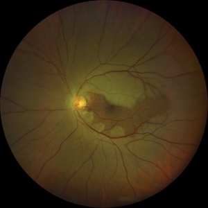
Oct 28 2020 by Fang Helen Mi
Wide-field Clarus photography showing diffuse retinal ischemia and edema, with sparing of the cilioretinal artery region.
Condition/keywords: central retinal artery occlusion (CRAO), cilioretinal sparing
-
 Central Retinal Vein Occlusion with Retinal Neovascularization
Jan 19 2022 by Olivia Rainey
Ultra-widefield fluorescein angiogram of a 56-year-old male with a Central Retinal Vein Occlusion with Retinal Neovascularization affecting his left eye. The patient presented on 1/19/2022 with scNLP vision in the left eye. The patient has good PRP, however areas of ischemia still remain untreated by laser. He also has severe neovascular glaucoma contributing to his poor vision.
Photographer: Olivia Rainey, OCT-C, COA
Imaging device: Optos California
Condition/keywords: central retinal vein occlusion (CRVO), FA early phase, fluorescein angiogram (FA), hemorrhage, ischemic CRVO, left eye, neovascular glaucoma, Optos, pan-retinal photocoagulation (PRP), retinal ischemia, retinal neovascularization, ultra-wide field imaging
-
 Central Retinal Vein Occlusion with Severe Retinal Ischemia
Jan 19 2022 by Olivia Rainey
Ultra-widefield fluorescein angiogram of a 56-year-old male with a Central Retinal Vein Occlusion with Severe Retinal Ischemia affecting his right eye. The patient presented on 1/19/2022, sc20/20-2 vision in the right eye. The patient has had a good response to Eylea with complete resolution of edema. The physician is considering PRP to ischemic periphery in the future and given the degree of ischemia in both eyes, she recommends that the patient's PCP check carotid Doppler US.
Photographer: Olivia Rainey, OCT-C, COA
Imaging device: Optos California
Condition/keywords: central retinal vein occlusion (CRVO), FA late phase, fluorescein angiogram (FA), ischemic CRVO, Optos, retinal ischemia, ultra-wide field imaging
-
 Patchy Ischemic Whitening in Sturge Weber Syndrome
Nov 18 2024 by Edward F. Hall, MD, FASRS
Left fundus photograph of a 44-year-old man showing patchy ischemic retinal whitening associated with Sturge-Weber Syndrome. The precise cause of this rare complication remains unclear, but it may be linked to choroidal vascular congestion and a compartment syndrome-like effect on the local retinal arteriolar circulation. OCT imaging confirmed inner retinal ischemia and thickening
Photographer: Karissa Kuhl
Imaging device: Optos
Condition/keywords: retinal ischemia, Sturge Weber Syndrome
-
 Multiple Myeloma with Cytomegalovirus Retinitis
Apr 5 2018 by Kim Barrett
Ultra-wide field fluorescein angiogram of a 77-year-old male with multiple myeloma. Patient's angiogram presented significant peripheral retinal ischemia and cystoid macular edema. Patient tested positive for polymerase chain reaction, confirming cytomegalovirus retinitis. Patient is being treated with intravitreal ganciclovir and his current vision is 20/200.
Photographer: Kim Barrett, COA
Imaging device: Optos
Condition/keywords: cystoid macular edema (CME), fluorescein angiogram (FA), fluorescein leakage, intravitreal ganciclovir, myeloma, peripheral ischemia, positive polymerase chain reaction (PCR), ultra-wide field imaging
-
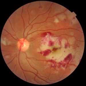
 Retinal Ischemia, Edema, and Hemorrhages on the Infero-Temporal Macula
Aug 26 2019 by Narciso F. Atienza, MD, MBA, FASRS, FPCS, FPAO.
47-year-old female who came in with blurring of vision of the right eye of 2 weeks duration. She is hypertensive with poor control, taking Amlodipine irregularly. Denies any cardiac problem non-diabetic. Vision upon presentation was 20/400 (OD), 20/20 (OS) colored fundus photo of the right eye showing areas of retinal ischemia, edema and hemorrhages on the infero-temporal macula extending to the arcade.
Photographer: Narciso F Atienza, Jr. MD, MBA
Imaging device: Topcon TRC
Condition/keywords: edema, hemorrhage, inferotemporal arcade, retinal ischemia
-
 Wyburn Mason Racemose Angiomatosis
May 22 2016 by Olivia Rainey
Color fundus montage of an 13-year-old female with arteriovenous malformation (Wyburn Mason Racemose Angiomatosis) affecting her right eye. The retinal arteriovenous malformation appears to be stable. She presented with NLP in the eye, strabismus, and peripheral retinal ischemia. She is at risk for neovascular complications; however, she is currently being treated with Sirolimus. Since she is on this systemically, there is no need to perform intraocular anti-VEGF injections or PRP laser. She also presented with optic atrophy affecting her left eye, secondary to chiasmal involvement of arteriovenous malformation. She has had a potential progressive visual field loss involving the temporal aspect of her visual field from the left eye. There is sector optic atrophy. Presumably, this is due to a compressive effect of her arteriovenous malformation on the nasal nerve fiber layer (corresponding to the temporal visual field) crossing to the right occipital cortex at the chiasm.
Photographer: Olivia Rainey
Imaging device: Topcon 50dx
Condition/keywords: arteriovenous malformation, color fundus photograph, color photo, montage, peripheral ischemia, Sirolimus
-
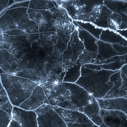
 Marked Retinal Ischemia in Patient with Mixed Connective Tissue Disease
Feb 26 2013 by Sharon Fekrat, MD FACS FASRS
Fluorescein angiogram of the right eye of a 27-year-old female with mixed connective tissue disease and marked retinal ischemia. Panretinal laser photocoagulation (PRP) has been performed for neovascularization elsewhere (NVE).
Condition/keywords: mixed connective tissue disease, retinal ischemia
-
---thumb.jpg/image-square;max$300,300.ImageHandler) BRVO Late FA 102 Degree ART
BRVO Late FA 102 Degree ART
Apr 18 2014 by Susanna S. Park, MD, PhD
Wide angle fluorescein angiography late transit view taken of the left eye of a 45-year-old woman with branch retinal vein occlusion showing peripheral retinal ischemia with staining and leakage of the affected retinal veins.
Photographer: Karishma Chandra, UC Davis Eye Center
Condition/keywords: branch retinal vein occlusion (BRVO), fluorescein leakage, wide angle imaging
-
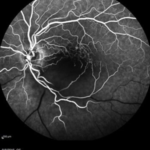 Central Retinal Artery Occlusion
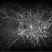
May 16 2017 by Olivia Rainey
Fluorescein angiogram of an 66-year-old female with a central retinal artery occlusion affecting her left eye.
Photographer: Olivia Rainey
Imaging device: Heidelberg Spectralis
Condition/keywords: 50 degrees, central retinal artery occlusion (CRAO), fluorescein angiogram (FA), left eye, mid phase, retinal ischemia
-
 Central Retinal Vein Occlusion
Sep 2 2012 by Hyung-Woo Kwak, MD
Multiple dense, dark, blotchy hemorrhages, cotton-wool spots, and pale optic disc are signs suggestive of retinal ischemia in CRVO.
Imaging device: Zeiss F450 plus
Condition/keywords: central retinal vein occlusion (CRVO)
-
 Diabetic Retinopathy
Oct 2 2013 by Jerald A. Bovino, MD
This patient with long standing diabetes has peripheral non-perfusion.
Condition/keywords: retinal ischemia
-
 Familial Exudative Vitreo-retinopathy
Jul 6 2021 by Akansha Sharma
Color photo montage of 21-year-old male with familial exudative vitreoretinopathy in an amblyopic eye.
Photographer: Dr. Akansha Sharma-Retina Foundation, Ahmedabad
Condition/keywords: familial exudative vitreoretinopathy (FEVR), retinal ischemia
-
 Familial Exudative Vitreo-retinopathy
Jul 6 2021 by Akansha Sharma
Infra-red capture of 5-year-old male with familial exudative vitro-retinopathy with disc pallor.
Photographer: Dr. Akansha Sharma-Retina Foundation, Ahmedabad
Condition/keywords: familial exudative vitreoretinopathy (FEVR), retinal ischemia
-
 Foveal Thinning Post Blunt Trauma
Aug 25 2018 by Dhaivat Shah
28-year-old male. Post blunt trauma with tennis ball. Fundus color photo shows large area of retinal thinning. Multi color image shows dull red color over fovea, depicting thinning. SD-OCT shows inner retinal ischemia and foveal thinning with early macular hole formation.
Imaging device: Spectralis
Condition/keywords: blunt trauma
-
 Hypertensive Retinopathy
Aug 24 2012 by Geoffrey G. Emerson, MD, PhD, FASRS
A 35-year-old man has headaches and decreased vision. The right eye measures 20/25 and the left eye measures 3/200. The blood pressure measures 180/110. This fluorescein angiogram shows dilated capillaries and capillary dropout in the central macula of the left eye.
Photographer: Geoffrey Emerson, MD, PhD, Retina Center, Minneapolis
Condition/keywords: hypertensive retinopathy, papilledema, retinal ischemia
-
 Ischemic Proliferative Diabetic Retinopathy
Aug 22 2012 by Edwin H. Ryan, MD
Fundus photograph of 35-year-old poorly-controlled diabetic woman, 6/200 vision.
Photographer: Edwin Ryan Jr. MD, VitreoRetinal Surgery, PA
Condition/keywords: retinal ischemia
-
 Ischemic Proliferative Diabetic retinopathy
Aug 22 2012 by Edwin H. Ryan, MD
Fluorescein angiogram of 35-year-old poorly-controlled diabetic woman, 6/200 vision.
Photographer: Edwin Ryan Jr. MD, VitreoRetinal Surgery, PA
Condition/keywords: retinal ischemia
 Loading…